In Hwa A. Lim und Charles A. Cantor, editors, Proc. 3. Int.
Conf. on
Bioinformatics and Genome Research, page 445,
(World Scientific, 1994).
On the Hypercube Structure of
the Genetic Code
Abstract
A representation of the genetic code as a six–dimensional Boolean hypercube is proposed. It is assumed here that this structure is the result of the hierarchical order of the interaction energies of the bases in codon–anticodon recognition. The proposed structure demonstrates that in the genetic code there is a balance between conservatism and innovation. Comparing aligned positions in homologous protein sequences two different behaviors are found: a)There are sites in which the different amino acids present may be explained by one or two “attractor nodes” (coding for the dominating amino acid(s)) and their one–bit neighbors in the codon hypercube, and b) There are sites in which the amino acids present correspond to codons located in closed paths in the hypercube. The structure of the code facilitates evolution: the variation found at the variable positions of proteins do not corresponds to random jumps at the codon level, but to well defined regions of the hypercube.
a Departamento de Física y Matemáticas, Universidad de las Américas, Puebla, Sta. Catarina Mártir, 72820 Cholula, Puebla, México
b Dirección General de Investigaciones, Universidad Veracruzana, Xalapa, Ver. 91000, México
c Humboldt–Universität zu Berlin, Institut für Theoretische Physik, Invalidenstraße 110, D–10099 Berlin, Germany
d Laboratoire de Physique et Mechanique des Milieux Heterogenes, Ecole Superieure de Physique et de Chimie Industrielles, 10 rue Vauquelin, F–75231 Paris Cedex 05, France
1 Introduction
The genetic code is the biochemical system for gene expression. It deals with the translation, or decoding, of information contained in the primary structure of DNA and RNA molecules into protein sequences. Therefore, the genetic code is both a physico–chemical and a communication system. Physically molecular recognition depends on the degree of complementarity between the interacting molecular surfaces (by means of weak interactions); informationally, a prerequisite to define a code is the concept of distinguishability. It is the physical indistinguishability of some codon–anticodon interaction energies that makes the codons synonymous, and the code degenerate and redundant [1].
In natural languages [2] as well as in the genetic code, the total redundancy is due to a hierarchy of constraints acting one upon another. The specific way in which the code departs from randomness is, by definition, its structure. It is assumed here that this structure is the result of the hierarchical order of the interaction energies of the bases in codon–anticodon recognition. As we shall see, it may be represented by a six–dimensional boolean hypercube in which the codons (actually the code–words; see below) occupy the vertices (nodes) in such a way that all kinship neighborhoods are correctly represented. This approach is a particular application to binary sequences of length six of the general concept of sequence–space, first introduced in coding theory by Hamming [3].
A code–word is next to six nodes representing codons differing in a single property. Thus the hypercube simultaneously represents the whole set of codons and keeps track of which codons are one–bit neighbors of each other. Different hyperplanes correspond to the four stages of the evolution of the code according to the Co–evolution Theory [4, 5, 6]. Hops within three of the “columns” (four–dimensional cubes), consisting of the codon classes NGN, NAN, NCN, and NUN, lead to silent and conservative amino acid substitutions, while hops in the same hyperplane (four–dimensional subspace belonging to any of the codon classes ANN, CNN, GNN or UNN) lead to non–conservative substitutions, frequently found in proteins. The proposed structure demonstrates that in the genetic code there is a good balance between conservatism and innovation. To illustrate the results several examples of the non–conservative variable positions of homologous proteins are discussed. Two different behaviors are found: a) There are sites in which the different amino acids present may be explained by one or two “attractor nodes” (coding for the dominating amino acid(s)) and their one–bit neighbors in the codon hypercube, and b) There are sites in which the amino acids present correspond to codons located in closed paths in the hypercube.
2 Codon–Anticodon Interaction
In his early paper Eigen [7] recognized that the optimization between stability and rate, that is always found for enzyme–substrate interactions, also applies to the codon–anticodon interaction. However, he attributes the codon s size to mechanistic coincidences: “codons with less than three bases would be very unstable (at least for A and U). Codons with more than three bases, especially for G and C, become too ‘sticky’ ”. This is certainly not a coincidence, but a requirement for the system to function as an efficient communication device. Three bases are needed for effectively binding the adapter to the messanger. Thus, the codon s size determines the range of codon–anticodon overall interaction strength within which recognition can occur444Interestingly enough, this feature of genetic communication system has, its counterpart in human communication. In a series of experiments on reading lists of words, performed by J.E. Karlin and J.R. Pierce (Pierce J.R., An Introduction to Information Theory, Dover Publications,Inv. N.Y. 1961), in which the subject “transmits” the information translating it into the new form, speech rather than print, by reading the list aloud, they concluded that: “It seems fairly clear that reading speed is limited by word recognition not by word utterance” (underlined in the original). See also [8]. Genetic translation rate is limited, among other things, by codon–anticodon recognition which in turn depends on base–pair lifetimes in a given structural situation. These life–times are influenced by the nature of the pairs: they are shorter for AT than for GC pairs [9].
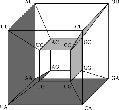
The four bases occurring in DNA (RNA) macromolecules define the corresponding alphabet X: {A, C, G, T} or {A, C, G, U}. Each base is completely specified by two independent dichotomic categorizations (Fig. 1):

(i) according to chemical type C : {R, Y}, where R: (A, G) are purines and Y:(C, U) are pyrimidines, and (ii) according to H–bonding, H : {W, S}, where W:(A, U) are weak and S:(C, G) strong bases. The third possible partition into imino/keto bases is not independent from the former ones.
Denoting by the chemical type and by the –bond category of the base , at position of a codon, our basic assumption says that the codon–anticodon interaction energy obeys the following hierarchical order:
This means, that the most important characteristic determining the codon–anticodon interaction is the chemical type of the base in the second position. The next most important characteristic is whether there is a weak or strong base in this position, then the chemical type of the first base and so on.
The bases are represented by the nodes of a 2–cube (Fig. 1). The first attribute is the chemical character and the second the hydrogen–bond character. Extending this association to base triplets, each codon is in a unique way associated with a codeword consisting of six attribute values (see Table 1).
0 0 0 0 1 1 A A C N 0 0 0 0 1 0 A A U N 0 0 0 0 0 0 A A A K 0 0 0 0 0 1 A A G K 1 0 0 0 0 1 U A G t 1 0 0 0 0 0 U A A t 1 0 0 0 1 0 U A U Y 1 0 0 0 1 1 U A C Y 1 1 0 0 1 1 C A C H 1 1 0 0 1 0 C A U H 1 1 0 0 0 0 C A A Q 1 1 0 0 0 1 C A G Q 0 1 0 0 0 1 G A G E 0 1 0 0 0 0 G A A E 0 1 0 0 1 0 G A U D 0 1 0 0 1 1 G A C D 0 1 1 0 1 1 G U C V 0 1 1 0 1 0 G U U V 0 1 1 0 0 0 G U A V 0 1 1 0 0 1 G U G V 1 1 1 0 0 1 C U G L 1 1 1 0 0 0 C U A L 1 1 1 0 1 0 C U U L 1 1 1 0 1 1 C U C L 1 0 1 0 1 1 U U C F 1 0 1 0 1 0 U U U F 1 0 1 0 0 0 U U A L 1 0 1 0 0 1 U U C L 0 0 1 0 0 1 A U G M 0 0 1 0 0 0 A U A I 0 0 1 0 1 0 A U U I 0 0 1 0 1 1 A U C I 0 0 1 1 1 1 A C C T 0 0 1 1 1 0 A C U T 0 0 1 1 0 0 A C A T 0 0 1 1 0 1 A C G T 1 0 1 1 0 1 U C G S 1 0 1 1 0 0 U C A S 1 0 1 1 1 0 U C U S 1 0 1 1 1 1 U C C S 1 1 1 1 1 1 C C C P 1 1 1 1 1 0 C C U P 1 1 1 1 0 0 C C A P 1 1 1 1 0 1 C C G P 0 1 1 1 0 1 G C G A 0 1 1 1 0 0 G C A A 0 1 1 1 1 0 G C U A 0 1 1 1 1 1 G C C A 0 1 0 1 1 1 G G C G 0 1 0 1 1 0 G G U G 0 1 0 1 0 0 G G A G 0 1 0 1 0 1 G G G G 1 1 0 1 0 1 C G G R 1 1 0 1 0 0 C G A R 1 1 0 1 1 0 C G U R 1 1 0 1 1 1 C G C R 1 0 0 1 1 1 U G C C 1 0 0 1 1 0 U G U C 1 0 0 1 0 0 U G A t 1 0 0 1 0 1 U G C W 0 0 0 1 0 1 A G G R 0 0 0 1 0 0 A G A R 0 0 0 1 1 0 A G U S 0 0 0 1 1 1 A G C S
In some of the hypercube directions single feature codon changes (one–bit code–word changes) produce synonymous or conservative amino acid substitutions in the corresponding protein (when the hops occur in three of the 4–cubes displayed as “columns” in Figs. 2 and 3) while in other directions lead to context dependent replacements which in general conserve only certain physical properties. However, if these properties are the only relevant ones in the given context, the substitution has little effect on the protein structure as well. These low–constraint sites facilitate evolution because they allow the transit between hypercube columns belonging to amino acids with very different physico–chemical properties (e.g. hydrophobic and hydrophilic amino acids, respectively).
3 Gray Code Structure of the Genetic Code
An –dimensional hypercube, denoted by , consists of nodes, each addressed by a unique –bit identification number. A link exists between two nodes of if and only if their node addresses differ in exactly one bit position. A link is said to be along dimension if it connects two nodes whose addresses differ to as the th bit (where the least significant bit is referred to as the 0th bit). is illustrated in Fig. 3. Two nodes in a hypercube are said to be adjacent if there is a link present between them. The (Hamming) distance between any two cube nodes is the number of bits differing in their addresses. The number of hops needed to reach a node from another node equals the distance between the two nodes. A –dimensional subcube in involves nodes whose addresses belong to a sequence of n symbols {0, 1, *} in which exactly of them are of the symbol “*” (i.e. they don t care symbol whose value can be 0 or 1).


The idea to propose a Gray Code representation of the Genetic Code goes back to Swanson [10] where this concept is explained in detail (see also [11]). A great number of different Gray Codes can be associated to the Genetic Code, depending of the order of importance of the bits in a code–word. In Table 1 our chosen Gray Code is displayed. It is constructed according to our main hypothesis
For example, the first two lines of the table differ in the last bit, corresponding to which is the least significant bit; the second and the third lines differ in the next least significant bit, i.e. , and so forth.
4 The Structure of Codon Doublets
This section is more mathematical than the rest of the paper, therefore it is suggested to non–mathematical readers to skip the details. This will not be an obstacle for the understanding of the rest of the paper.
In a pioneering paper Danckwerts and Neubert [12] discussed the symmetries of the sixteen codon doublets in terms of the Klein–4 group of base transformations. Here their result will be recast in a form of a decision–tree (Fig. 4), and their analysis will be extended to the doublets.

They found the following structure for the set of doublets:
Starting from AC generate the set:
The sets and consist of four–fold and less than four–fold degenerate doublets, respectively.
They showed that: “ and are invariant by operating with on , but no operation on leaves or invariant. Thus carries more information555see also [8] than and is therefore more important for the stability of and than . A change of with respect to its hydrogen bond property does not change the resulting amino acids if all doublets of either or are affected.
Reversing supposition and conclusion, and may be defined as those doublet sets of 8 elements which are invariant under the –transformation. Then experience shows that and are fourfold and less than fourfold degenerate respectively.”
Thus, the third base degeneracy of a codon does not depend on the exact base , but only on its –bond property (weak or strong).
The above results can be simply visualized as a decision–tree (Fig. 4). As can be seen from this figure, the redundancy of a codon is determined only by the H–bond character of and : SSN codons (with 6 H–bonds in ) belong to while WWN codons (with 4 H–bonds in ) belong to . However, for codons WSN and SWN (with 5 H–bonds in ) it is not possible to decide unless one has more information about the second base: WCN and SUN belong to while WGN and SAN belong to . In all cases at most three attributes are necessary to determine the redundancy of a codon up to this point, of course, the non–degenerate codons (UAG for Methionine and UGG for Thryptophan) will require the specification of the six attributes.
From the decision rules obtained from Fig. 4 it is clear that there are branches where the refinement procedure cannot continue (the branches which end in ) because no matter which base occupies the third codon position the degeneracy cannot be lifted. This imposes a limit to the maximum number of amino acids which can be incorporated to the code without recurring to a “frozen accident” hypothesis. Our proposal generalizes the “2–out–of–3” hypothesis of Lagerkvist [8], which refers only to codons in the SSN class.
The sixteen doublets can be represented as the vertices of a four–dimensional hypercube (Fig. 5). As can be seen from this figure, the sets and are located in compact regions. Notice that this figure differs from the one introduced by Bertman and Jungck [13], which considered as basic transformations and instead of and as we did. Since the operator changes two bits we do not consider it as basic.
Let s consider now the structure of the set of doublets. Exactly as before, define the following sets:
where consists of the doublets ending in a strong base (NS), and of the doublets ending in a weak base (NW). Then
Notice that the operator acting on has the same form as the operator acting on above, except that b acts as the third base instead of the first.
The sets and are invariant under the (1, b)–transformations. Then experience shows that the 32 codons in the class , with in or constitute a complete code, codifying for the 20 amino acids and terminator signal (stop–codon) if allowance is made for deviating codon–assignments found in Mitochondria [14]. For the codons in this is true in the universal code; for codons in AUA should codify for M instead of I and UGA for W instead of stop signal. Both changes have been observed in Mitochodria. This more symmetric code has been considered more similar to an archetypal code than the universal code [14]. Only after the last attribute was introduced the universal code was obtained with the split of AUR into AUA (I) and AUG (M); and UGR into UGG (W) and UGA (t). It has been speculated that primordial genes could be included in a 0.55–kb open reading frame [15]. The same authors calculated that with two stop codons this open reading frames would have appeared too frequently. From the present view the assignment of UGA to a stop codon was a late event that optimized this frequency (this interpretation differs from the one proposed in [15] and [16] which assume a primordial code with three stop codons). Other deviations of the universal code most likely also occurred in the last stages of the code s evolution.
In the same way as before the sixteen doublets can be represented as the vertices of a four–dimensional hypercube (fig. 6). The sets and are also located in compact regions. Codons with in are frequently used in eukaryots. In contrary, codons with in are frequently used in prokaryots. The described structure of the code allows a modulation of the codon–anticodon interaction energy [17].

5 Results
Besides the results mentioned in the last section which refer to codon doublets, to further illustrate the significance of proposed approach we are going to consider several examples. In the first example (Fig. 7) we discuss the alignment studied by the method of hierarchical analysis of residue conservation of Livingstone and Barton (Fig. 2 of [18]). In position 11 appear the following amino acids: R, W, H, G, D, which according to their approach have no properties in common. In Fig 2 this cluster of amino acids is shown. By looking at the Atlas of amino acid properties [19] we see that, from the properties proposed by Grantham [20] (composition, polarity and volume), apparently the only requirement for the amino acids at this site is to maintain a certain degree of polarity. From this observation we may conclude that most probably it is an external site. Simply by looking at such a diverse set of amino acids one can hardly realize that they have clustered codons. This clustering facilitates the occurrence of mutations that in the course of evolution were fixed, in view of the low physico–chemical requirements at the site.

As a second example (Fig. 3) we consider site 33 of the alignment of 67 SH2 domains, Fig 6 of [9]. We can see from Fig. 3 that the cluster around the codon CAC (H) explains, by one–bit changes, the amino acids R, Q, L, H, D. Furthermore, a second cluster around the codon AGC (S) explains the amino acids R, N, S, T. Finally, a silent change from AGC (S) to UCC (S) accounts for the minor appearance of the small neutral amino acids A, T, P. In a similar way, the variation of the hypervariable region of immunoglobulin kappa light FR1 at position 18 can be explained (Fig. 7. Finally, by looking to the residue frequencies in 226 globins displayed in Table 3 of the paper by Bashford et al. [21] it is seen that there are variable positions in which one or two residues predominately occur and the rest are only marginally represented, and others in which the frequencies are more evenly distributed among the amino acids present. As it can be easily shown, the first class of positions may be associated at the codon level with one (or two) attractor node(s) and its one–bit neighbors, and the second one with closed trajectories in the hypercube. The corresponding figures are not included because of lack of space.
6 Concluding Remarks
The present approach goes beyond the usual analyses in terms of single base changes because it takes into account the two characters of each base and therefore it represents one–bit changes. Besides, the base position within the codon is also considered. The fact that single bit mutations occur frequently is expected from probabilistic arguments. However, one could not expect, a priori, that a cluster of mutations would correspond at the amino acid level to a cluster of amino acids fixed by natural selection. We have found that this situation presents itself for many positions of homologous protein sequences of many different families (results not included). The structure of the code facilitates evolution: the variations found at the variable positions of proteins do not corresponds to random jumps at the codon level, but to well defined regions of the hypercube.
Acknowledgement
This work received economical support from: Proyecto CONACyT No. 1932–E9211. We thank professors Werner Ebeling and Michael Conrad for encouraging comments.
References
- [1] Crick, F. (1966). Codon–anticodon pairing: the wobble hypothesis. J. Mol. Biol., 19: 548–555.
- [2] Harris, Z. (1988). Language and Information. Columbia University press, New York.
- [3] Hamming, R. W. (1950). Bell Syst. Tech. J., 29: 147–160; See also: Ebeling W. and Feistel R. (1982), Physik der Selbstorganisation und Evolution. Akademie–Verlag, Berlin; Ebeling W., Feistel R. and Jiménez–Montaño, M. A. (1977); On the theory of stochastic replication and evolution of molecular sequences. Rostocker Physikalische Manuskripte, Heft 2:105–127.
- [4] Dillon, L. (1978). The genetic mechanism and the origin of life. Plenum Press, New York.
- [5] Wong, J. (1975). A co–evolution theory of the genetic code. Proc. Nat. Acad. Sci. USA, 72: 1909–1912.
- [6] Wong, J. (1976). The evolution of a universal genetic code. Proc. Nat. Acad. Sci. USA, 73: 2336–2340.
- [7] Eigen, M. (1971). Selforganization of matter and the evolution of biological macromolecules. Naturwiss., 58: 465–523.
- [8] Lagerkvist, U. (1978). “Two out of three”: an alternative method for codon reading. PNAS, USA, 75: 1759–1762. Also (1981) Unorthodox codon reading and the evolution of the genetic code. Cell 23: 305–306.
- [9] Guéron M., Charretier E., Kochoyan M. and Leroy J. L. (1990). Applications of imino proton exchange to nucleic acid kinetics and structures. in Frontiers of NMR in Molecular Biology, 225–238. Alan R. Liss, Inc.
- [10] Swanson, R. (1984). A unifying concept for the amino acid code. Bull. Math. Biol., 46 187–204.
- [11] Jiménez–Montaño,M.A.,(1994). On the syntactic structure and redundancy distribution of the genetic code. BioSystems, 32: 11–23.
- [12] Danckwerts H.J. and Neubert D. (1975). Symmetries of genetic code–doublets. J. Mol. Evol., 5: 327–332.
- [13] Bertman M. O.and Jungck J. R.(1979). Group graph of the genetic code. The J. of Heredity, 70: 379–384.
- [14] Jukes T. H. (1983). Evolution of the amino acid code. in: Evolution of genes and proteins. M. Nei and R. K. Koehn Eds. Sinauer Associates Inc. Sunderland, Mass 191–207.
- [15] Naora H, Miyahara K. and Curnow R. N. (1987). Origin of noncoding DNA sequences: molecular fossils of genome evolution. Proc. Natl. Acad. Sci. USA, 84: 6195–6199.
- [16] Brentani R. R. (1990). Complementary hydropathy and the evolution of interacting polypeptides. J. Mol. Evol., 31: 1239–243.
- [17] Grosjean H., Sankoff D., Min Jou W., Fiers W. and Cedergren R. J. (1978). Bacteriophage MS2 RNA: A correlation between the stability of the codon–anticodon interaction and the choice of code words. J. Mol. Evol., 12: 113–119; Distribution of the genetic code. BioSystems, 32: 11–23.
- [18] Livingstone C.D., and G. J. Barton (1993). Protein sequence alignments: a strategy for the hierarchical analysis of residue conservation. CABIOS, 9: 745–756.
- [19] Nakai K., Kidera A., and Kanehisa M. (1988). Cluster analysis of amino acid indices for prediction of protein structure and function. Prot. Eng., 2: 93–100.
- [20] Grantham R.(1974) Amino acid difference formula to help explain protein evolution. Science, 185: 862–864.
- [21] Bashford D., Chothia C. and Lesk A.M. (1987) Determinants of a protein fold. J. Mol. Biol., 196: 199–216.