Anomalous properties of heat diffusion in living tissue caused by branching artery network. Qualitative description
Abstract
We analyze the effect of blood flow through large arteries of peripheral circulation on heat transfer in living tissue. Blood flow in such arteries gives rise to fast heat propagation over large scales, which is described in terms of heat superdiffusion. The corresponding bioheat heat equation is derived. In particular, we show that under local strong heating of a small tissue domain the temperature distribution inside the surrounding tissue is affected substantially by heat superdiffusion.
I Introduction. Living tissue as a heterogeneous medium
Blood flowing through vessels forms paths of fast heat transport in living tissue and under typical conditions it is blood flow that governs heat propagation on scales about or greater than one centimeter (for an introduction to this problem see, e.g., CH80 ; SE70 ). Blood vessels make up a complex network being practically a fractal. The larger is a vessel, the faster is the blood motion in it and, so, the stronger is the effect of blood flow in the given vessel on heat transfer. Blood flow in capillaries practically does not affect heat propagation whereas blood inside large vessels moves so fast that its heat interaction with the surrounding cellular tissue is ignorable CH80 . Thus, there should be vessels of a certain length that are the smallest ones among the vessels wherein blood flow affects heat transfer remarkably. The value of can be estimated as BOOK (see also CH80 ; SE70 ):
| (1) |
where is the temperature diffusivity of the cellular tissue determined by its thermal conductivity , specific heat , and density , the value is the blood perfusion rate (the volume of blood going through tissue region of unit volume per unit time), and the factor is logarithm of the mean ratio of the individual length to radius of blood vessels forming peripheral circulation. For the vascular networks made up of the paired artery and vein trees where all the vessels are grouped into the pairs of the closely-spaced arteries and veins with opposite blood currents the coefficient accounts for the counter-current effect 111Initially the factor was phenomenologically introduced in the bioheat equation to take into account a certain renormalization of the blood perfusion rate caused by the counter-current effect C80 ; W87a ; W87b . Its theoretical estimate was later obtained independently by Weinbaum et al. WXZE97 and Gafiychuk & Lubashevsky BOOK (announced for the first time in we1 ).. For the vascular networks where the artery and vein trees are arranged independently of each other the factor should be set equal to unity, . In particular, for the typical values of the ratio –40 Mch89 , the thermal conductivity W/cmK, the heat capacity J/g K, and the density g/cm3 of the tissue as well as setting the blood perfusion rate min-1 from (1) we get the estimates mm and .
In the mean-field approximation the effect of blood flow on heat transfer is reduced to the renormalization of the temperature diffusivity, , WJ85 and the appearance of the effective heat sink CH80 ; BOOK ; WXZE97 in the bioheat equation:
| (2) |
Here is the tissue temperature field averaged over scales about , the parameter is the blood temperature insider the systemic circulation arteries, and the summand called below the temperature generation rate is specified by the heat generation rate as . The renormalization of the temperature diffusivity is mainly determined by the blood vessels of lengths about and due to the fractal structure of vascular networks the renormalization coefficient is practically a constant of unity order, BOOK .
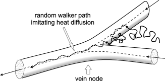
Let us imitate the temperature evolution in terms of random walks whose concentration is . Then the part of the vein tree made up of vessels whose lengths exceed or are about the scale forms the system of traps. In fact, blood streams going through the vein tree merge into greater streams at the nodes (Fig. 1). Therefore an effective random walker after reaching the boundary of one of these veins inevitably will be moved by blood flow into the internal points of large veins. Then, due to relatively fast blood motion inside these veins it will be carried away from the tissue region under consideration, which may be described in terms of the walker trapping or, what is the same, the heat sink BOOK . Since the mean distance between these veins is determined mainly by the shortest ones, i.e. by the veins of length the mean time during which a walker wanders inside the cellular tissue before being trapped is BOOK
| (3) |
In obtaining the given expression we have assumed the vascular network to be embedded uniformly in the cellular tissue, so the tissue volume falls per one vein (and artery, respectively) of length . Whence it follows, in particular, that the rate at which the walkers are being trapped by these vein, i.e. the rate of their disappearance is estimated as , leading together with expression (1) to the heat sink of intensity in the bioheat equation (2). The characteristic spatial scale of walker diffusion in the cellular tissue before being trapped is , i.e.
| (4) |
The scale gives us also the mean penetration depth of heat penetration into the cellular tissue from a point source or, what is the same, the widening of the temperature distribution caused by heat diffusion in the cellular tissue. It is the result obtained within the mean-field approximation.
Beyond the scope of the mean-field theory we meet several phenomena. One of them is the temperature nonuniformities caused by the vessel discreteness Bi86 which can be described assuming the heat sink in equation (2) to contain a random component BOOK . Another is fast heat transport over scales exceeding substantially the length caused by the effect of blood flow through the artery tree. The latter phenomena is the main subject of the present paper.
II Fast heat transport with blood flow through large artery tree
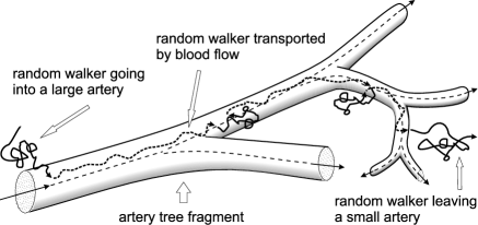
Let at a certain time a random walker wandering in the cellular tissue get a boundary of a large artery, i.e. an artery of length exceeding . It should be noted that such an event is of low probability and cannot be considered within the standard mean-field approximation because the relative number of large arteries is small. Due to the direction of the blood motion from larger arteries to smaller ones as well as the high blood flow rate in the large arteries the walker will be transported fast to one of the arteries of length (Fig. 2). The blood flow rate in small vessels is not high enough to affect the walker motion essentially and it has inevitably to leave this artery and wander in the cellular tissue until being trapped by the veins of length . Thereby a certain not too large number of random walkers generated, for example, inside a cellular tissue neighborhood of a point can be found during the time inside a cellular tissue neighborhood of a point at a distance much larger than , i.e. . The given effect may be regarded as anomalously fast heat diffusion in living tissue, i.e. heat superdiffusion.
Dealing with heat transfer in living tissue we may confine our consideration to the peripheral vascular networks typically embedded uniformly into the cellular tissue, at least at the first approximation M77 . The latter statement means, in particular, the fact that for a fixed peripheral vascular network the vessel collection comprising all the arteries of length meets the condition of the volume approximately falling per each one of these arteries. Therefore as is seen in Fig. 2 a walker going into a large artery of length at initial time during the time before being trapped by the veins can be found equiprobably at each point of the given artery neighborhood of size . In other words, this walker makes a large jump of length that exceeds substantially the mean-field diffusion length .
In what follows we will analyze the temperature distribution averaged over all the possible realizations of the vascular network embedding. This enables us to regard a walker entering a large artery of length as a random event whose probability is independent of the walker initial position. Thereby, the probability for a walker to make a large jump over the distance is also independent of the spatial coordinates . It should be noted that for a fixed realization of the vascular network embedding the probability depends essentially on the spatial coordinates and the heat transfer in living tissue on large scales has to exhibit substantial dependence on the specific position of the heat sources.
Now we estimate the value of assuming the heat sources to be localized inside a domain of size . Two different factors determine the value of . First, it is the process of walker trapping by an artery of length going through the domain . If blood flow in this artery has practically no effect on heat diffusion. On the average a random walker during the time travels the distance in the cellular tissue until being trapped by the veins of length . So for a walker to enter this artery and, thus, to leave the domain with blood flow in it the walker, on one hand, should be located at initial time inside a cylindrical neighborhood of the given artery whose radius is about and the volume is . On the other hand, it has to avoid being trapped by the veins of length . The probability of the latter event is about . Indeed, a vein of length may be treated as a trap of cylindrical form. Thereby in qualitative analysis the walker trapping can be described in terms of two-dimensional random walks in the plane perpendicular to the artery under consideration where the trapping veins are represented by small circular regions. Their density is about which directly leads to the latter estimate. Therefore the total number of walkers leaving the domain with blood flow through the given artery per unit time is
| (5) |
In obtaining (5) we have taken into account expression (3). Since the trapped walkers spread uniformly over a region of size the resulting density of the walker transition rate to a point spaced at a distance about from the domain is
| (6) |
It should be noted that the transition rate , as it must, does not depend on the local value of blood perfusion rate.
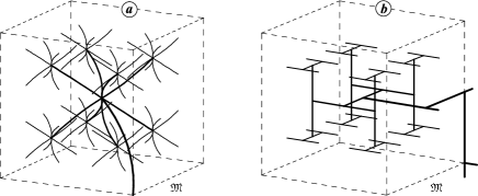
At the second step we should average the obtained transition rate over the possible realizations of the vascular network embedding. Let us adopt a simplified model for the vascular network shown in Fig. 3 where the vessel lengths and of the neighboring hierarchy levels and are related as . Figure 3 demonstrates a more adequate model for the peripheral artery tree which, however, within the framework of the present qualitative analysis may be reduced to the former one by combining three sequent two-fold nodes into one effective four-fold node at all the levels. In this case the cubic domain of volume falls per each artery of level . Let us now consider individually three characteristic forms of the domain , a ball or a cube of size (), an infinitely long cylinder of radius (), and a plane layer of thickness (). For the ball or the cube, i.e. a region bounded in three dimensions the probability that an artery of level passes through the domain is about
For the infinitely long cylinder
and for the plane layer . Multiplying by the corresponding values of we get the result of averaging the walker transition rate over the possible realizations of the vascular network embedding. The obtained result is written as
| (7) |
where the value actually plays the role of the dimension of the space inside which the temperature field can be considered. At the next step we should sum the terms (7) over all the levels of the large artery tree. However due to the strong increase of the terms (7), , as the level number increases the arteries of length mainly contribute to the value of . So the term describing the fast heat transport with blood flow through large arteries from the domain (located near the origin of the coordinate system) can be written as
| (8) |
where is the region containing the peripheral vascular network as a whole and the integration in the three dimensional space over the domain allows for all its three considered types.
Expression (8) together with the mean-filed bioheat equation (2) enables us to write the following equation governing the anomalous heat transfer in living tissue
| (9) |
where we have added directly the value in order to cut off the spatial scales smaller than the length and ignored the difference between the effective temperature diffusivity and the true one of the cellular tissue. Equation (9) is the desired governing equation of the anomalous fast heat diffusion in living tissue for the averaged temperature field. It should be noted that the second term on the right-hand side of equation (9) depends weakly on the blood perfusion rate. Therefore, for the nonuniform distribution of the blood perfusion rate equation (9) holds also.
Anomalous temperature distribution under local strong heating
Hyperthermia treatment as well as thermotherapy of small tumors of size about or less 1 cm is related to local strong heating of living tissue up to temperatures about 45C or higher values. In this case the tissue region heated directly, for example, by laser irradiation is also of a similar size. Due to the tissue response to such strong heating the blood perfusion rate can grow tenfold locally whereas in the neighboring regions the blood perfusion rate remains practically unchanged Song84 . The feasibility of such nonuniform distribution of the blood perfusion rate may be explained applying to the cooperative mechanism of self-regulation in hierarchically organized active media BOOK ; SIAM . Therefore in the region affected directly the blood perfusion rate can exceed the blood perfusion rate in the surrounding tissue substantially. In this case the characteristic length of heat diffusion into the surrounding tissue is about
| (10) |
giving us also the minimal size of the region wherein the tissue temperature increase is mainly localized. In the neighboring tissue the blood perfusion rate keeps up a sufficiently low value , which makes the heat propagation with blood flow through large arteries considerable. Indeed, let us estimate the temperature increase caused by this effect using the obtained equation (9). The temperature increase at a point spaced at the distance from the region (of size ) affected directly, i.e. the tail of the temperature distribution is mainly determined by the anomalous heat diffusion and, so, is estimated by the expression
| (11) |
As seen from (11) for a sufficiently local and strong heating of the tissue, i.e. when and the temperature increase at not too distant points such that can be considerable. Otherwise the anomalous heat diffusion is ignorable.
Acknowledgements.
This work was supported by STCU grant #1675 and INTAS grant #00-0847.References
- (1) M. M. Chen and K. R. Holmes, “Microvascular contributions in tissue heat transfer”. Ann. N. Y. Acad. Sci., 335, 137–154 (1980).
- (2) Heat Transfer in Medicine and Biology. Analysis and Applications, A. Shitzer and R. C. Ebergard, Editors (Plenum, New York, 1970).
- (3) V. V. Gafiychuk and I. A. Lubashevsky. Mathematical Description of Heat Transfer in Living Tissue, (VNTL Publishers, Lviv, 1999); e-print: adap-org/9911001,9911002.
- (4) G. I. Mchedlishvili. Microcirculation of Blood. General Principles of Control and Disturbances (Nauka Publishers, Leningrad, 1989) (in Russian).
- (5) S. Weinbaum and L. M. Jiji. “A new simplified bioheat equation for the effect of blood flow on local average tissue temperature”, ASME J. Biomech. Eng. 107, 131–139 (1985).
- (6) S. Weinbaum, L. X. Xu, L. Zhu, and A. Ekpene. “A new fundamental bioheat equation for muscle tissue: Part I–Blood perfusion term”, ASME J. Biomech. Eng. 119, 278–288 (1997).
- (7) J. W. Baish, P. S. Ayyaswamy, and K. R. Foster. “Small-scale temperature fluctuations in perfused tissue during local hyperthermia”, ASME J. Biomech. Eng. 108, 246–250 (1986).
- (8) B. B. Mandelbrot, The Fractal Geometry of Nature (Freeman, New York, 1977).
- (9) C. W. Song, A. Lokshina, I. G. Rhee, M. Patten, and S. H. Levitt. “Implication of blood flow in hyperthermia treatment of tumors”. IEEE Trans. Biom. Eng., BME-31 (1), 9–15 (1984).
- (10) I. A. Lubashevsky and V. V. Gafiychuk. “Cooperative mechanism of self-regulation in hierarchical living systems”, SIAM, J. Appl. Math. 60(2), 633-663 (2000).
- (11) J. C. Chato. “Heat transfer to blood vessels”, ASME J. Biom. Eng. 102, pp. 110–118 (1980).
- (12) E. H. Wissler. “Comments on the new bioheat equation proposed by Weinbaum and Jiji”, ASME J. Biom. Eng. 109, pp. 226–233 (1987).
- (13) E. H. Wissler. “Comments on Weinbaum and Jiji’s discussion of their proposed bioheat equation ”, ASME J. Biom. Eng. 109, pp. 355–356 (1988).
- (14) I. A. Lubashevsky, A. V. Priezzhev, V. V. Gafiychuk, and M. G. Cadjan. “Free-boundary model for local thermal coagulation”. In: Laser-Tissue Interaction VII, S. L. Jacques, Editor, Proc. SPIE 2681 81–91 (1996).