Electromechanical Limits of Polymersomes
Abstract
Self-assembled membranes of amphiphilic diblock copolymers enable comparisons of cohesiveness with lipid membranes over the range of hydrophobic thicknesses . At zero mechanical tension the breakdown potential for polymersomes with is compared to for liposomes with . Nonetheless, electromechanical stresses at breakdown universally exhibit a dependence, and membrane capacitance shows the expected strong -dependence, conforming to simple thermodynamic models. The viscous nature of the diblock membranes is apparent in the protracted post-poration dynamics.
pacs:
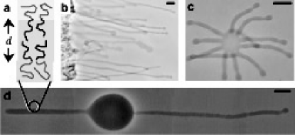
PACS Numbers: 82.70.Uv, 87.68.+z, 68.65.-kA primary task for any biological membrane is to separate inside from out, partitioning ions and other solutes that generate a transmembrane potential . Electrically excitable cells, particularly neurons, control and propagate this potential for purposes of signaling and inter-communication [1]. Efforts to exploit and better understand such phenomena have most recently motivated the semi-patterned growth of nerve cells on circuits [2] as well as the generation of artificial networks with soft nanotubes pulled from vesicles [3]. Further opportunities in such directions now seem possible with wholly synthetic block copolymers that, analogous to lipids in water, self-assemble into membranes, minimizing interfacial exposure of hydrophobic segments (Fig. 1) and thereby generating vesicles termed polymersomes [4].
Physical limits of self-assembled lipid membranes impose important constraints on electrochemical signals. Indeed, the operating range of biomembrane excitations, such as the action potential of a neuron, is generally less than over time scales of milliseconds. At the same time, a finite resting tension is exerted on most cell membranes, including neurons [5], through substrate adhesion and additional cellular mechanisms. From optical trap techniques and other methods, is now generally believed to be in the range of to [6], though additional transient stresses on cells can readily magnify such tensions [7]. For example, electrocompressive stresses arise with a voltage drop across a membrane and have long been recognized as coupled, most simply, to lateral tensions through an integrated form of the Lippmann equation [8]:
| (1) |
where is the capacitance per unit area of the membrane, and is an equivalent tension. Alternative descriptions of electromechanical coupling have been put forward and include a thermodynamic study of block copolymer lamellae perforation that was specialized in its comparisons to lipid membranes [9]. Direct tests of lipid membrane cohesion by Needham and Hochmuth [10] have clearly demonstrated the electromechanical limits of lipid membranes via the combination of micropipette aspiration and electroporation techniques. From these and related tests it has become clear that transmembrane potentials higher than and mechanical tensions higher than are not sustainable in lipid membranes [5, 10]. Clearly, a hydrophobic thickness of only for such membranes is a determinant of electromechanical stability.
Formation of polymersomes in dilute aqueous solutions has been described elsewhere [4]. Generally, the phase behavior of amphiphiles composed of strongly segregating segments is determined by the hydrophilic fraction and mean molecular weight [11]. Lipids provide initial guidance in the synthesis [12] of biomimetic super-amphiphiles; Table I shows for vesicle-forming phospholipids and diblock copolymers, although the latter are much higher in and somewhat more polydisperse. For the copolymers used here, the hydrophilic segment of polyethylene oxide (PEO) forms a highly hydrated brush [13] while aggregation is driven by a hydrophobic block of either polyethylethylene (PEE) or its unsaturated homologue polybutadiene (PBD). As will be reported elsewhere [14], scaling of with (as well as elastic properties) is consistent with the hydrophobic core behaving as a fluid-like melt.
When small pieces of our copolymers are added to water, vesicular tubes spontaneously sprout and grow (Fig. 1(b)). Vesicle formation processes - kinetics, yields, and morphologies - exhibit temperature dependences that likely stem from hydration-facilitated melting of PEO [12]. The vesicles formed exhibit a wide variety of shapes that range from multi-armed stars to hundreds of micron-long axon-like tubes (Fig. 1) as well as flaccid spheroids. Transformations between morphologies are easy to achieve through osmotic adjustments of the external solution [4] and thus reveal the semi-permeable nature of the copolymer membranes. Our first generation polymersomes composed of a PEO-PEE diblock copolymer (designated OE7 in Table I) possess a core thickness and have already been shown to be mechanically tough [4]. In this Letter, we elaborate the more general electromechanical responses of polymersomes including two new covalently-crosslinkable PEO-PBD copolymers of similar block fraction but higher molecular weight (OB16 and OB18 in Table I). Specific comparisons are made with our own measurements of the highly representative lipid stearoyl-oleoyl-phosphatidylcholine (SOPC), which are comparable to the results reported elsewhere [10, 15]. Thus, for the first time we can thoroughly study the cohesiveness of membranes as a function of their hydrophobic core thickness over the range .
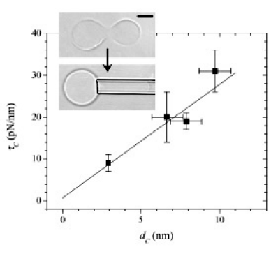
Flaccid polymersomes were progressively aspirated into a micropipette (Fig. 2 inset) at stress rates of up to the point of rupture. Values of the lysis tension for SOPC as well as the three polymersomes studied are shown in Table II. The results generally show that increasing leads to an increase in stability, consistent with general ideas of meso-phase stability for strongly segregated copolymers [11]. As such, the hydrophobic core is expected to behave as a dense fluid. Assuming incompressibility, the thickness at rupture is given by , where is the area strain at rupture as measured directly from images of aspirated vesicles. For lipids, is very small, typically [15], so that . For polymersomes, however, ranges from for OE7 to for OB18. Thus, polymersomes should thin considerably. A simple linear fit of vs. is found to intersect the -axis at essentially zero tension (Fig. 2). Moreover, the slope of this fit defines a lysis stress . Such a stress is typical of cavitation pressures for homogeneous fluids [16] suggesting that lysis of membranes can occur through nucleation of water-filled cavities inside the hydrophobic core.
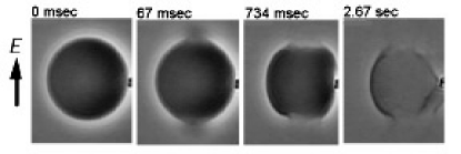
Electromechanical experiments were performed to determine the breakdown potential at different membrane tensions. Once again a flaccid vesicle was aspirated to a prescribed, subcritical tension while being manipulated to a position between two platinum wire electrodes separated by . A square-shaped pulse was applied across the electrodes at intervals that varied from seconds to minutes depending on the post-poration dynamics of the vesicle (see below). The applied potential was increased at discrete intervals until membrane rupture was observed. A typical electroporation event at low applied tension is shown in Fig. 3. The electrodes were arranged parallel to the pipette, generating an electric field as illustrated, and the suction pressure was sufficient to hold the vesicle in the micropipette. A first image taken at zero applied field ( ) demonstrates the integrity of the membrane by showing a phase-dense vesicle interior (sucrose solution) against a phase-light exterior (equi-osmolar glucose). Dramatic rupture within 1-2 video frames of the applied pulse was invariably indicated by a ’jet’ of released sucrose; such jets always occurred at focal point(s) on the membrane where the surface normal is parallel/antiparallel to . However, the dynamics of pore formation differed considerably from one membrane system to another. Membranes composed of OB16 (Fig. 3) typically showed two very large, antipodal pores that grew to many microns in size over seconds. Continued growth often completely rendered the OB16 polymersomes, despite the low tensions. Phospholipid vesicles, in contrast, showed more rapid and even reversible pore dynamics at vanishing tension, as described by others [17, 18]. Polymersomes of the smallest copolymer, OE7, exhibited liposome-like dynamics whereas polymersomes composed of the largest copolymer, OB18, were far more protracted in their poration dynamics. With OB18, encapsulated sucrose was invariably lost over periods of tens of seconds, with weak, barely visible jets implying relatively small but sustained pores.
Kinetic diversity in membrane poration is understood at a first level in terms of the interplay between edge energy, characterized by a line tension that tends to close the pore, and the work done by any dynamic membrane tensions that tend to force the pore open. A balance between these two energies gives the metastable pore size above which the pore will grow - provided sufficient tension is maintained. Sandre et al. [18] recognized the important role of viscosities in vesicle pore dynamics. In the limit of vanishing tension, the rate of pore closure was postulated to scale as where is the surface viscosity. Although is expected - from simple bending energy considerations of a hydrophilic pore - to increase in linear proportion to , separate measurements of lateral diffusion of copolymers (Lee et al. (unpublished)) suggest that increases strongly with and is at least ten-fold greater in the present polymersomes compared to lipid membranes. Moreover, the of both OB16 and OB18 exceed the relevant entanglement molecular weights ( to ), so that the polymeric nature of the present systems provides a clear basis for the slowed pore dynamics.
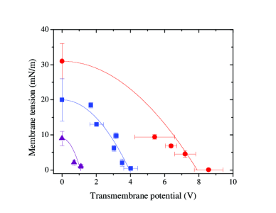
The transmembrane potential has been calculated previously for a spherical shell [19] as where is the radius of the vesicle and is the angle between the membrane surface normal and . The charging time is given by , where and are internal and external solution resistivities, respectively. In this work, is orders of magnitude smaller than the pulse duration used. To assess coupling of the mechanical and electrical stresses as suggested by Eq. (1), polymersomes are aspirated to a set tension and subsequently subjected to electrical stresses as explained above. At each imposed tension, is determined and the resulting points are plotted in the plane (Fig. 4). The data show that the higher the membrane tension, the lower the breakdown potential. For OB18, the results are remarkable: this thick membrane can transiently withstand the potential of a battery.
Fitting the rupture data () for each self-assembled membrane system by Eq. (1) readily generates an amphiphile-specific estimate of at rupture (Table II). Within this phase space spanned by and , the area under each parabolic segment provides a phenomenological measure of the range of electromechanical function or robustness. For SOPC, this robustness is very small (see Table II); for polymersomes, in contrast, the robustness can be orders of magnitude larger. Fitted values of polymersome are nonetheless very low compared to those reported for lipid membranes, [19]. This is qualitatively consistent with the enhanced thickness of polymersome membranes since , where is the hydrophobic dielectric constant. On the other hand, at increases considerably with (see Eq. (1) and Fig. 4). By definition, the surface charge at rupture should then be independent of . Indeed, Table II indicates that varies by only a factor of about two within this structurally diverse set of membrane systems studied.
Polymersomes made from OB16 and OB18 offer the further possibility of crosslinking the membrane core. Free radical polymerization in solution proves highly effective [20] with dramatic increases in the robustness of the membranes. Simple osmotic rupture tests - where large vesicles are observed to burst after suspension in a sufficiently hypotonic solution - indicate that of cross-linked membranes are of order . Thus . Using such values in Eq. (1) and assuming values for listed in Table II, for the cross-linked OB’s are estimated to exceed for pulses.
Electrically excitable cells, particularly neurons, control and propagate electrochemical potentials for signaling purposes [1], but the transmembrane voltages employed are always constrained by physical limits of a self-assembled lipid membrane system. The results here with a series of self-assembling biomimetic diblock copolymers considerably expand these limits, and clearly indicate the overall electromechanical robustness of hyperthick membranes. As might be expected, and both increase with while decreases and hardly changes. Though a microscopic theoretical treatment of membrane electroporation is lacking, the capacitance and insulation properties of such self-assemblies provide a more physically reliable basis for microelectronics applications such as sensor platforms and those that might exploit conducting copolymer segments. In addition, drug delivery applications could benefit through formulations of robust copolymer vesicles that reversibly reseal or not when challenged by sufficient membrane tensions and/or voltages.
ACKNOWLEDGMENTS HAE and HB thank R. Parthasarathy for discussions and comments. HAE was supported by NIH/NHLBI T32HL-07954 training grant. DED gratefully acknowledges discussions with D.A. Hammer and E.A. Evans. Funding was provided by NSF-MRSEC at the U. of Minnesota and U. of Pennsylvania, and NASA.
REFERENCES
- [1] B. Alberts et al., Essential Cell Biology (Garland Publishing, Inc. New York, 1997).
- [2] P. Fromherz and A. Stett, Phys. Rev. Lett. 75, 1670 (1995); M.P. Maher et al., J. Neurosci. Meth. 87, 45 (1999).
- [3] E.A. Evans et al., Science 273, 933 (1996); A. Karlsson et al., Nature 409, 150 (2001).
- [4] B.M. Discher et al., Science 284, 1143 (1999); B.M. Discher et al., Curr. Opin. Coll. Interface Sci. 5, 125 (2000).
- [5] R.M. Hochmuth et al., Biophys. J. 70, 358 (1996).
- [6] J. Dai and M.P. Sheetz, Biophys. J. 77, 3363 (1999); E. Evans and A. Yeung, Biophys. J. 56, 151 (1989)
- [7] R. Simson et al., Biophys. J. 74, 514 (1998); H. Ra et al., J. Cell Sci. 12, 1425 (1999).
- [8] G. Lippmann, Ann. Phys. 149, 546 (1873)
- [9] R.R. Netz and M. Schick, Phys. Rev. E 53, 3875 (1996).
- [10] D. Needham and R.M. Hochmuth, Biophys. J. 55, 1001 (1989).
- [11] M.W. Matsen and M. Schick, Curr. Opin. Coll. Interface Sci. 1, 329 (1996); D.A. Hajduk et al., J. Phys. Chem. B 102, 4269 (1998)
- [12] M.A. Hillmyer and F.S. Bates, Macromolecules 29, 6994 (1996).
- [13] Y-Y.Won et al., J. Phys. Chem. B 104, 7134 (2000).
- [14] H. Bermudez et al., (unpublished).
- [15] K. Olbrich et al., Biophys. J. 79, 321 (2000); W. Rawicz et al., ibid. 79, 328 (2000). Note that we focus on pure phospholipids; cholesterol is known to stiffen and thereby toughen membranes [10], but cholesterol does not, on its own, self-assemble into a membrane. In contrast, the elasticity of all the pure amphiphilic self-assemblies here differ by less than two-fold [14].
- [16] F.R. Young, Cavitation (McGraw-Hill, New York, 1989).
- [17] D. Zhelev and D. Needham, Biochim. Biophys. Acta 1147, 89 (1993).
- [18] O. Sandre et al., Proc. Nat. Acad. Sci. 96, 10591 (1999).
- [19] K.S. Cole, Membrane, Ions and Impulses (University of California Press, California, 1972).
- [20] Y-Y. Won et al., Science 283, 960 (1999).