Electrostatically induced undulations of lamellar DNA–lipid complexes
Abstract
We consider DNA-cationic lipid complexes that form lamellar stacks of lipid bilayers with parallel DNA strands intercalated in between. We calculate the electrostatically induced elastic deformations of the lipid bilayers. It is found that the membranes undulate with a periodicity that is set by the DNA interaxial distance. As a consequence the lamellar repeat distance changes resulting in a swelling or compression of the lamellar stack. Such undulations may be responsible for the intermembrane coupling between DNA strands in different layers as it is observed experimentally.
pacs:
68.10.-mFluid surfaces and fluid-fluid interfaces and 64.70.MdTransitions in liquid crystals1 Introduction
Electrostatic adsorption of polyelectrolytes onto oppositely charged surfaces, such as lipid membranes, has been the subject of intense experimental and theoretical research in the last decade. Of particular interest is the spontaneous complexation of DNA with both cationic and neutral lipids due to their possible application to gene therapy Felgner97 ; Crystal95 . These so-called ”lipoplexes” show a diversity of equilibrium and metastable structures Sternberg94 ; lasic97 ; Raedler97 ; fang97 ; salditt97 ; koltover98 ; Artzner98 ; Zantl98 ; huebner99 ; Koltover99 . For example, it has been shown through X-ray diffraction analysis Raedler97 ; salditt97 ; Koltover99 that DNA molecules and lipids can form lamellar complexes with DNA intercalated in between lipid bilayers. Another complex formed for sufficiently flexible membranes is an inverse hexagonal lipid structure with DNA inside the water regions koltover98 .
Several theoretical studies help to understand many of the phenomena observed for lipoplexes dan96 ; dan97 ; bruinsma98b ; May97 ; dan98 ; bruinsma98 ; golubovic98 ; ohern98b ; Harries98 ; Schiessel98 ; menes98 ; ohern98 ; ohern99 ; may00 ; menes00 , however many more remain to be elucidated. May et al. may00 studied in detail the phase behavior of aqueous mixtures of DNA, cationic lipid and neutral lipid. Their model is based on the two thermodynamically stable structures found experimentally koltover98 and predicts the phase diagram as a function of the DNA/lipid composition and the elastic properties of the lipid bilayers. Another problem that was considered theoretically is the dependence of the interaxial spacing of DNA rods in lamellar complexes on the DNA/lipid composition. Bruinsma bruinsma98 presented an analytical approach that is applicable to lipoplexes with weakly charged bilayers. The numerical study of Harries et al. Harries98 predicts the interaxial spacing also for higher charge densities. According to both studies the isoelectric point of the lipoplex (the point at which the anionic charges of the DNA balance the cationic charges of the lipids) is unstable to further adsorption of DNA or lipids. The formation of say an isoelectric complex is driven by the release of the small counterions that were ”condensed” on the highly charged DNA and on the charged bilayer before complexation. A lipoplex close to the isoelectric point is very susceptible to the uptake of further cationic lipids or DNA – if available – since this will be accompanied by the release of the corresponding counterions into the lipoplex111A similar instability is also expected for the complexation of charged spheres and oppositely charged polyelectrolytes. A single highly charged chain will wrap around a single sphere forming a complex that is beyond the isoelectric point (”overcharging”) and this effect is driven by the release of counterions from the wrapped chainpark99 . On the other hand, a chain in a solution of highly charged spheres will complex more spheres than necessary to be isoelectric, and this effect is driven by the release of counterions of the complexed spheresSchiessel00 .. The theoretical predictions show good agreement with a recent experimental study Koltover99 .
A different approach to the problem of the distance between DNA strands was given by Dan dan96 . In this study the preferred distance was predicted to be the result of two competitve mechanisms, electrostatic repulsion between the strands and their membrane-induced attraction due to the perturbation of the lipid packing in the membrane close to the adsorbed DNA. The model assumes DNA strands adsorbed on a single membrane as it was investigated experimentally by Fang and Yang fang97 using atomic force microscopy experiments. They found that the distance between DNA strands was about 5 nm, a distance that was predicted by Dan within her model dan96 .
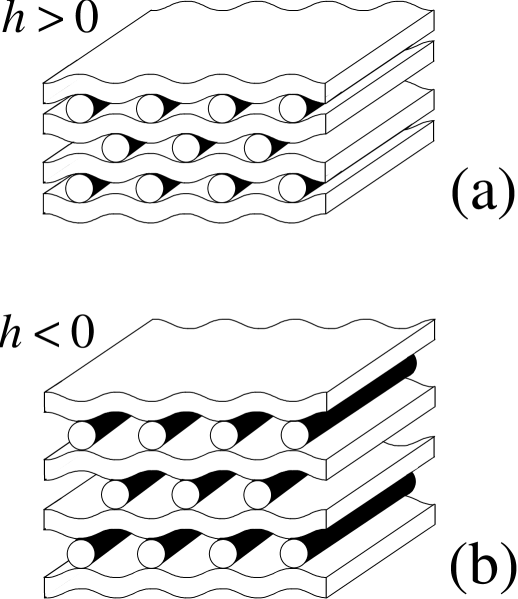
In that model it is assumed that the membrane (that is supported by a solid surface) is locally perturbed close to the DNA in such a way that the monolayer thickness is slightly increased dan96 . It should be expected that for lamellar lipoplexes one also has perturbations. Since the membranes are allowed to undergo shape changes freely (no supporting layer) one might expect undulations leading to a compression or sweeling of the whole lamellar stack as depicted in Fig. 2. Such undulations might lead to an intermembrane coupling between DNA rods in different layers – resulting in a 3D ordering of the DNA rods. Lipoplexes with a 3D rectangular ordering of the DNA molecules were indeed observed experimentally Artzner98 .
Most experiments on lamellar lipoplexes indicate that such a type of perturbation of the membranes around the DNA molecules – if present – is smallRaedler97 ; salditt97 . Undulations of the membranes should lead to a lamellar repeat distance that is larger or smaller than the sum of the bilayer thickness and the diameter of the DNA molecule (including a hydration shell). Considering complexes at the isoelectric point and changing the ratio of charged to neutral lipids it was observed that the lamellar repeat distance stays always close to a value that indicates flat membranesRaedler97 ; salditt97 . Thus even though the lipid dilution experiments lead to a considerable increase of the interaxial spacing between DNA rods, the undulations remain too small to be non-ambiguously detected. On the other hand, for more flexible membranes where detectable membrane undulations could be expected the system switches to the inverse hexagonal phase instead koltover98 .
Recently Subramanian et al. Subu00 studied the complexation of the anionic polypeptide poly-glutamic acid with a mixture of cationic (DDAB) and neutral (DLPC) lipids by means of small angle X-ray scattering and neutron scattering. It was observed that the lipid organizes in a multilamellar phase with the polypeptide chains intercalated in between the membranes. Compared to the DNA complexes discussed above, the polypeptides do not show any in-plane ordering even though it is assumed that they are in the -helical state. As for the DNA lipoplexes a ”lipid dilution” experiment was performed for isoelectric polypeptide lipoplexes. Contrary to the outcome for the DNA complexes, a considerable increase of the lamellar spacing was found when the cationic lipids were diluted by neutral ones. For high lipid dilution the spacing saturated at a constant value of 60Å which coincides with the equilibrium value of pure DLPC membranes. Subramanian et al. Subu00 suggested that this behavior could be due to a ”pinching mechanism” including membrane undulations similar to the ones depicted in Fig. 2 (case ). The pinching sites are formed due to the electrostatic interaction between the negatively charged poly-glutamic acid and the cationic DDAB lipids. Away from the pinched regions the properties of the lipoplex are dominated by the properties of the pure DLPC membranes. Whether it is possible to have pinches in a lipoplex was studied by one of the authors Schiessel98 . By comparing the gain in electrostatic free energy with the bending energy of forming a pinch, the parameter range was estimated at which pinching can be expected. It was shown that this effect should occur if the line charge density of the rods is sufficiently high and the membranes are sufficiently flexible, a situation that might be fulfilled for the polypeptide lipoplex considered in Ref. Subu00 .
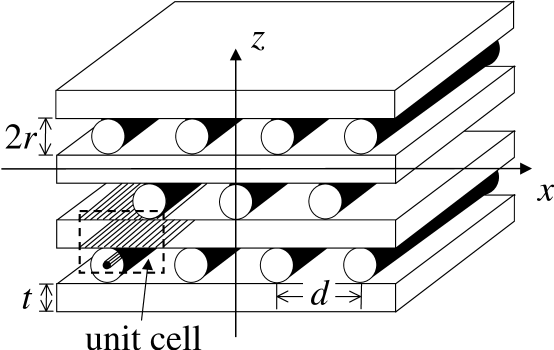
A different approach to the pinching problem is taken in the present study. We start out with a perfectly flat lamellar lipoplex as depicted in Fig. 1. The DNA rods are assumed to be ordered within a 3D rectangular lattice as it was observed by Artzner et al. Artzner98 . Our goal is to calculate how the electrostatic interaction between the negatively charged ”rods” and the positively charged membranes modifies the conformation of the membranes. We show that there are in principle two possibilities, namely a compression of the lamellar stack as depicted in Figure 2 () or an expansion as depicted in the same Figure (case ).
In the next section we introduce the model system and calculate its electrostatic and bending free energies for arbitrary but small periodic undulations of the membranes. By minimizing the free energies of the undulation with respect to its Fourier components we show in Sect. 3 that the electrostatic interaction usually favors a compression of the lamellar complex – at least if the underlying assumptions of our model are fulfilled. These assumptions are discussed in Sect. 4 where we also present some conclusions.
2 Free energy of model lipoplex
The aim of the following calculation is to determine the electrostatic contribution to the undulations of a lamellar stack of membranes with DNA molecules intercalated in between. Our model system consists of two constituents, the membranes and the DNA molecules. The membranes have a uniform thickness and carry positive charges on both sides. The surface charge density is given by on each side of the bilayer and is assumed to be uniform. In our model the membranes are perfectly transparent for the electric field lines, i.e., we have a homogeneous dielectric constant throughout the lipoplex. The bilayers are flexible with a bending rigidity . The DNA molecules are modelled as infinitely long rigid rods of radius . For simplicity, we assume the negative charges of the DNA molecules to be located along their middle axis with the linear charge density . Following the experimental observation of a lamellar stack with DNA forming smectic arrays we arrange the components of our model in the following way (cf. Fig. 1). All membranes are parallel to the -plane with their midplanes at the positions The DNA rods are aligned in the -direction. The interhelical spacing between neighboring DNA molecules is constant and is denoted by . Excluded volume requires that . The rods in one layer are located at , in the neighboring layers they are displaced by , i.e., they are at the positions etc. Furthermore, the rods are assumed to be always attached to the two neighboring membranes.
The electrostatic interaction between the charges is calculated within the Debye-Hückel approximation. In this approximation the potential is determined by with the appropriate boundary conditions. Here denotes the Debye screening length that is given by where is the bulk salt concentration and is the Bjerrum length ( is the unit charge, is the thermal energy and the dielectric constant; in an aqueous solution). The total electrostatic contribution to the free energy of the system is given by the sum of the (screened) electrostatic interactions and the translational entropy of the counterions Verwey ; Goldstein90 :
| (1) |
The integration extends over all charged surfaces of the system with being the corresponding charge densities.
We ask the following question: How are the membranes deformed by the electrostatic interaction? In order to answer this question we will calculate the induced undulations of the membrane up to the first order in the deformation amplitude.
Consider the membrane at . Electrostatics induces a deformation around the flat state, . Due to the symmetry the deformation profile is of the form
| (2) |
where the hat denotes summation over odd only. This undulation leads to the following curvature energy (per area ):
| (3) |
In order to calculate the electrical free energy, Eq. 1, we compute first the electrical potential induced by the charges on the upper surface of the membrane (note that is translational invariant in -direction). At that charged surface, i.e., at , we have the boundary condition which is here of the form
| (4) |
(up to terms of the order ). By expanding up to first order in the amplitudes we find the following form of the potential above the membrane ()
| (5) |
Each fulfills the Debye-Hückel equation separately. They can be expanded in Fourier series where with . The coefficients follow from the boundary condition at the membrane together with the fact that due to symmetry . We find that only the coefficients are non-vanishing and are given by . This leads to (for ):
| (6) | |||||
The total potential induced by the membrane at is the sum of the contributions of the upper charged boundary, , cf. Eq. 6, and of the lower one, : . Using we find:
| (7) | |||||
Furthermore, the potential induced by the line charge of the rod has the form
| (8) |
where is the distance from the line and is a modified Bessel function with for and for . The total electrical potential is the sum of the potential that follows from all membranes and the potential that is due to all the rods: .
We calculate now the total electrostatic contribution to the free energy per unit cell. A unit cell has the width (in –direction) and a height that corresponds to the (average) distance between neighboring layers (for the case of a flat membrane – depicted in Fig. 1 – this height equals ). According to Eq. 1, we obtain the total electrostatic energy (per unit cell) by integrating the total potential over all charged surfaces that lie within this cell. The unit cell in Fig. 1 contains three charged surfaces, , and . is a stripe of the upper surface of one bilayer that is uniformly charged with the density . is the corresponding lower charged surface. is the surface carrying the charges of one rod; we assume this to be the surface of a cylinder with radius and charge density . It follows that the electrostatic energy (per area) has three contributions: The inter-(and intra-) membrane interaction , the membrane-rod interaction and the interaction between the rods . Thus
| (9) |
Here we made use of the identity .
We start by calculating the change of the membrane–membrane interactions induced by their undulations (up to first order in ). The position of the midplane of the th membrane is given by
| (10) |
with and . Fig. 2 shows schematic views of lamellar structures that are compressed – case (a) with – and swollen – case (b) with . Denote the contribution of th membrane to the potential by . Then
| (11) | |||||
Equation 11 shows that a swelling of the system () decreases the membrane-membrane interaction whereas a compression () is unfavorable. Note that we neglected terms of second order in the . As can be seen from Eq. 3 terms of the form lead to a renormalization of the bending constant, . It can be shown that ( is the Gouy-Chapman length)Winter88 ; Lekker89 ; Kiometzis89 ; Fogden90 ; Pincus90 ; Higgs90 ; Duplantier90 ; Harden92 ; Andelman95 . For a wide range of parameters one has . In the following we use the bare bending rigidity , keeping in mind that it has to be replaced by when is comparable to .
We estimate now the contribution of the membrane–rod attraction. Consider the rod at the position and . The rod is located within an infinite stack of membranes. This can be accounted for by simply summing twice over the contributions of all the membranes that are located above the rod, i.e. . From Eq. 7 follows that is given by (note that )
| (12) | |||||
The contribution of the first term of Eq. 12 to is of the form:
| (13) | |||||
The second term of Eq. 12 leads to the following expression:
| (14) |
The total membrane–DNA contribution favors a compression of the lamellar stack – thus constituting a competing mechanism to the membrane-membrane repulsion.
We are left with the calculation of the interaction energy between the rods. We focus here one two important cases. Case 1: and (”vertical screening”): In this case the interaction between rods in different layers is negligible compared to the rod-rod interaction within the same layer. Then it is sufficient to sum over the contributions of all rods to the left and to the right of the given rod :
| (15) |
Case 2: and (weak screening): In this case all rods contribute to the interaction energy. After some algebra we arrive at
| (16) |
As expected, in both cases the repulsive rod-rod interaction favors swelling.
3 Undulations in isoelectric complexes
We consider first the lamellar complex in equilibrium with a solution of free DNA strands. We ask: What is the interaxial distance between the DNA strands in the lipoplex that minimizes the electrostatic free energy of the complex? As pointed out by Bruinsma bruinsma98 counterion release will control ; here, however, we determine the equilibrium spacing for the case when there is no counterion release, i.e., we assume the rods being below the Manning threshold. We also neglect entropic changes due to the adsorption of free DNA strands into the lipoplex. We will show that, as a result of the geometry, such a system will equilibrate at the isoelectric point – if the electrostatic interaction is sufficiently long-ranged. In the following we only account for the contributions independent of the ’s and treat the contribution of membrane bending afterwards as a perturbation.
Let us first consider the case of high ionic strength where (strong screening). Then the free energy per area is given by
| (17) |
i.e., by the membrane-rod attraction, Eq. 13; other terms are negligible. It follows that the minimum is at . Excluded volume interaction between the rods will lead to . Clearly, in the case of strong screening as a result of the short range of the electrostatic interaction the lipoplex is equilibrated far from the isoelectric point. The resulting complex is ”overcharged” by the DNA rods. (A similar situation occurs for the adsorption of rods on an oppositely charged surface, cf. Ref. Nguyen00 ).
We discuss next the two cases introduced above. Case 1: and (vertical screening): From Eqs. 13 and 15 we find (up to terms of the order )
| (18) |
is minimized for which corresponds to the isoelectric point of the complex, i.e., the point at which the charges of the cationic lipids and of the DNA are exactly balanced. Case 2: and (weak screening): From Eqs. 13 and 16 follows
| (19) |
Again the free energy is minimized at the isoelectric interhelical spacing .
Thus in the limiting case (no salt, no screening) the lipoplex is forced to be at the isoelectric point. This is an artefact of our model which does not account for counterions that would be present in lipoplexes with an excess of DNA molecules or cationic lipids. This holds for both cases, for Case 1 corresponding effectively to decoupled two-dimensional layers and for Case 2 which is truly three-dimensional. Therefore our theory is only applicable to isoelectric lipoplexes where all counterions are expected to be released.
We consider now the undulations occuring in lipoplexes in general and then focus again on the isoelectric point. The change of the total electrostatic free energy as a function of the deformation follows from the Eqs. 11, 13–16:
| (20) |
The change of the total free energy due to bending is given by the sum of the electrical contribution and the bending energy , Eq. 3. Minimizing with respect to the amplitudes leads to with
| (21) |
Thus the deformation modes decrease rapidly with increasing . Now we are in the position to calculate the deformation of the membranes. Inserting into Eq. 2 we find the following deformation profile
| (22) |
where the polynomial expression is valid for (the continuation outside this interval follows from the symmetry of the configuration). A good approximation for all values of (relative error smaller than ) is given by .
The coefficient at the isoelectric point of the complex is given by
| (23) |
In the case of vertical screening we find a positive (and -independent) value of and thus a positive value of , , corresponding to a compression of the isoelectric lamellar stack. Interestingly, in the case of weak screening the undulations disappear. In fact, as long as the vertical screening is operative the membrane–membrane repulsion is smaller than the membrane-rod attraction in the isoelectric lipoplex and therefore we find a compression of the lamellar stack. For weak screening, the rod-rod repulsion between different layers cancels this net attraction, cf. Eq. 20. Let us consider typical values for , , and , say Å (DNA), Å2, (water) and . For these values we find (in the case of vertical screening) Å, i.e., the undulations are rather small.
Finally, we estimate how the undulations of the isoelectric complex disturb the interaxial spacing between the rods and in turn move the complex away from its isoelectric point. We consider the case of vertical screening (Case 1, , ). In this case the amplitudes of the undulations depend strongly on the interhelical distance, namely , cf. Eq. 23. Inserting into Eq. 20 (, ) we find two correction terms to Eq. 18, namely from the membrane-membrane repulsion and from the membrane-rod attraction. Evidently, the mem-brane-membrane repulsion favors smaller values of that lead to smaller undulations, whereas the membrane-rod interaction is enhanced for larger undulations, i.e., larger values of are favorable. At the isoelectric point the correction term from the membrane-rod interaction exceeds the other term and as a result the interhelical distance is slightly increased, , with
| (24) |
In that sense the undulations lead to an effective repulsion between the DNA strands proportional to (as long as one is close enough to the isoelectric point). Undulations are one of several mechanisms that might be responsible for the increase of the interhelical spacing with increasing salt concentration as it is observed experimentally (cf. Ref. Koltover99 for details).
4 Discussion
The main idea of the preceding analysis is to give a simple estimate of the role of the electrostatic interactions in a lamellar lipoplex. In many instances some of the underlying assumptions are not fulfilled. But even in this case our model might give an idea about what the contributions of the electrostatics to the overall conformation might be.
One severe approximation is the assumption of a transparent membrane. The lipid bilayer represents a low dielectric slab that – depending on its thickness – might screen most of the electrical field so that charges (say of phosphate group on a DNA molecule) are not ”seen” on the other side of the membrane. As a rule of thumb, a membrane that is much thinner than the distance of a charge from the membrane might be considered to be transparent for this charge whereas a thicker membrane (of thickness ) is opaque and can be approximated by an infinitely thick slab. In that case the effect of the low dielectric lipid can be accounted for by the use of an appropriate image charge – which is a simple task for a flat membrane but difficult to handle for an undulating one. The two cases and are elaborated in some detail in Footnote 2 in Ref. Schiessel98 . The typical thickness of a lipid membrane is Å which is of the order of the diameter of the DNA rod (Å), i.e., one is in the crossover regime between the two cases. In any case, the presence of the low-dielectric lipid will lead to a modification of the simple situation discussed in this paper. It should be expected that the partial confinement of electrical field lines emanating from the DNA rods by the neighboring membranes favors a swelling of the lamellar stack.
Another feature not considered in this study is the demixing of neutral and charged lipids within the bilayers. There might be at least three effects. (i) The electrostatic attraction drives the cationic lipids towards the DNA rods, resulting in a depletion of charged lipids in the membrane parts in between two neighboring rods in the same layer. This reduces the membrane-membrane repulsion between neighboring membranes resulting in an increase of the compression of the lipoplex. This effect should be important if the average mole fraction of cationic lipids in the bilayers is low. (ii) For the opposite limit of highly charged membranes their surface charge density might exceed the surface charge density of the rods. In this case an enhancement of neutral lipids close to the DNA is expected that allows for a better matching of the two charge densities. These two effects were indeed observed in the numerical study by Harries et al. Harries98 (cf. Fig. 7 in that paper). (iii) Finally, membrane undulations may also affect the charge densities on each side of the bilayer and vice versa. A depletion of cationic lipids on one side of the membrane will be accompanied by an enhancement on the other side due to the symmetry of the arrangement of DNA rods, cf. Fig. 1. This might lead to a spontaneous curvature of the bilayer of either sign which in turn affects the membrane undulations discussed in our study. It is clear that the competition of these three effects can lead to a rather complex behavior of the charge density profile of the lipids along the -direction. In our model we do not account for these effects. We expect that our approach has to be modified especially when a large fraction of the lipids is neutral. In that case the description of local, highly charged pinches Schiessel98 might be more appropriate (cf. the discussion of the polypeptide lipoplex Subu00 given in the Introduction of our paper).
Recently it has been possible to non-ambiguously detect undulations in a lipoplexRaedlerpp . From a careful analysis of the data on the lipoplex presented in Ref. Artzner98 if was possible to construct an electron density map revealing its high resolution structure. The structure shows undulations with an amplitude of a few Angstrom leading to a compression of the lamellar structure as depicted in Fig. 2a. Furthermore, the amplitudes show a sharp increase for larger interhelical spacings. Both observations are in qualitative and semi-quantitative agreement with the results of our model calculation, cf. Eq. 23. It is worth noting that the lipids of these lipoplexes form a lipid-gel phase Artzner98 . In this phase the lipid bilayers show a high compressibility allowing the DNA-induced deformations to cross nearly unperturbed through the bilayer (as implicitely assumed in our model). The induced undulations might be the prevailing mechanism for the interlayer coupling that leads to the rectangular columnar superlattice of the DNA strands observed for this class of lipoplexes.
Acknowledgement: We wish to thank J. O. Rädler for sharing experimental results prior to publication. We would like to acknowledge useful conversations with A. Ben-Shaul and S. May.
References
- (1) P.L. Felgner, Sci. Am. 276, 102 (1997).
- (2) R.G. Crystal, Science 270, 404 (1995).
- (3) B. Sternberg, F. Sorgi, L. Huang, FEBS Lett. 356, 361 (1994).
- (4) D.D. Lasic, H. Strey, M.C.A. Stuart, R. Podgornik, P.M. Frederik, J. Am. Chem. Soc. 119, 832 (1997).
- (5) J.O. Rädler, I. Koltover, T. Salditt, C.R. Safinya, Science 275, 810 (1997).
- (6) Y. Fang, J. Yang, J. Phys. Chem. B 101, 441 (1997); J. Phys. Chem. B 101, 3453 (1997).
- (7) T. Salditt, I. Koltover, J.O. Rädler, C.R. Safinya, Phys. Rev. Lett. 79, 2582 (1997); Phys. Rev. E 58, 889 (1998).
- (8) I. Koltover, T. Salditt, J.O. Rädler, C.R. Safinya, Science 281,78 (1998).
- (9) F. Artzner, R. Zantl, G. Rapp, J.O. Rädler, Phys. Rev. Lett. 81, 5015 (1998).
- (10) R. Zantl, F. Artzner, G. Rapp, J.O. Rädler, Europhys. Lett. 45, 90 (1999).
- (11) S. Huebner, B.J. Battersby, R. Grimm, G. Cevc, Biophys. J. 76, 3158 (1999).
- (12) I. Koltover, T. Salditt, C.R. Safinya, Biophys. J. 77, 915 (1999).
- (13) N. Dan, Biophys. J. 71, 1267 (1996).
- (14) N. Dan, Biophys. J. 73, 1842 (1997).
- (15) R. Bruinsma, J. Mashl, Europhys. Lett. 41, 165 (1998).
- (16) S. May, A. Ben-Shaul, Biophys. J. 73, 2427 (1997).
- (17) N. Dan, BBA-Biomembranes 1369, 34 (1998).
- (18) R. Bruinsma, Eur. Phys. J. B 4, 75 (1998).
- (19) L. Golubović, M. Golubović, Phys. Rev. Lett. 80, 4341 (1998).
- (20) C.S. O’Hern, T.C. Lubensky, Phys. Rev. Lett. 80, 4345 (1998).
- (21) D. Harries, S. May, W.M. Gelbart, A. Ben-Shaul, Biophys. J. 75, 159 (1998).
- (22) H. Schiessel, Eur. Phys. J. B 6, 373 (1998).
- (23) R. Menes, P. Pincus, R. Pittman, N. Dan, Europhys. Lett. 44, 393 (1998).
- (24) C.S. O’Hern, T.C. Lubensky, Phys. Rev. E 58, 5948 (1998).
- (25) C.S. O’Hern, T.C. Lubensky, J. Toner J., Phys. Rev. Lett. 83, 2745 (1999).
- (26) S. May, D. Harries, A. Ben-Shaul, Biophys. J. 78, 1681 (2000).
- (27) R. Menes, N. Grønbech-Jensen, P.A. Pincus, Eur. Phys. J. E 1, 345 (2000).
- (28) S. Park, R.F. Bruinsma, W.M. Gelbart, Europhys. Lett. 46, 454 (1999).
- (29) H. Schiessel, R. Bruinsma, W. M. Gelbart, preprint
- (30) G. Subramanian, R.P. Hjelm, T.J. Deming, G.S. Smith, Y. Li, C.R. Safinya, J. Am. Chem. Soc. 122, 26 (2000).
- (31) E.J.W. Verwey, J.Th.G. Overbeck, Theory of the Stability of Lyophobic Colloids (Elsevier, Amsterdam,1948).
- (32) R.E. Goldstein, A.I. Pesci, V. Romero-Rochin, Phys. Rev. A 41, 5504 (1990).
- (33) S.A. Safran, Thermodynamics of Surfaces, Interfaces, and Membranes (Addison-Wesley, Reading, MA, 1994).
- (34) M. Winterhalter, W. Helfrich, J. Phys. Chem. 92, 6865 (1988).
- (35) H.N.W. Lekkerkerker, Physica A 159, 319 (1989).
- (36) M. Kiometzis, H. Kleinert, Phys. Lett. A 140, 520 (1989).
- (37) A. Fogden, D.J. Mitchell, B.W. Ninham, Langmuir 6, 159 (1990).
- (38) P. Pincus, J.-F. Joanny, D. Andelman, Europhys. Lett. 11, 763 (1990).
- (39) P.G. Higgs, J.-F. Joanny, J. Phys. France 51, 2307 (1990).
- (40) B. Duplantier, Physica A 168, 179 (1990).
- (41) J.L. Harden, C. Marques, J.-F. Joanny, D. Andelman, Langmuir 8, 1170 (1992).
- (42) D. Andelman in: Structure and Dynamics of Membranes. Eds. R. Lipowsky and E. Sackmann (North-Holland, 1995).
- (43) T.T. Nguyen., A.Yu. Grosberg, B.I. Shklovskii, J. Chem. Phys. 113, 1110 (2000).
- (44) J.O. Rädler, unpublished results