Nondiffusive heat transfer in muscle tissue. Preliminary results
Abstract
We present preliminary experimental data that enable us to suggest that heat transfer in cellular tissue under local strong heating is a more complex phenomenon than a simple heat diffusion. Namely, we demonstrate that under local strong heating of a muscle tissue heat transfer in it exhibits substantial anisotropy unexplained in the context of the standard diffusion model. The observed temperature dynamics is also characterized by nonlinear behavior as well as by a certain repeat reversibility. The latter means that the time variations in the temperature of a cellular tissue undergoing repeated acts of heating go in the same way at least approximately. We explain the observed anomalous properties of heat transfer by suggesting the flow of the interstitial liquid to appear due to nonuniform heating which, in turn, affects the heat transfer. A possible mechanism responsible for this effect is discussed.
I Introduction
During the last two decades the description of heat transfer in living tissue (bioheat transfer) and mathematical modeling of temperature distribution in it under local strong heating have attracted attention due to several reasons. First, this problem is an important part of planning and optimizing hyperthermia treatment and thermotherapy of tumors where the precise control over the tissue temperature is essential (see, e.g., (I1, ; I2, ; I3, )). Second, what is interesting from the standpoint of fundamental physics, living tissue is a complex medium in which highly branching blood vessels form a fractal network of fast heat transport I4 , endowing heat propagation in it with anomalous properties BLG . Third, living tissue is an active medium, in particular, it responds to strong heating, which causes the blood perfusion rate to grow locally by tenfold Song84 . In this case one meets also the basic problem of how a natural hierarchical system comprising a huge amount of elements can respond properly to changes in its state when none of the elements possesses the total information required to govern the system as a whole LGenv95 ; gaf1 .
By now a number of significant steps towards developing the bioheat transfer theory have been made. In particular, Chen & Holmes CH80 actually stated the microscopic description of the problem. Baish et al. Bi86 , Lagendijk et al. LD1 ; LD2 , Roemer et al. RD1 ; RD2 , and Gafiychuk & Lubashevsky BLG studied the effect of the vessel discreteness on the temperature field. Weinbaum et al. WJ85 ; WXZE97 and Lubashevsky & Gafiychuk BLG took into account sophisticated details of heat interaction between different vessels, namely, the counter-current effect, Chato C80 and Weinbaum & Jiji WJ85 , that renormalizes the kinetic coefficients of heat propagation in the tissue. Lubashevsky & Gafiychuk BLG ; LGenv95 ; gaf1 proposed a hierarchical model for the tissue response to local heating. There is also an approach by Lagendijk et al. (see, e.g., Lag and references therein) that dealing with individual vessels tackles the bioheat transfer problem numerically.
All these models and approaches are based on the assumption that the cellular tissue is a passive medium and heat propagation in it obeys Fick’s law, i.e. the heat flux in the cellular tissue is proportional to the temperature gradient . Namely, and the thermal conductivity is isotropic or, at least, is slightly anisotropic. Indeed, according to the experimental data summarized in GMLM95 , for example, the value of the temperature diffusivity ( and are the specific heat and density of the tissue) is approximately the same for different tissues, , 1.3, 1.4, 1.5, 1.2 cm2/s for human muscle, kidney, spleen, liver, and blood, respectively. We note that models developed in food engineering also regard the cellular tissue as a passive medium (see, e.g., FE ). This assumption is justified by a number of self-consistent results obtained in a variety of experimental works devoted to measuring thermal characteristics of perfused and nonperfused biological tissues (see, e.g., GMLM95 and references therein, and, also, ExC ; V1 ; V2 ; V3 ; GrE ). However, measuring thermal characteristics of living tissue is a difficult task because of its complex structure. So, interpretation of the obtained experimental data and verification of the corresponding mathematical models are related problems which are far from being completed (see, e.g., LX ; Var ), concerning especially strong heating. Nevertheless, up to now all the peculiarities of heat transfer in living tissue are assumed to be due to the convective transport with blood.
During hyperthermia treatment the tissue temperature grows up to the upper boundary of the tissue surviving, i.e. up to C, moreover, during a thermotherapy course based on the thermal tissue coagulation the temperature can attain values about 100 ∘C. So the question of whether the cellular tissue undergoing so strong heating can be described in the framework of the passive medium model is not evident. Therefore the thermal properties of cellular tissue under local strong heating deserve individual investigations to clarify if the physical background of the bioheat transfer theory has to be modified. In particular, the fact that heat propagation in cellular tissue can exhibit anomalous properties was noted in HDE .
In this paper we present preliminary experimental data that have enabled us to doubt whether cellular tissue, at least some of its types, is in fact a passive medium and heat propagation in it does obey Fick’s law.
II Temperature dynamics in the tissue bulk under strong local heating
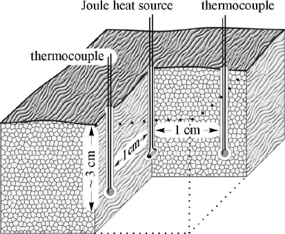
First, we studied time variations of the temperature inside a fragment of cow haunch muscle not undergone freeze after death and cut out so that muscle fibers be parallel to its surface (Fig. 1). A small resistance of size mm was embedded into the tissue at a depth of 3 cm through this free surface oriented horizontally. Passing a current through it we got a local heat source of controlled power. Thermocouples measuring the temperature were located at a distance of 1 cm from the resistance along the fibers and in the transverse direction.
We studied the heating dynamics for various values of the initial tissue temperature from 0 ∘C to 30 ∘C and various specimens of muscle, the results were practically identical. Fig. 2 demonstrates typical time variations of the tissue temperature obtained for two values of the heat source power equal to 1.2 W ( mA, O) and 2.7 W ( mA, O). The time origin, , is the beginning of heating and the vertical dashed line, min, represents the heating tune-out. All the shown curves were obtained for tissue regions undergone heating for the first time because under strong heating irreversible transformations of the living tissue structure can occur. It should be noted that the curves corresponding to heating power of 1.2 W were obtained for the tissue with the initial temperature C, so in Fig. 2 they are shifted down by 1.4 ∘C to make the comparison more easy.
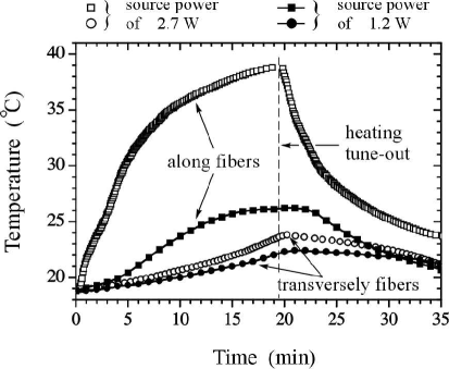
As seen in Fig. 2 the time variations of the tissue temperature along the fibers and in the transverse direction exhibit substantially different features. In fact, for the longitudinal direction the characteristic time of the temperature variations can be estimated as min from the -fold decrease after the heating tune-out. The same estimate will be get if we apply to the point where the curve describing the temperature increase under 1.2 W heating transfers from the concave to convex form. For the temperature increase under 2.7 W heating the latter time seems to be even shorter. The analogous curves describing the temperature variations in the transverse direction are characterized by a substantially longer time scale . In particular, the temperature increase curves remain concave up to the heating tune-out. So, if we related the time scales to the temperature diffusivities along the fibers and transversely them with the expression then we would have been to set that . From our point of view the latter estimate contradicts the physical properties of heat transfer in cellular tissue because it does not contain any regions where the heat conductivity takes so different values that could give rise to this estimate.
Under strong heating the cellular tissue coagulates and changes its properties, however, on the time scales under consideration it is essential only for temperatures exceeding C J . Therefore, this effect seems not to be substantial enough to play a key role in the heat propagation through the cellular tissue in the given case. Indeed, if we again assume the tissue temperature to be governed by the diffusion equation with, may be, an anisotropic diffusivity, then the stationary temperature field caused by a -source will be scaled up as when all the spatial scales are reduced by the factor , i.e. . So, under 2.7 W heating the tissue temperature should attain values about 60 ∘C at points being at a distance of 0.5 cm from the heat source along the fibers and at a substantially shorter distance in the transverse direction. Thus the region where the cellular tissue has to coagulate during the heating is of the relative volume not exceeding 10% with respect to the 11 cm3 region actually tested in the experiment. Under 1.2 W heating the region of the coagulated tissue is still less and even seems not to form at all because of finite dimensions of the resistance.
Fig. 2 also shows that heat propagation in cellular tissue under strong heating must be nonlinear. At least, the change of the heating power from 1.2 W to 2.7 W, i.e. by 2.3 times, corresponds to the temperature increase by 2.7 times at the points distant from the heat source along the fibers and only by 1.4 times at the points in the transverse direction.
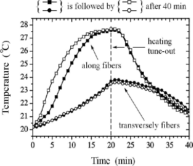
Fig. 3 demonstrates our test of the repeat irreversibility of the heating process showing if irreversible transformations of the tissue structure caused by strong heating are essential in the case under consideration. Namely, to answer the question of whether heat transfer in cellular tissue exhibiting such anomalous properties can go in the same way under repeated acts of heating we tested the temperature dynamics for the source power of 1.2 W. The obtained curves present time variations of the tissue temperature at the points shown in Fig. 1 during two successive processes of heating of the same tissue region that are separated in time by 40 min. We see not too significant change in the temperature variations only at the point distant from the heat source along the fibers that occurs at the stage of direct heating. During the repeated heating the temperature increase is characterized solely by a faster growth rate. So the mechanism responsible for these anomalous properties of heat transfer seems to be of reversible nature, at least, at first approximation.
We assume that these heat transfer anomalies are due to motion of the interstitial liquid induced by heating, which is demonstrated in the next section.
III Temperature dynamics in a tissue layer under strong surface heating
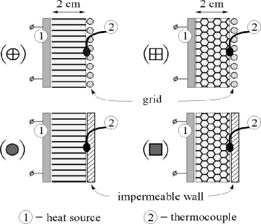
Following actually a standard way of measuring the tissue heat conduction we studied heat propagation through a tissue layer of thickness of 2 cm (Fig. 4). One of its surfaces was kept up at high temperatures about 100 ∘C whereas the temperature growth at the opposite surface was recorded by a thermocouple. The tissue layer was oriented vertically to avoid the gravitation effect. We studied four arrangements distinguished by the fiber orientation with respect to the walls bounding the layer and by the wall permeability to the interstitial liquid. In one case we used the impermeable walls to suppress the induced motion of the interstitial liquid, in the other we applied a grid to make it as free as possible. The distance between the walls was controlled by a certain small fixed force pressing the system so that there were no air-gaps between the tissue and the walls.
It should be noted that in the given situation heat propagation through the cellular tissue differs substantially from the case considered in the previous section. In fact, when the thickness of the tissue layer is much less than the other dimensions and its surface is heated uniformly, the temperature field can practically vary solely along the direction perpendicular to the layer surface. Under such conditions the interstitial liquid flow can appear only in the same direction and inevitably has to be affected by the properties of the opposite layer surface. For local heating of the cellular tissue at internal points distant far from its surface the structure anisotropy should give rise to the interstitial liquid flow of partly swirling form. In this case the effect of the surface properties seems to be weakened.
Fig. 5 exhibits the obtained results. When the tissue layer is bounded by the impermeable walls and the interstitial liquid has no way to move the evolution of tissue temperature is to be governed by the conventional heat diffusion and to depend weakly on the fiber orientation. Exactly such a behavior was observed. Then assuming the tissue temperature to obey the diffusion equation we can directly estimate the corresponding temperature diffusivity . Namely, in the given case the tissue temperature at one boundary is fixed and equal approximately to 100 ∘C and at the other we may impose the zero flux condition on the temperature field. The initial temperature is 20 ∘C and according to Fig. 5 the tissue temperature attains values about 50 ∘C at latter boundary in 10 min. Then fitting the solution of the diffusion equation subject to the appropriate conditions to these data we get the estimate cm2/s. The latter corresponds well to the available data for the tissue temperature diffusivity within the accuracy of our measurements.

In the tissue layer bounded by the grid heat transfer exhibits another behaviour. When the muscle fibers were parallel to the layer boundaries the temperature growth turned out to be of the same form as for the impermeable walls. By contrast, when the tissue fibers were perpendicular to the boundaries the temperature growth deviated substantially from the previous dependence (Fig. 5). We can explain this effect only admitting an essential flux of the interstitial liquid to appear due to the nonuniform heating of the cellular tissue. It is likely that the fiber arrangement gives rise to long channels in the muscle tissue running along the fibers. So, when there are no substantial obstacles to motion of the interstitial liquid nonuniform heating of the tissue is able to induce the interstitial liquid flux affecting remarkably heat transfer. To partly justify the given assumption we note that for the latter fiber orientation the blood efflux through the grid was visually strong in comparison with the other cases when the measured tissue temperature attained the values about 33 ∘C.
In order to make this phenomenon more evident the next section presents its certain visualization.
IV Mass propagation caused by nonuniform heating
If nonuniform heating of cellular tissue causes the interstitial liquid to flow in it affecting substantially heat propagation itself then the interstitial liquid flux can be directly visualized through the induced propagation of a certain substance dying the tissue. Indeed, diffusion coefficients of simple molecules in cellular tissue (of order 10-5 cm2/s or even less) are much less than the temperature diffusivity, so mass propagation in cellular tissue is to be essentially accelerated by this interstitial liquid flux.
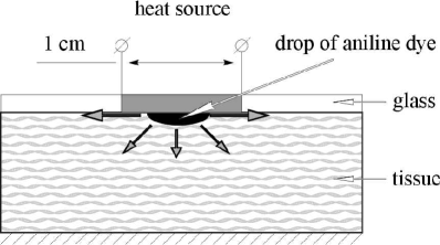
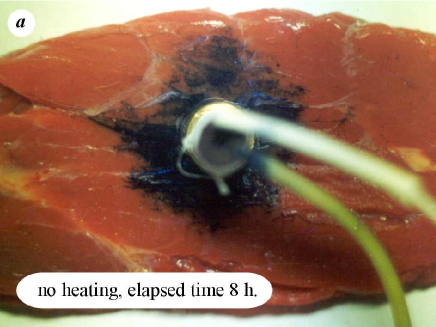
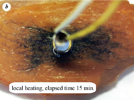
Figure 6 illustrates the experiment showing the effect of nonuniform heating on propagation of aniline dye in the cellular tissue. We put an aniline dye drop in a lunula on the horizontal tissue surface and pressed down the tissue with a glass plate to make the tissue surface plane and, thereby, to exclude the influence of the surface relief. A metallic disc of diameter of 1 cm was embedded into the glass plate placed so that this disc be right over the drop. An ohmic resistance was attached to the external disc surface and passing a current through it we locally heated the tissue. The colour patterns formed by the aniline propagation are represented in Fig. 7. Figure exhibits the colour pattern appeared in the tissue without heating after a lapse of 8 hours. As is seen, it is roughly of symmetrical form and the muscle fibers have no substantial effect on the aniline propagation in this case. The opposite situation is shown in figure for the aniline propagation under a strong local heating. Already after 15 min the colour pattern attained the previous one in size and got a substantially anisotropic form, the pattern dimension along the fibers is more that twice its mean size in the transverse direction. Moreover, in this figure it is clearly seen as aniline dry propagates inside small grooves on the tissue surface whereas without heating such grooves practically have no effect.
V Discussion and hypotheses
The presented preliminary experimental data enable us to suggest that heat transfer in cellular tissue under local strong heating is a more complex phenomenon than a simple heat diffusion. On one hand, the observed strong anisotropy in the heat propagation along the muscle fibers and in the transverse direction cannot be explained keeping in mind only the heat conduction through the cellular tissue. Indeed, otherwise, we should assume that the temperature diffusivity along the fibers, , is fully three times greater than the corresponding value in the transverse direction, (), whereas the cellular tissue contains no elements differing from one another so remarkably in the heat characteristics. On the other hand, the fiber arrangement endows the muscle tissue with a significant anisotropy for motion of the interstitial liquid through the tissue as through a porous medium. Therefore, if the nonuniform temperature field can cause the interstitial liquid to flow then heat propagation in the muscle tissue will be able to exhibit substantial anisotropy in properties. We think that in the presented experiments exactly this phenomenon, i.e., the interstitial liquid flux induced by nonuniform heating and, in turn, its effect on heat transfer was observed. Obviously, it should be nonlinear, which corresponds to the data presented in Fig. 2.
The experimental data shown in Fig. 3 enable us to assume that heat transfer in cellular tissue under such conditions is characterized by the repeat reversibility. The latter means that the time variations in the temperature of a cellular tissue undergoing repeated acts of heating go approximately in the same way. So a possible microscopic mechanism responsible for the induced flux of the interstitial liquid is also of the reversible nature, at least, it is not due to the tissue thermal coagulation or another thermal damage of the tissue. For example, it could be implemented through the liquid redistribution between the tissue cells and the intercellular space due to the change in the osmotic equilibrium induced by heating. In the given case an additional amount of the interstitial liquid appearing in the heated region has to give rise to its flow from this region.
We point out again that the interstitial liquid flux induced by heating seems to be the integral feature of heat transfer in cellular tissue. Therefore, only under special conditions heat propagation in cellular tissue is reduced solely to diffusion. Moreover, mass and heat propagation have to be related with each other through this flux, which is illustrated in Figs. 5 and 7.
Concluding the paper we would like to note that we observed anomalous properties of heat transfer in muscle tissue where the fibers endow the tissue with well developed anisotropy. There are organs, for example, kidney and liver whose cellular tissue does not possess significant anisotropy on the average regarding such an organ as a whole. For rigorously isotropic media the effect of a similar liquid flux induced by heating is likely to be depressed. However, on smaller scales these organs are certainly to exhibit local anisotropy with random orientation in space. Therefore, local strong heating again has to induce the interstitial liquid flux that, may be, changes its direction randomly in different regions of a given organ. In this case heat transfer is also to exhibit anomalous properties, which cannot be described, in general, by the conventional diffusion equation (see, e.g., I92 ).
Acknowledgements.
These investigations were support in part by the Russian State Programme “Integration”, Grant 0075.References
- (1) An Introduction to the Practical Aspects of Clinical Hyperthermia, S. B. Field and I. W. Hand eds. (London, Taylor & Francis, 1990).
- (2) Thermal Dosimetry and Treatment Planning, M. Gautherie ed. (Springer, Berlin, 1990).
- (3) Laser-Induced Interstitial Thermotherapy, G. Müller and A. Roggan, eds. (SPIE Optical Engineering Press, Bellingham, 1995).
- (4) Heat Transfer in Medicine and Biology, Analysis and Application, A. Shitzer and R. C. Eberhart eds. (Plenum, New York, 1985) v. 1,2.
- (5) V. V. Gafiychuk and I. A. Lubashevsky. Mathematical Description of Heat Transfer in Living Tissue, (VNTL Publishers, Lviv, 1999); e-print: adap-org/9911001, 9911002. The first announcement of the obtained results was also presented in BLG1 ; BLG2 for the continuum description of living tissue and in BLG3 ; BLG4 for the effect of the vessel discreteness.
- (6) C. W. Song, A. Lokshina, J. G. Rhee, M. Patten, and S. H. Levitt. “Implication of blood flow in hyperthermia treatment of tumors”, IEEE Trans. Biom. Eng. BME-31, n. 1, pp. 9–16 (1984).
- (7) I. A. Lubashevsky and V. V. Gafiychuk. “A simple model of self-regulation in large natural hierarchical systems”, J. Env. Syst. 23(3), pp. 281–289 (1995).
- (8) I. A. Lubashevsky and V. V. Gafiychuk “Cooperative mechanism of self-regulation in hierarchical living systems”, SIAM J. Appl. Math. 60, pp. 633–663 (2000); adap-org/9808003.
- (9) M. M. Chen and K. R. Holmes. “Microvascular contributions in tissue heat transfer”, Ann. N.Y. Acad. Sci. 335, pp. 137–154 (1980). It should be noted, however, that the first attempt to describe heat transfer in living tissue was made in the paper by Pennes, H. H. “Analysis of tissue and arterial blood temperatures in the resting human forearm”, J. Appl. Phys. 1, pp. 93–122 (1948).
- (10) J. W. Baish, P. S. Ayyaswamy, and K. R. Foster. “Small-scale temperature fluctuations in perfused tissue during local hyperthermia”, ASME J. Biomech. Eng. 108, pp. 246–250 (1986).
- (11) J. J. W. Lagendijk. “The influence of blood flow in large vessels on the temperature distribution hyperthermia”, Phys. Med. Biol. 27, pp. 17–23 (1982).
- (12) J. Grezee and J. J. W. Lagendijk. “Temperature uniformity during hyperthermia: the impact of large vessels”, Phys. Med. Biol. 37(6), pp. 1321–1337 (1992).
- (13) R. B. Roemer. “Thermal dosimetry”, in: Thermal Dosimetry and Treatment Planning, M. Gautherie ed. (Berlin, Springer, 1990), pp. 119–214.
- (14) Z. P. Chen, R. B. Roemer. “The effects of large blood vessels on temperature distributions during simulated hyperthermia”, J. Biomech. Eng. 114(4), pp. 473–481 (1992).
- (15) S. Weinbaum and L. M. Jiji. “A new simplified bioheat equation for the effect of blood flow on local average tissue temperature”, Trans. ASME J. Biom. Eng. 107, pp. 131–139 (1985).
- (16) S. Weinbaum, L. X. Xu, L. Zhu, and A. Ekpene. “A new fundamental bioheat equation for muscle tissue: Part I–Blood perfusion term”, ASME J. Biomech. Eng. 119, pp. 278–288 (1997).
- (17) J. C. Chato. “Heat transfer to blood vessels”, ASME J. Biom. Eng. 102, pp. 110–118 (1980).
- (18) A. N. T. J. Kotte, G. M. J. van Leeuwen, and J. J. W. Lagendijk. “Modelling the thermal impact of a discrete vessel tree”, Phys. Med. Biol. 44, pp. 57–74 (1999).
- (19) K. Giering, O. Minet, I. Lamprecht,and G. Müller. “Review of thermal properties of biological tissue”, in: Laser-Induced Interstitial Thermotherapy, G.J. Müller and A. Roggan eds. (SPIE Optical Engineering Press, Bellingham, 1995), pp. 45–65.
- (20) R. P. Singh and D. R. Heldman. Introduction to Food Engineering, 2-nd edition (Academic Press, New York, 1993).
- (21) H. Arkin, K. R. Holmes, and M. M. Chen. “A technique for measuring the thermal conductivity and evaluating the “apparent conductivity” concept in biomaterials”, J. Biomech. Eng. 111(4), pp. 276–282 (1989).
- (22) J. W. Valvano, J. T. Allen, and H. F. Bowman. “The simultanious measurment of thermal conductivity, thermal diffusivity and perfusion in small volumes of tissue”, J. Biomech. Eng. 106, pp. 192–197 (1984).
- (23) G. T. Anderson, J. W. Valvano, and R. R. Santos. “Self-heated thermistor measurements of perfusion”, IEEE Trans. Biomed. Eng. 39(9), pp. 877–885 (1992).
- (24) D. Y. Yuan, J. W. Valvano, and G. T. Anderson. “Measurement of thermal conductivity, thermal diffusivity, and perfusion”, Biomed. Sci. Instrum. 29, pp. 435–442 (1993).
- (25) J. Grezee, J. Mooibroek, C. K. Bos, and J. J. W. Lagendijk. “Interstitail heating: experiments in artificial perfused bovine tongues”, Phys. Med. Biol. 36(6), pp. 823–833 (1991).
- (26) H. Arkin, L. X. Xu, and K. R. Holmes. “Recent developments in modeling heat transfer in blood perfused tissues”, IEEE Trans. Biomed. Eng. 41(2), pp. 97–107 (1994).
- (27) M. C. Kolios, A. E. Worthington, M. D. Sherar, and J. W. Hunt. “Experimental evaluation of two simple thermal models using transient temperature analysis”, Phys. Med. Biol. 43(11), pp. 3325–3340 (1998).
- (28) S. J. Jacques. “Laser-tissue interections: photochemical, photothermal, and photomechanical”, Surg. Clin. North Am. 72(3), 531–558 (1992).
- (29) K. Mitra, S. Kumar, A. Vedavarz, and M. K. Moallemi. “Experimental evidance of hyperbolic heat conduction waves in processed meat”, J. Heat Transfer 117, pp. 568–573 (1995).
- (30) M. B. Isichenko. “Percolation, statistical topography, and transport in random media”, Rev. Mod. Phys. 64, n. 4, pp. 961–1043 (1992).
- (31) I. A. Lubashevsky, A. V. Priezzhev, V. V. Gafiychuk, and M. G. Cadjan. “Free boundary model for local thermal coagulation”, in: Laser-Tissue Interaction VII, S. L. Jacques, ed., Proc. SPIE 2681, pp. 81–91 (1996).
- (32) I. A. Lubashevsky, A. V. Priezzhev, V. V. Gafiychuk, and M. G. Cadjan. “Local thermal coagulation due to laser–tissue interaction as irreversible phase transition”, J. Biomed. Opt. 2(1), pp. 95–105 (1997).
- (33) I. A. Lubashevsky, A. V. Priezzhev, V. V. Gafiychuk, and M. G. Cadjan. “Dynamic free boundary model for laser thermal tissue coagulation”, in: Laser-Tissue Interaction and Tissue Optics II, H. J. Albrecht, G. Delacrétaz, T. H. Meier, R. W. Steiner, and L. O. Svaasand, eds., Proc. SPIE 2923, pp. 48–57 (1996).
- (34) I. A. Lubashevsky, A. V. Priezzhev, and V. V. Gafiychuk. “Effective interface dynamics of heat diffusion limitted thermal coagulation”, J. Biomed. Opt. 3(1), pp. 102–111 (1998).