[
Role of Secondary Motifs in Fast Folding Polymers: A Dynamical Variational Principle
Abstract
A fascinating and open question challenging biochemistry, physics and even geometry is the presence of highly regular motifs such as -helices in the folded state of biopolymers and proteins. Stimulating explanations ranging from chemical propensity to simple geometrical reasoning have been invoked to rationalize the existence of such secondary structures. We formulate a dynamical variational principle for selection in conformation space based on the requirement that the backbone of the native state of biologically viable polymers be rapidly accessible from the denatured state. The variational principle is shown to result in the emergence of helical order in compact structures.
]
A fundamental problem in every day life is that of packing with examples ranging from fruits in a grocery, clothes and personal belongings in a suitcase, atoms and colloidal particles in crystals and glasses, and amino acids in the folded state of proteins. The simplest problem in packing consists of determining the spatial arrangement that accomodates the highest packing density of its constituent entities with the result being a crystalline structure. Besides packing considerations, dynamical effects play a significant role when rapid packing/unpacking is entailed, as in the formation of amorphous glasses where crystallization is dynamically thwarted or in the more familiar suitcase problem.
Fast packing has been recognized as a central issue for biopolymers, such as proteins, since the early work of Levinthal [1]. Further, the native conformations display extremely regular motifs, such as -helices or -sheets. In this Letter we postulate a direct connection between the dynamics of rapid folding and the emergence of secondary motifs in the native state conformations. In fact, an intuitive approach to rapid and reproducible folding might be to create neat patterns of lower dimensional manifolds than the physical space and bend and curl them into the final folded state. For proteins, secondary structures such as -helices and -sheets are indeed patterns in low dimensions.
There are two key aspects distinguishing a protein from a generic heteropolymer: the specially selected sequence of amino acids and the three-dimensional structure that it folds reversibly into. For a given target native structure, the selection mechanism in sequence space is the principle of minimal frustration [2]. The chosen sequences are such that their target native states are reached through a funnel-like landscape [3] which facilitates the harmonious fitting together of pieces to form the whole.
The three-dimensional structure impacts on the functionality of the protein and a fascinating issue is the elucidation of the selection mechanism in conformation space that picks out certain viable structures from the innumerable ones with a given compactness. Earlier studies have shown that there is a direct link between viable native conformations and high designability [4]. Recently [5], it was observed that the natural folds of proteins have a much larger density of nearby structures than generic (artificial) conformations of the same character and that the exceedingly large geometrical accessibility of natural proteins may be related to the presence of secondary motifs.
The realization that proteins have secondary structures arose with early crystallographic studies and the brilliant deduction of Pauling et al. [6] of the ability of an -helix of the correct pitch to accomodate hydrogen bonds, thus promoting its stability. Inspired by the findings of Pauling, helix-coil transition models have been used to study the thermodynamics of helix formation [7]. The models encompass features that ensure the helical nature of the low-energy states by assuming first that that monomers can be in a helical state and by then introducing co-operative interactions that favor helical regions. It is interesting to note, however, that the number of hydrogen bonds is nearly the same when a sequence is in an unfolded structure in the presence of a polar solvent or in its native state rich in secondary structure content [8]. It has also been suggested that the -helix is an energetically favorable conformation for main-chain atoms but the side-chain suffers from a loss of entropy [8, 9]. Nelson et al. [10] have shown both numerically and experimentally that non-biological oligomers fold reversibly like proteins into a specific three-dimensional structure with high helical content driven only by solvophobic interactions. Recent studies have attempted to explain the emergence of secondary structure from geometrical principles rather than invoking detailed chemistry. Despite the concerted efforts of several groups, a simple general explanation remains elusive. In particular, the work of Yee et al. [11], Hunt et al. [8], and Socci et al. [12] have shown that compactness alone can only account for a small secondary structure content. These facts are also corroborated by the recent study of the kinetics of homopolymer collapse, where no evidence was found for the formation of local regular structures [13].
We propose a selection mechanism in structure space in the form of a variational principle postulating that, among all possible native conformations, a protein backbone will attain only those which are optimal under the action of evolutionary pressure favouring rapid folding. Our goal is to elucidate the role played by the bare native backbone independent of the selection in sequence space and hence of the (imperfectly-known) inter-amino-acid potentials. We therefore choose to employ a Go-like model [14] with no other interaction that promotes or disfavours secondary structures. The model is a sequence-independent limiting case of minimal frustration [2] which, for a given target native state conformation, favours the formation of native contacts – the energy of a sequence in a conformation is simply obtained as the negative of the number of contacts in common with the target conformation. We will consider two non-consecutive amino acids to be in contact if their separation is below a cutoff Å(the results are qualitatively similar when slightly different values of in the range Åare chosen).
The energy of structure in the Go model is given by
| (1) |
where the sum is taken over all pairs of amino acids, is the target structure, is the contact map of structure :
| (2) |
where is the distance of amino acids and .
The polypeptide chain is modelled as a chain of beads subject to steric constraints [15, 5]. We adopted a discrete representation similar to the one of Covell and Jernigan [15], in which each bead occupies a site of an FCC lattice with lattice spacing equal to 3.8 Å. Such a representation is able to describe the backbone of natural proteins to better that 1 Å rmsd per residue (equal to the best experimental resolution) and preserves typical torsional angles. All discretized structures were subject to a suitable constraint: any two non-consecutive residues cannot be closer than Å due to excluded volume effects and the distance between consecutive residues can fluctuate between 2.6 Å 4.7 Å. Such constraints were determined by an analysis of the coarse-grainings of several proteins of intermediate length ( residues). In order to enforce a realistic global compactness for a backbone of length , the number of contacts in all the target structures considered was chosen [16] to be around while, locally, no residue was allowed to make contact with four or more consecutive residues.
In order to assess the validity of the variational principle, it is necessary to evaluate the typical time, , taken to fold into a given target structure, , followed by a selection of the structures , that have the smallest folding times. To do this, an initial set of ten conformations was generated by collapsing a loose chain starting from random initial conditions. In each case, we modified the random initial conformation by using Monte Carlo dynamics: we move up to 3 consecutive beads to unoccupied discrete positions that do not violate any of the physical constraints and accept the moves according to the standard Metropolis rule. The energy is given by eq. (1), while the temperature for the MC dynamics was set to 0.35. This value was chosen in preliminary runs so that it was higher than the temperature [2] below which the sequence is trapped in metastable states but comparable to the folding transition temperature so that conformations with significant overlap with the native state are sampled in thermal equilibrium.
For each structure, as a measure of the folding time we took the median over various attempts (typically 41) of the total number of Monte Carlo moves necessary to form a pre-assigned fraction of native contacts, typically 66%, starting from a random conformation. Our results were unaltered on increasing this fraction to 75%; indeed, this fraction could be progressively increased towards 100% with successive generations without increase in the computational cost since better and better folders are obtained.
A new generation of ten structures is created by “hybridizing” pairs of structures of the previous generation ensuring that structures with small folding times are hybridized more and more frequently as the number of generations, , increases [17]. To do this, each of the two distinct parent structures to be hybridized, and are chosen with probability proportional to , where is the index of the current generation (initially equal to 1), is the median folding time. Then, a hybrid map is created by taking the union of the two parent maps:
| (3) |
Because it is not guaranteed that corresponds to a three-dimensional structure obeying the same physical constraints as and , the corresponding hybrid is constructed by taking one of the two parent structures (or alternatively a random one) as the starting conformation and carrying out MC dynamics favouring the formation of each of the contacts in the union map (i.e. using eq. (1) with substituted by ). The dynamics is carried out starting from a temperature of and then decreasing it gradually over a sufficiently long time (typically thousands of MC steps) to achieve the maximum possible overlap with the union map, while simultaneously maintaining the realistic compactness. The resulting hybrid structure is typically midway between the two parent structures, in that it inherits native contacts from both of them. We adopted the following definition in order to obtain an objective and unbiased way to quantitatively estimate the presence of secondary content: a given residue, was defined to belong to a secondary motif if, for some , one of these conditions held:
| (4) | |||||
| (5) | |||||
| (6) | |||||
| (7) |
The former [latter] identifies the presence of helices and parallel [anti-parallel] sheets in natural proteins, which can be identified by the visual inspection of contact matrices and appears as thick bands parallel or orthogonal to the diagonal.
The upper plot of Fig. 1 shows the decrease of the typical folding time over the generations for chains of length 25, while the middle panel shows the accompanying increase in the number of residues in secondary motifs (secondary content). The bottom panel shows a milder decrease of the contact order (i.e. a larger number of short-range contacts) as the generations evolved, in agreement with the experimental findings of Plaxco et al.[18]
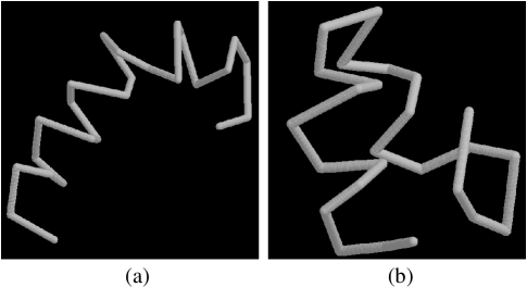
One of the optimal structures of length 25 is shown in Figure 2a. Due to the absence of any chirality bias in our structure space exploration, the helix does not have a constant handedness. The signature of the secondary motifs in the optimal structures is clearly visible in the contact maps of Figure 3, which are not sensitive to structure chirality. Strikingly, the variational principle selects conformations with significant secondary content as those facilitating the fastest folding. The correlation of the emergence of secondary structures with decrease of folding times is shown in the plot of Fig. 4. We verified that the hybridization procedure is not biased towards low contact order by iterating it for various generations and hybridizing the structures at random. Even after dozens of generations, the generated structures had secondary contents of about 1/3-1/4 of the true extremal structures.
The very high secondary content in optimal conformations was found to be robust against changes in chain length or compactness of the target structure. On requiring that the structure be more compact, bundles of helices emerge [see Fig. 2b] along with an increase in contact order, signalling the presence of some longer range contacts, which are necessitated in order to accomodate the shorter radius of gyration. It is noteworthy that our calculations lead predominantly to -helices and not sheets, a fact accounted for by the demonstration that steric overlaps and the associated loss of entropy lead to the destabilization of helices in favor of sheets [9], the appearance of such sheets only in sufficiently long proteins [19] and the much slower folding rate of -sheets compared to -helices [20]. It is remarkable that the same requirement of rapid folding is sufficient to lead to a selection in both sequence and structure space underscoring the harmony in the evolutionary design of proteins. The results and strategies presented here ought to be applicable in protein-engineering contexts, for example by ensuring optimal dynamical accessibility of the backbone of proteins. A systematic collection of the rapidly-accessible structures of various length should also lead to the creation of unbiased libraries of protein folds.
Acknowledgements This work was supported by INFM, INFN sez. di
Trieste, NASA, NATO and The Donors of the Petroleum Research Fund
administered by the American Chemical Society. We thank F. Seno and A.
Trovato for useful discussions.
REFERENCES
- [1] C. Levinthal, J. Chim. Phys. 65, 44 (1968).
- [2] J. D. Bryngelson and P. G. Wolynes, Proc. Natl. Acad. Sci. USA 84, 7524-7528 (1987); J. D. Bryngelson, J. N. Onuchic, J. N. Socci and P. G. Wolynes, Proteins: Struc. Funct. Genet. 21, 167-195 (1995).
- [3] P. E. Leopold, M. Montal and J. N. Onuchic, Proc. Natl. Acad. Sci. USA 89, 8721-8725 (1992); P. G. Wolynes J. N. Onuchic and D. Thirumalai, Science 267, 1619-1620 (1995); J. N. Onuchic, Z. Luthey Schulten and P. G. Wolynes, Ann. Rev. Phys. Chem. 48, 545-600 (1997); K. A. Dill and H. S. Chan, Nature Structural Biology 4, 10-19 (1997).
- [4] H. Li, R. Helling, C. Tang and N. Wingreen, Science 273, 666-669 (1996); N. E. G. Buchler and R. A. Goldstein, Proteins: Struc. Funct. Genet. 34, 113-124 (1999); C. Micheletti, A. Maritan, J. R. Banavar and F. Seno, Phys. Rev. Lett. 80, 5683 (1998); C. Micheletti, A. Maritan and J. R. Banavar, J. Chem. Phys. 110, 9730 (1999).
- [5] C. Micheletti, J. R. Banavar, A. Maritan and F. Seno,Phys. Rev. Lett. 82, 3372-3375 (1999).
- [6] L. Pauling, R. B. Corey and H. R. Branson, Proc. Nat. Acad. Sci. 37, 205-208 (1951).
- [7] B. H. Zimm and J. Bragg, J. Chem. Phys., 31, 526 (1959); O. B. Ptitsyn and A. M. Skvortsov, Biophys. 10, 1007 (1965); I. M. Lifshitz, A. Y. Grosberg and A. R. Khokhlov, Rev. Mod. Phys., 50, 683 (1978).
- [8] N. G. Hunt, L. M. Gregoret and F. E. Cohen, J. Mol. Biol. 241, 214-225 (1994).
- [9] R. Aurora, T. P. Creamer, R. Srinivasan and G. D. Rose, J. Mol. Biol. 272, 1413-1416 (1997).
- [10] J. C. Nelson, J. G. Saven, J. S. Moore, and P. G. Wolynes, Science 277, 1793-1796 (1997).
- [11] D. P. Yee, H. S. Chan, T. F. Havel and K. A. Dill, J. Mol. Biol. 241, 557-573 (1994).
- [12] N. D. Socci, W. S. Bialek, and J. N. Onuchic, Phys. Rev. E 49, 3440-3443 (1994).
- [13] A. Halperin and P. M. Goldbart, Phys. Rev. E, in press (cond-mat/9905306).
- [14] N. Go, Macromolecules 9, 535-541 (1976).
- [15] D. G. Covell and R. Jernigan Biochem. 29, 3287 (1990).
- [16] The native state structures of monomeric proteins of length between 50 and 200 show an excellent correlation of this form, when two non-consecutive amino acids along the sequence are defined to be in contact when they are within 6.5 Å of each other.
- [17] J. H. Holland, Adaptation in natural and artificial systems, MIT press ed. (1992).
- [18] K. M. Plaxco, K. T. Simons and D. Baker, J. Mol. Biol. 277, 985-994 (1998).
- [19] A. P. Capaldi and S. E. Radford, S. E., Curr. Op. Str. Biol. 8, 86-92 (1998).
- [20] V. Muñoz, E. R. Henry, J. Hofrichter and W. A. Eaton, Proc. Natl. Acad. Sci. USA, 95, 5872 (1998).