[
Charge localization and phonon spectra in hole doped La2NiO4
Abstract
The in-plane oxygen vibrations in La2NiO4 are investigated for several hole-doping concentrations both theoretically and experimentally via inelastic neutron scattering. Using an inhomogeneous Hartree-Fock plus RPA numerical method in a two-dimensional Peierls-Hubbard model, it is found that the doping induces stripe ordering of localized charges, and that the strong electron-lattice coupling causes the in-plane oxygen modes to split into two subbands. This result agrees with the phonon band splitting observed by inelastic neutron scattering in La2-xSrxNiO4. Predictions of strong electron-lattice coupling in La2NiO4, the proximity of both oxygen-centered and nickel-centered charge ordering, and the relation between charged stripe ordering and the splitting of the in-plane phonon band upon doping are emphasized.
pacs:
PACS numbers: 75.60.Ch, 74.25.Kc, 71.45.Lr, 71.38.+i]
There is currently great interest in the importance of charge localization and ordering tendencies in a variety of doped transition metal oxides: including nickelates, bismuthates, cuprates, and manganites[1, 2, 3, 4, 5, 6, 7]. Recent experiments have suggested nanoscale coexistence of charge and spin ordering, as well as related multiscale dynamics [1, 2, 3, 4, 5, 6, 7, 8, 9, 10]. The cuprates have been widely investigated, both theoretically and experimentally, as this inhomogeneity may be related to high-temperature superconductivity[11, 12, 13, 14, 15, 16, 17, 18].
The nickelates are considered strong electron-lattice (e-l) coupling systems, which helps stabilize charge-ordering in the form of ”stripe” phases [19, 20]. For the commensurate 1/3 doping case of La1.67Sr0.33NiO4, it has been shown in optical absorption and Raman scattering experiments that new phonon modes appear when the temperature is lowered below the stripe-ordering temperature (=240 K); this is a signature of the stripe formation [8, 9, 10]. Until now, only the temperature dependence and the apical oxygen (Ni-O(2)) vibrations have been investigated. However, the doping dependence and the in-plane oxygen vibrations (Ni-O(1) stretching modes) are also very important for the properties of the quasi-two dimensional nickelate materials. In this study, we use an inhomogeneous Hartree-Fock (HF) plus random-phase approximation (RPA) numerical method for a two-dimensional (2D), four-band Peierls-Hubbard model, to interpret the inelastic neutron scattering spectra. This reveals specific signatures of the stripe patterns in the in-plane oxygen phonons.
Our main results are: (i) There is agreement between the results from our multiband model including electron-electron and e-l interactions and the inelastic neutron scattering spectra for the in-plane oxygen vibrations with various commensurate hole-doping concentrations; (ii) The theoretical results predict new vibrational modes (“edge modes”) which are associated with oxygen motions near localized holes or in the vicinity of stripes; (iii) The e-l coupling strength, at which the best agreement between our model and the inelastic neutron scattering data is achieved, is close to the transition from an oxygen-centered stripe phase to a nickel-centered one. This suggests that the nickelates may be in a mixed state of both stripe phases, and sensitive to temperature, pressure and magnetic field.
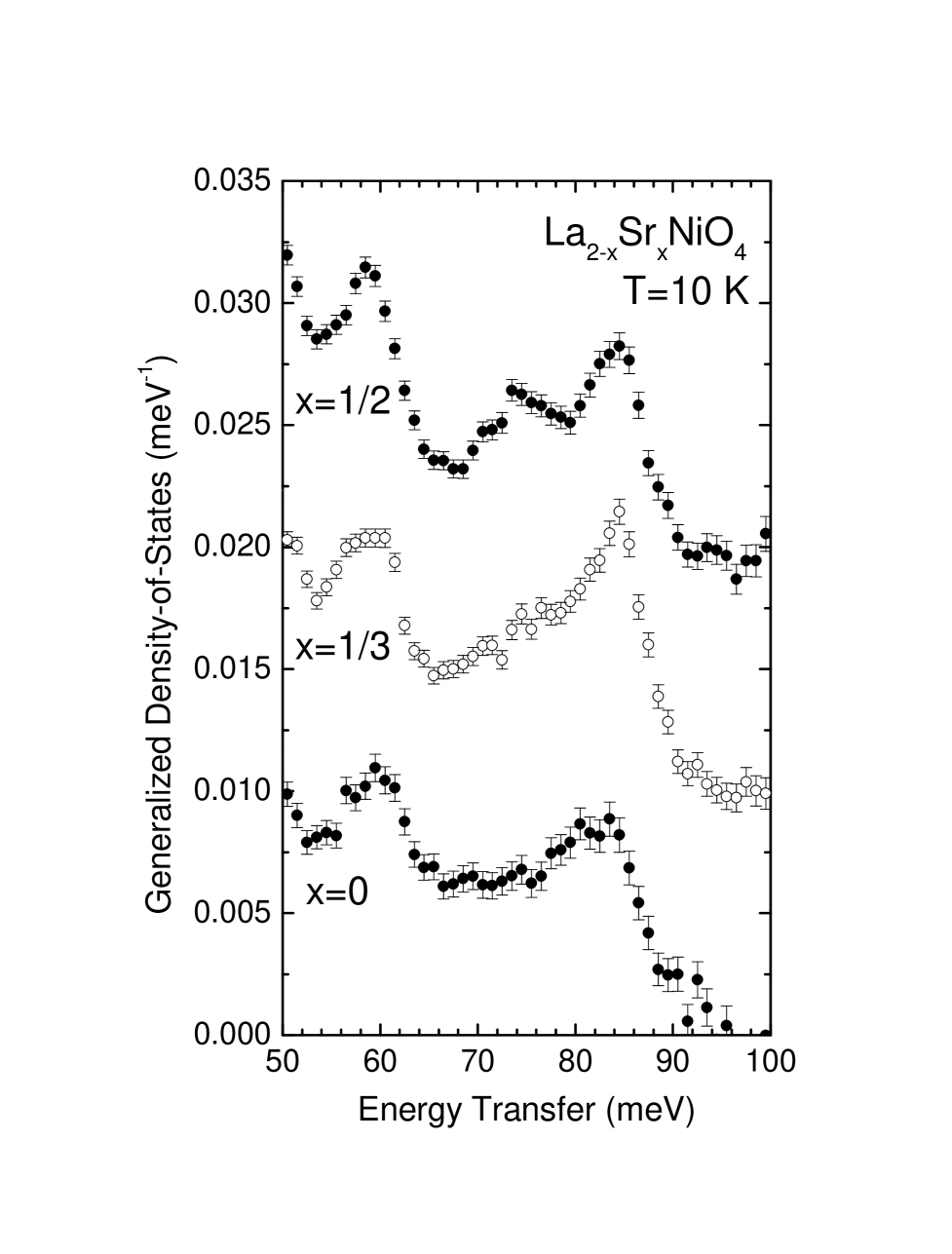
The inelastic neutron scattering spectra were measured on polycrystalline La2-xSrxNiO4 for various doping concentrations, 0, 1/8, 1/4, 1/3, 1/2. Time-of-flight neutron scattering measurements were performed on the Low Resolution Medium Energy Chopper Spectrometer at Argonne National Laboratory’s Intense Pulsed Neutron Source. For all measurements, an incident neutron energy of 120 meV was chosen and data were summed over all scattering angles from . Detailed information on the experiment, as well as the preparation of the samples used, can be found in Ref. [21]. The focus is on the (neutron-scattering-weighted) generalized density-of-states (GDOS) of phonons and particular attention is given to the in-plane oxygen vibrations, i.e., the Ni-O(1) stretching modes.
Figure 1 shows the experimental GDOS for several hole concentrations at =10 K for phonon modes in the range from 50-100 meV. From the analysis of lattice dynamical shell model calculations, it is known that the in-plane Ni-O(1) oxygen stretching modes are separated in frequency from other vibrations, and the phonon intensity above 65 meV is associated entirely with these vibrations. For the low doping samples ( and , not shown), there is little change in the 81 meV phonon band [21], but for the and samples, a new peak appears around 75 meV along with a slight hardening of the main band. This feature is interpreted as a splitting of the 81 meV Ni-O(1) stretching band into two phonon bands centered at approximately 83 meV and 75 meV.
In order understand this dependence of the Ni-O(1) stretching modes on hole concentration and its possible reflection of stripe ordering, we have performed a calculation of the phonon spectrum in a minimal Peierls-Hubbard model in 2D. Due to the strong e-l coupling expected in the nickelates, we resort to modeling with an inhomogeneous HF plus RPA numerical approach [22, 23, 24]. This has proven to be a very robust method for studying charge localization and stripe formation, especially when electron-lattice coupling is strong, obviating subtle many-body effects and quantum fluctuations [25].
We use a 2D four-band extended Peierls-Hubbard model of a doped NiO2 plane, which includes both electron-electron and e-l interactions [23, 24]. Here, for nickelate oxides, besides the orbital used in the cuprate oxide models [23], the Ni orbitals must be included to account for the higher spin state () at half-filling (i.e. undoped). Our model Hamiltonian is [25]:
| (1) | |||||
| (2) |
where creates a hole with spin at site in orbital (Ni , , or O ). The Ni-O hopping has two values: between and and between and . The O-site electronic energy is , and Ni-site energies are and for , with the crystal-field splitting on the Ni site. describes the electron correlations in the Ni orbitals,
| (3) | |||||
| (4) | |||||
| (5) |
The electron-electron interactions include the on-site Ni Coulomb repulsions () as well as the Hund interaction () at the same Ni site to account for the high spin situation. (The interplay of the two orbitals can also lead to pseudo Jahn-Teller distortions, but these are not our focus here). We emphasize that, due to the large spin at the nickel site, Hund’s rule leads to ferromagnetic exchange coupling , and ,with the Pauli matrix. For the e-l interaction, we consider that the Ni-O hopping is modified linearly by the O-ion displacement as , where the () applies if the bond shrinks (stretches) with positive . For the lattice terms, we study only the motion of O ions along the Ni-O bonds—other oxygen (or Ni) distortion modes can readily be included if necessary. It is known that for the nickelate oxides the e-l coupling is stronger than in cuprate oxides [19, 20], and is therefore likely to play an even more decisive role in the formation, localization, and nature of stripe phases. We adopt the following representative paremeters for the nickelate materials [20]: , , , , and (all in units of ). In real oxides [20, 26], is estimated to be in the range 1.3 eV 1.5 eV. The electron-lattice coupling strength is varied to achieve a best fit to the neutron scattering data; we find . The commensurate doping cases are examined in a 4x4 unit supercell for , 1/2 and a 3x3 unit supercell for . Periodic boundary conditions are used.
The densities-of-states (DOS) of in-plane phonons were calculated from our model at , 1/3, 1/2 and are shown in Fig. 2. For the undoped case, as the ground state is spatially homogeneous, only one oxygen phonon band appears, centered around 80.5 meV. When holes are added into the NiO2 plane at , the ground state is found to be a stripe pattern with more holes accumulating along the (1,1,0) direction, forming an antiphase domain wall within the original antiferromagnetic background. This is consistent with many neutron, optical and Raman scattering experiments [4, 8, 9, 10]. Interestingly, we find that a new phonon band appears centered at 75 meV. In addition, there is a hardening of the main phonon band (to 83 meV) corresponding to an overall splitting of 8 meV. From examination of the eigenvectors, the main character of this band is local oxygen vibrations in the vicinity of the stripe, i.e. having the nature of localized ”edge modes” (see [25] for more details). For the higher doping , the charge-ordering takes on a commensurate checker-board pattern. A similar splitting of the 85 meV phonon band into two phonon bands around 83 and 75 meV is again found. At this half doping, the checker-board ground state is in essence a commensurate charge-density-wave (CDW) system with the nature of an ordered binary alloy.
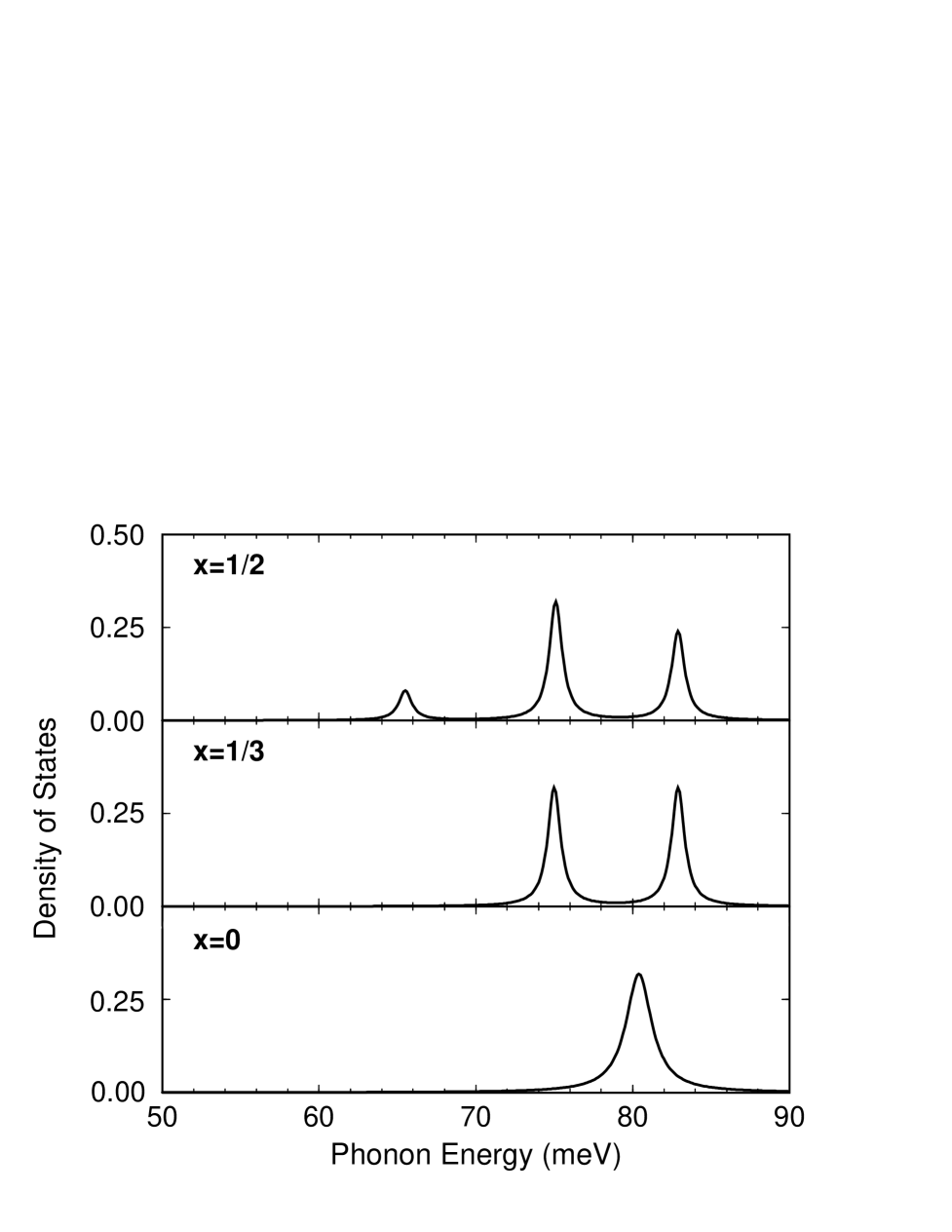
The above results are in agreement with the GDOS data obtained from inelastic neutron scattering, which are shown in Fig. 1. In addition to the splitting energy, even the slight hardening of the main band observed experimentally is accounted for in the model. Besides the new phonon modes centered at 75 meV appearing for the 1/3 and 1/2 doping, another low intensity phonon mode around 65 meV is also predicted in our model at (Fig. 2). The signature for these modes are weak, however, so that they may be difficult to detect in the current experiment. In so far as we have included only a small subset of the possible oxygen displacement patterns and wavevectors in the model (whereas the neutron scattering experiment samples all wavevectors and polarizations), the relative intensities and widths of the bands obtained by experiment and theory cannot be usefully compared.
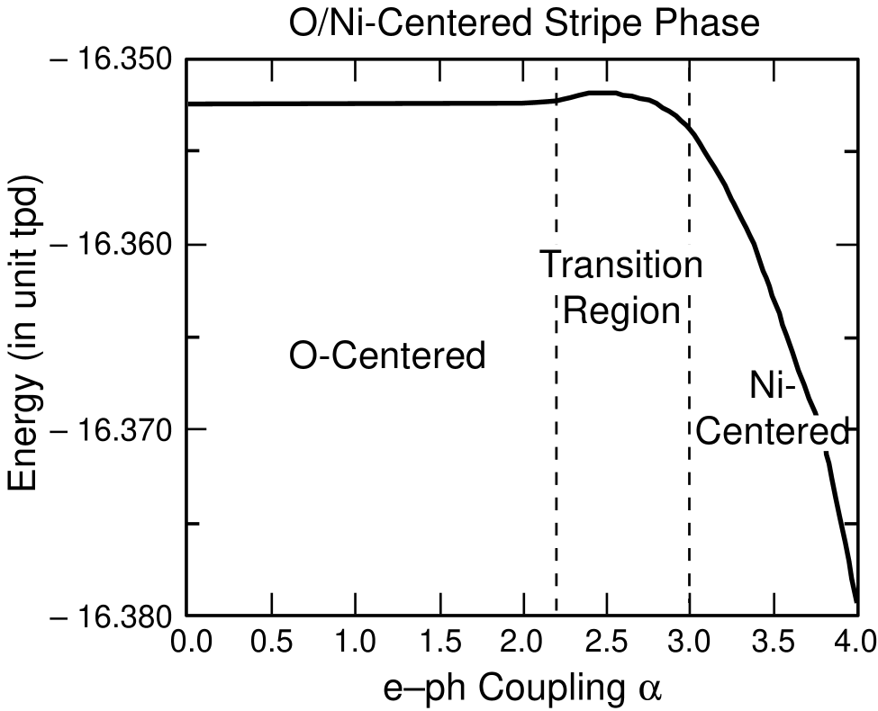
We emphasize that the excellent agreement between our model and the GDOS experimental data is achieved by varying the e-l coupling strength to match the positions of the phonon bands. As noted, the choice of best fits the data; our model calculations predict a variation of the phonon splitting with which is quite large ( 7 meV). Most strikingly, as illustrated in Fig. 3, an O-centered stripe is found as the ground state at small , while for a larger , a Ni-centered stripe is found as the ground state[25]. The transition region includes , where the best agreement between Fig. 2 based on our model and Fig. 1 on the inelastic neutron scattering spectra is achieved. It has been suggested from various experimental data that stripe formation for can not be simply assigned as Ni-centered or O-centered [19], but is also dependent on temperature. Our comparison of theory and experiment provides a possible explanation on the sensitivity of stripe formation; they suggest that La1.67Sr0.33NiO4 may be in a mixed stripe phase state, and also in a region of sensitivity to temperature, pressure, magnetic field, etc.
In conclusion, we have made a study of oxygen breathing lattice vibrations in La2-xSrxNiO4 via inelastic neutron scattering compared with predictions of a 2D four-band model, including both electron-lattice and electron-electron interactions. The in-plane oxygen vibrations above 65 meV were thoroughly investigated. The splitting of the in-plane 81 meV band upon doping into two subbands centered around 75 meV and 83 meV is observed experimentally and predicted theoretically, and interpreted in terms of new localized phonon modes (“edge modes” at charge localized stripes). The excellent agreement between the experiment and the model strongly supports the view that strong electron-lattice coupling in this kind of material plays a decisive role on the charge localization and mesoscopic stripe formation. For the doping at , at which stripes are found both in experiments and our model, our results suggest that there may be a mixed state of O- and Ni-centered stripe phases, and sensitivity to temperature, pressure, and magnetic field. Our model calculations also predict distinctive dispersion of the phonon bands, as well as inhomogeneous magnetoelastic coupling along the boundaries between charge-rich and magnetic nanophase domains [25]. These predictions require additional experiments for their confirmation and consequences.
We have benefitted from valuable discussions with Dr. J. T. Gammel. This work is supported (in part) by the U. S. Department of Energy under contract W-7405-Eng-36 with the University of California. This work has benefited from the use of the Intense Pulsed Neutron Source at Argonne National Laboratory. This facility is funded by the U. S. Department of Energy, BES-Materials Science, under contract W-31-109-Eng-38.
REFERENCES
- [1] J. M. Tranquada, D. J. Buttrey, V. Sachan, and J. E. Lorenzo, Phys. Rev. Lett. 73, 1003 (1994).
- [2] J. M. Tranquada et al., Phys. Rev. B 54, 7489 (1996).
- [3] C. H. Chen, S.-W. Cheong and A. S. Cooper, Phys. Rev. Lett. 71, 2461 (1994).
- [4] S.-W. Cheong et al., Phys. Rev. B 49, 7088 (1994); S.-H. Lee and S.-W. Cheong, Phys. Rev. Lett. 79, 2514 (1997).
- [5] K. Nakajima et al., J. Phys. Soc. Jpn. 66, 809 (1997).
- [6] J. M. Tranquada et al., Nature(London) 375, 561 (1995); J. M. Tranquada, J. D. Axe, N. Ichikawa, A. R. Moodenbaugh, Y. Nakamura, and S. Uchida, Phys. Rev. Lett. 78, 338 (1997).
- [7] A. P. Ramirez, J. Phys.: Condens. Matter, 9, 8171 (1997).
- [8] G. Blumberg, M. V. Klein, and S.-W. Cheong, Phys. Rev. Lett. 80, 564 (1998).
- [9] T. Katsufuji, T. Tanabe, T. Ishikawa, Y. Fukuda, T. Arima, and Y. Tokura, Phys. Rev. B 54, R14320 (1996).
- [10] K. Yamamoto, T. Katsufuji, T. Tanabe, and Y. Tokura, Phys. Rev. Lett. 80, 1493 (1998).
- [11] J. Zaanen and O. Gunnarsson, Phys. Rev. B 40, 7391 (1989); D. Poilblanc and T. M. Rice, Phys. Rev. B 39, 9749 (1989); H. J. Schulz, J. Physique 50, 2833 (1989); K. Machida, Physica C 158, 192 (1989); K. Kata, K. Machida, H. Nakanishi and Fujita, J. Phys. Soc. Jpn. 59, 1047 (1990); J. A. Verges et al., Phys. Rev. B 43, 6099 (1991); M. Inui and P. B. Littlewood, Phys. Rev. B 44, 4415 (1991); J. Zaanen and A. M. Oles, Ann. Physik 5, 224 (1996).
- [12] V. J. Emery and S. A. Kivelson, Physica C 209, 597 (1993).
- [13] V. J. Emery and S. A. Kivelson, Nature(London) 374, 434 (1995).
- [14] A. H. Castro Neto and D. Hone, Phys. Rev. Lett. 76, 2165 (1996).
- [15] S. R. White and D. J. Scalapino, Phys. Rev. B 60, R753 (1999); S. R. White and D. J. Scalapino, Phys. Rev. B 55, R14701 (1997).
- [16] Yu. A. Krotov, D. H. Lee, and A. V. Balatsky, Phys. Rev. B 56, 8367 (1997).
- [17] J. Zaanen, M. L. Horbach, and W. van Saarloos, Phys. Rev. B 53, 8671 (1996).
- [18] A. Bianconi et al., Phys. Rev. Lett. 76, 3412 (1996).
- [19] P. Wochner, J. M. Tranquada, D. J. Buttrey, and V. Sachan, Phys. Rev. B 57, 1066 (1998).
- [20] J. Zaanen and P. B. Littlewood, Phys. Rev. B 50, 7222 (1994).
- [21] R. J. McQueeney, J. L. Sarrao, and R. Osborn, Phys. Rev. B 60, 80 (1999).
- [22] I. Batistic and A. R. Bishop, Phys. Rev. B 45, 5282 (1992).
- [23] K. Yonemitsu, A. R. Bishop, and J. Lorenzana, Phys. Rev. Lett. 69, 965 (1992).
- [24] K. Yonemitsu, A. R. Bishop, and J. Lorenzana, Phys. Rev. B 47, 12059 (1993).
- [25] Ya-Sha Yi, Z. G. Yu, A. R. Bishop, and J. T. Gammel, Phys. Rev. B 58, 503 (1998).
- [26] J. van Elp, P. Kuiper, and G. A. Sawatzky, Phys. Rev. B 45, 1612 (1992).