Excitonic Properties and Optical Signatures in Quasi-1D Metal-Halide Perovskites with Tunable Octahedral Connectivity
Abstract
Reducing the dimensionality of metal-halide perovskites enhances quantum and dielectric confinement, enabling tunable excitonic properties. In one dimension, the arrangement of metal-halide octahedra in chains with corner-, edge-, or face-sharing connectivity allows for additional structural flexibility. This not only expands material design possibilities but also reflects quasi-one-dimensional motifs that arise during perovskite formation but are poorly understood. Using first-principles many-body perturbation theory within the and Bethe-Salpeter Equation framework, we provide a comprehensive picture of how one-dimensional confinement, octahedral connectivity and dielectric screening affect optical absorption and exciton photophysics in these materials. Our calculations reveal that increasing octahedral connectivity leads to increased exciton binding and complex, anisotropic optical signatures. However, in experimental compounds, pronounced dielectric screening effects can shift exciton binding energies by several hundred meV, altering these trends. These findings offer insights and design principles for excitonic properties, and aid the interpretation of optical experiments on one-dimensional perovskites.
Metal-halide perovskites are formidable materials with record-breaking efficiencies in solar cell applications [1, 2]. Among the most attractive properties of these materials are their facile fabrication and immense compositional tunability. Their chemical formula, ABX3, can accommodate a monovalent cation at the A site (e.g., Cs+, CH3NH3+, CH(NH2)2+), a divalent metal at the B site (e.g., Pb2+, Sn2+) and a halide at the X site (e.g., I-, Br-, Cl-) [3]. Dimensional reduction can be achieved by incorporating a larger A-site molecule, yielding quasi-two dimensional (2D) structures in which large aromatic or aliphatic organic spacer cations separate sheets of corner-sharing metal-halide octahedra [4]. These quasi-2D materials exhibit tunable quantum and dielectric confinement effects because their layered sublattice structure resembles a quantum-well heterostructure, leading to excitonic features that persist at room temperature [5]. By changing the organic spacer [6, 7, 8, 9, 10, 11] and the number of inorganic layers [12, 13], band gaps, exciton binding energies, and other optoelectronic properties of these materials can be tailored for a wide range of applications, from photovoltaics [14] to spin-optoelectronics [15, 16].
Further enhancement of confinement affects and structural tunability can be achieved in quasi-1D organic-inorganic perovskites. In these materials metal-halide octahedra can arrange with corner-, edge- and face-sharing connectivity [17, 18, 19], and in various geometries, for example, in linear or zigzag chains, depending on octahedral connectivity and the type of organic cation [20, 21]. Since the first reported quasi-1D perovskite by Mitzi et al. [22], these materials have attracted increasing attention, not least because quasi-1D structural motifs have been reported to occur as intermediate phases during the formation of perovskite thin fil [23, 24]. Given their tunable structural, chemical, and photophysical properties, quasi-1D perovskites show potential for a wide range of applications [21]. For example, in 2018, Mao et al. fabricated (2,6-dmpz)3Pb2Br10 which contained a network of edge- and corner-sharing octahedra and had a photoluminescence quantum yield of 12% [25]. Yuan et al. reported C4N2HPbBr4 with an edge-sharing octahedral network which showed strong quantum confinement and the formation of self-trapped excitons [18]. Furthermore, Yu et al. observed a high photoluminescence quantum efficiency of 60% in (C4N2H12)3(PbBr5)2·4DMSO that exhibited a quasi-1D structure with corner-sharing octahedral connectivity [26]. Despite their significant potential, experimental and computational research on quasi-1D perovskites remains limited compared to their quasi-2D and 3D counterparts. This has been partly due to a lack of systematic and reliable methods for fabricating these low-dimensional structures, although recent advancements in characterization and fabrication [27, 28, 29, 30, 31] have opened new areas for applications of these materials [32, 33].
Given these recent advancements, an increasing number of computational studies have focused on exploring the relationship between the low-dimensional structures, the role of octahedral connectivity, and optoelectronic properties of quasi-1D halide perovskites [34, 35, 36]. In particular, Kamminga et al. and Deng et al. investigated the relationship between dimensionality, octahedral connectivity and electronic structure using first-principles Density Functional Theory (DFT). These studies showed that increasing connectivity - from corner-sharing (one shared iodine) to edge-sharing (two shared iodines) to face-sharing (three shared iodines) - leads to an increase in the band gap, indicating considerable tunability of band gaps through chemical substitution and confinement effects [37, 38, 39].
Despite the insights provided by these computational studies, significant gaps in understanding the photophysical properties of quasi-1D perovskites remain. To the best of our knowledge, most recent studies rely on DFT, which neglects the pronounced excitonic effects critical in low-dimensional materials. Excited-state methods, such as Green’s function-based many-body perturbation theory in the and Bethe-Salpeter Equation (BSE) framework, are essential to accurately capture these excitonic features [40, 41, 42, 43, 44, 45, 46, 47]. Moreover, while literature commonly associates corner-sharing octahedral structures with smaller band gaps compared to edge- or face-sharing motifs [48], recent calculations indicate that quantum confinement effects [49], organic cation screening [9], and organic-inorganic interactions [50, 51, 52, 53] significantly influence band gaps and exciton binding energies. These findings suggest that edge- and face-sharing structures do not necessarily exhibit larger band gaps in all cases, highlighting the need for systematic investigations using methodologies that fully capture dielectric screening, confinement effects and excitonic phenomena.
In this Letter, we use the and BSE approaches combined with group theory and electrostatic modeling to investigate the electronic structure, optical absorption, and excitonic effects in quasi-1D halide perovskites with corner-, edge-, and face-sharing connectivity. To isolate the effect of octahedral connectivity from other structural characteristics, we created minimal model structures inspired by known quasi-1D perovskite structures. Our calculations reveal that corner-sharing structures exhibit excitonic finestructures and optical absorption onsets qualitatively similar to their 2D and 3D analogues, albeit with significantly enhanced exciton binding energies and symmetry-induced finestructure splitting between excitonic states polarized along and perpendicular to the 1D perovskite chain directions. As connectivity increases from corner- to edge- and face-sharing, group-theoretical analysis indicates substantial changes in the character of low-energy excitations, leading to distinct optical signatures at the absorption onset, accompanied by increases in both band gaps and exciton binding energies. Importantly, however, we observe entirely reversed trends in the exciton binding energies when considering experimentally synthesized structures, which we find to be rooted in pronounced confinement and dielectric screening effects. Based on these results, we propose strategies for systematically tuning the photophysical properties of quasi-1D halide perovskites by controlling octahedral connectivity, organic spacer dimensions, and the dielectric environment provided by the organic moieties.
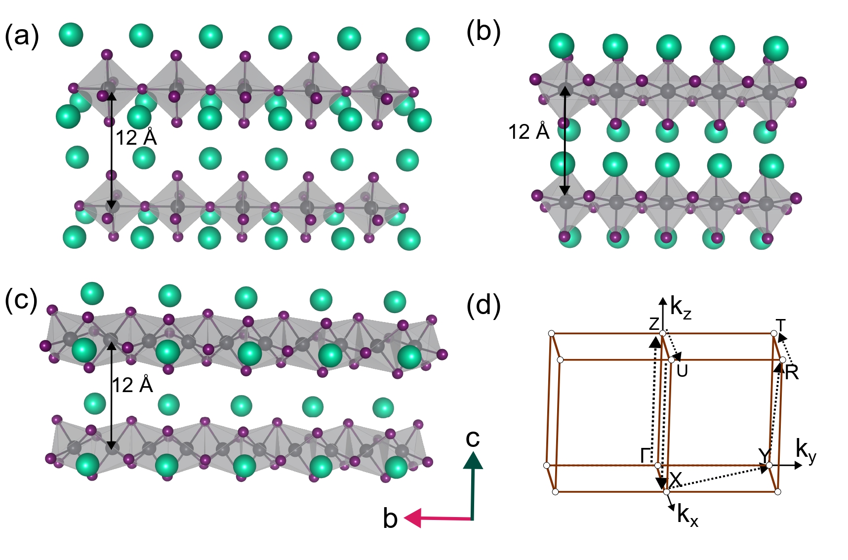
We begin by defining model structures with corner-, edge-, and face-sharing octahedral connectivity, shown in Fig. 1 (a), (b) and (c). We included Cs cations in these structures for charge compensation to avoid the appearance of spurious states below the conduction band minimum (CBM) as reported in Ref. 38. The resulting chemical formulas for the corner-, edge- and face-sharing geometry are Cs3PbI5, Cs2PbI4 and Cs2Pb2I6, respectively. The chain direction is [010], i.e., along the b-lattice vector. The corresponding Brillouin zone of the model structures is shown in Fig. 1 (d), where the direction of the 1D chain corresponds to to Y in reciprocal space. Details on the construction and structural parameters of these model structures can be found in the Supporting Information (SI).
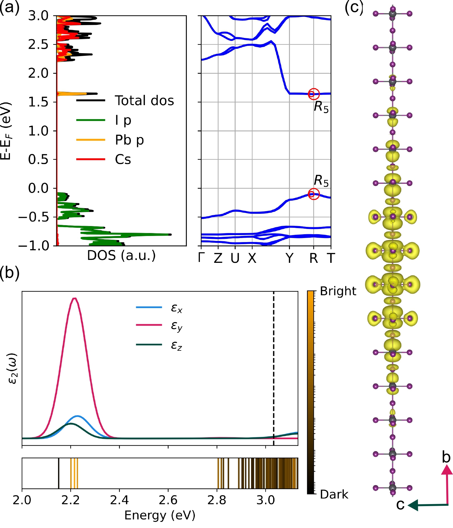
The electronic and excitonic properties of the corner-sharing model structure are shown in Fig. 2. The projected density of states (DoS), calculated using DFT with the PBE functional and including spin-orbit coupling (SOC) self-consistently (Fig. 2 (a)) is reminiscent of that of 3D \chAPbX3 halide perovskites, with the valence band maximum (VBM) derived from X p states and the conduction band minimum (CBM) comprised of Pb p states. Contrary to 3D \chAPbX3 and most known quasi-2D halide perovskites, we observe that electronic states derived from the A-site cation hybridize with the Pb p bands at energies slightly above the CBM, because of Cs-Cs interactions between neighboring periodic cells that are absent in quasi-2D or 3D structures. However, as the chain-chain distance is increased, these Cs-derived states are found at higher energies (Fig. S1).
The low-dimensional character of the structure is apparent from the bandstructure where dispersion is observed along the direction of the 1D chain of corner-sharing octahedra and little electronic interaction is present along the directions perpendicular to the chain. The band gap is direct at the high-symmetry point R. As shown in Table 1, using a “one-shot” G0W0 approach, in which DFT-PBE+SOC eigenvalues are corrected perturbatively by constructing the zeroth-order self-energy from PBE eigenvalues and eigenfunctions, leads to an opening of the band gap by 1.29 eV, similar to reports for 3D and quasi-2D perovskites [54, 55, 56, 57, 58] (see SI for computational details and convergence tests).
| Model | Experimental | |||||
| Corner | Edge | Face | Corner | Edge | Face | |
| EDFT | 1.73 | 1.90 | 2.54 | 1.92 | 2.19 | 1.53 |
| EGW | 3.03 | 3.57 | 4.03 | 2.98 | 3.22 | 3.16 |
| Ex | 881 | 1082 | 1276 | 541 | 476 | 416 |
| 2.57 | 2.44 | 2.35 | 3.68 | 3.86 | 4.06 | |
| din-chain | 32.24 | 20.79 | 20.95 | – | – | – |
| OSb/OSd | 1.1x10⁶ | 7.5x10² | 2.4x10⁵ | – | – | – |
We then use the BSE approach to calculate the imaginary part of the dielectric function as shown in Fig. 2 (b) (see SI for computational details and convergence tests). In line with its 1D nature, the corner-sharing structure has a large exciton binding energy of 881 meV (Table 1) double the value that was reported for the equivalent quasi-2D model structure [59]. The imaginary part of the dielectric function has its largest intensity along the direction of the chain. The onset of the absorption spectrum is composed of four states - one dark and three non-degenerate bright - originating from direct transitions at the high-symmetry -point R, in line with expectations based on group theory (details in SI). The excitonic state with in-chain polarization has the largest intensity. The corresponding exciton wavefunction, shown in Fig. 2(c), has an in-chain spatial extent of 32.24 Å (Table 1) and minimal extension in the out-of-chain direction (Fig. S2).
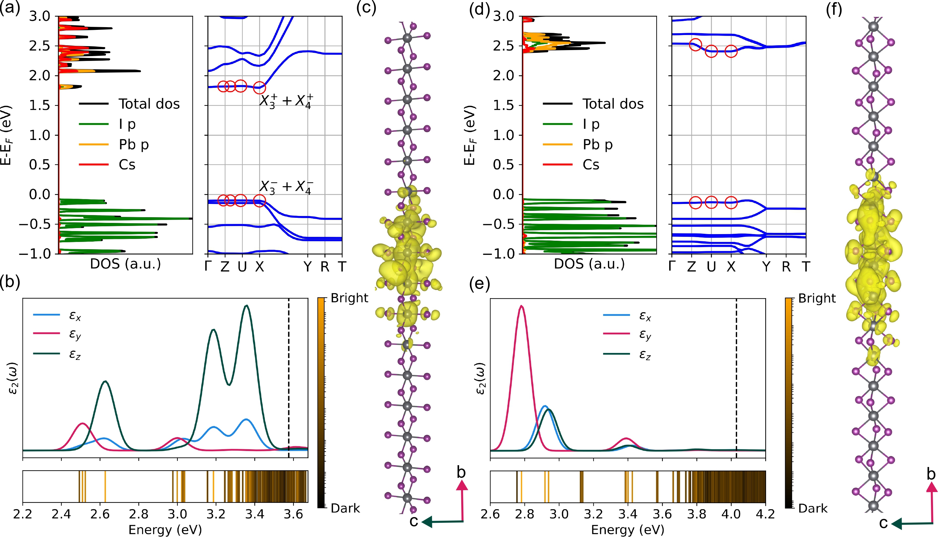
We now turn to the edge- and face-sharing geometries, shown in Fig. 3. The bandstructure of the edge-sharing structure, Fig. 3(a), shows little dispersion along the out-of-chain directions, to Z to U to X, and large electronic interactions along the in-chain direction, with the lowest electronic transition from VBM to CBM occurring at the high-symmetry point X. The position of the band gap is at a different -point than in the corner-sharing structure due to differing orbital interactions between the I and Pb orbitals at the points X and R (Fig. S3). The DFT-PBE+SOC (Table 1) band gap of the edge-sharing model structure is 1.90 eV, with a G0W0 correction of 1.67 eV. The exciton binding energy of 1082 meV is significantly higher than that of the corner-sharing structure. Here, given the different point group of the high symmetry point X, group theory predicts that the onset of the absorption is composed of four bright states (Table S2) one of which has a two orders of magnitude lower oscillator strength than the other three but is significantly brighter than the lowest-energy dark state in the corner-sharing structure (Table 1). Fig. 3 (b) shows that the onset of the imaginary part of the dielectric function originates from the in-chain direction, while, contrary to the corner-sharing geometry, the direction of the highest intensity is the out-of-chain c-direction. This is because in the edge-sharing structure, out-of-chain (axial) I p orbitals contribute significantly more to the VBM than edge-connecting (equatorial) I p orbitals (Fig. S3). This out-of-chain intensity is also present when the distance between the chains is increased (Fig. S4). Additionally, we note that excitonic absorption in the edge-sharing geometry has a more complex signature compared to the corner-sharing structure, arising from transitions between the relatively flat bands that comprise the band edges. The exciton wavefunction, shown in Fig. 3(c), is much more localized in the in-chain direction as compared to the corner-sharing structure. We find that 99% of the exciton wavefunction extents 20.79 Å in the in-chain direction (Table 1).
In the face-sharing structure, the lowest-energy VBM to CBM transition is located at the high-symmetry point U (Fig. 3(d)). Analogous to the two other cases, the reciprocal-space direction corresponding to the in-chain direction exhibits the largest electronic dispersion, albeit significantly smaller than that of the corner- and edge-sharing structures. The band gap, calculated to be 2.54 eV with DFT-PBE+SOC and increased by 1.48 eV with G0W0 (Table 1), is the largest among the three connectivity motifs. Consequently, the exciton binding energy of 1276 meV is larger than that of the corner- and the face-sharing structure too. The imaginary part of the dielectric function (Fig. 3 (e)) has the highest intensity in the in-chain direction, with a complex excitonic signature arising from the flat bands that comprise the VBM and CBM. Given the low-symmetry space group of the face-sharing structure, the point group of the high-symmetry point U was not identified. Regardless of that, we observe that the onset of the dielectric function is composed of four states, one dark and three bright with relative oscillator strength similar to the corner-sharing structure (Table 1). We note that while the exciton binding energy of the first bright state is larger than that of the edge-sharing structure, the in-chain exciton extent is similar (Table 1).
We now turn our attention to the optoelectronic properties of experimentally synthesized quasi-1D structures with corner-, edge-, and face-sharing octahedral connectivity, comparing them to our previously discussed model systems. For this comparison, we select three experimentally realized materials: corner-sharing (\ch(HSC(NH2)2)3PbI5) [60], edge-sharing (\ch(C6H10N2)(PbI4)2H2O) [61], and face-sharing (\ch(C2H7N2)6PbI3) [62] shown in Fig. S5. The experimental structures are optimized with DFT-PBE keeping the inorganic Pb-I network and the lattice vectors fixed, allowing only the molecular A site to relax. Then we calculate the DFT@PBE+SOC and @PBE+SOC band gaps and the +BSE exciton binding energies as before (see Table 1). We find that while the corner-sharing geometry has the smallest band gap, similar to our calculations for the model structures, the energetic ordering of the band gaps of the edge- and face-sharing structures is reversed: the face-sharing structure’s band gap is 80 meV smaller than that of the edge-sharing structure (Table 1). Additionally, we find that the exciton binding energies unexpectedly show an inverted trend as compared to the model structures. Specifically, the face-sharing structure exhibits the smallest exciton binding energy, followed by the edge- and the corner-sharing structure. Moreover, the exciton binding energies of the edge- and face-sharing experimental structures are more than a factor of two smaller than those predicted for the corresponding model systems.
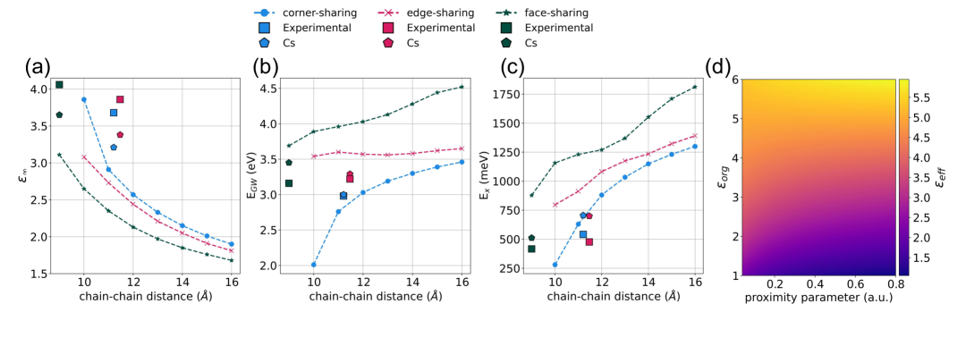
To understand the origin of these reversed trends, we systematically studied how the chain-chain distance and dielectric screening by the organic moieties influence the static dielectric constant (), the band gap, and the exciton binding energy (), as shown in Figures 4(a)-(c). For this, we calculated the dependence of these quantities on chain-chain distance using our model structures. Figure 4(a) demonstrates that decreases as the chain-chain distance increases, resulting in a monotonic increase in exciton binding energies for all three geometries (Figure 4(c)). For the corner- and face-sharing geometries, this increase closely follows the expected and the relationship for the band gap and the exciton binding energy (Fig. S6 and S8). Deviations are observed for the edge-sharing geometry which arise from more complex trends in its band gaps (Fig. S7), reflecting competing effects: reduced dielectric screening at larger distances tends to increase the band gap, while enhanced orbital hybridization between Pb-derived conduction-band states and Cs-derived states lowers the conduction band minimum (Fig. S9).
Additionally, Fig. 4 includes the three experimental structures (denoted "Experimental") and three equivalent structures in which the molecular A site was replaced by Cs (denoted "Cs"), to isolate the effect of organic spacer screening. In the corner-sharing case, the results for the experimental structure are close to the predictions of the model structure at an equivalent chain-chain difference, indicating minimal structural distortion and only slight differences upon substituting the molecular A site with Cs. This similarity arises primarily due to the inherently small dielectric constant contribution of the molecular site in this case. In contrast, the edge-sharing experimental structure substantially deviates from its model counterpart at the same chain-chain distance, exhibiting a significantly smaller band gap (by about 0.3 eV) and an exciton binding energy reduced by roughly 400 meV. This discrepancy partly results from enhanced dielectric screening by the molecular A site. Indeed, replacing this molecular site with Cs notably reduces the dielectric constant, increasing both the band gap and exciton binding energy. The face-sharing case is similar to the edge-sharing scenario. Replacing the molecular A site with Cs leads to a significant increase of the band gap and the exciton binding energy, but does not fully explain the deviation from the results for the model structures. Since the Pb-I bond lengths in these two structures are by construction the same as in the corresponding models, the remaining differences can be attributed to differences in symmetry between model and experimental structures (see SI).
Finally, to predict how the dielectric properties of the organic and inorganic sublattices influence the overall dielectric response of these 1D materials, and thus their band gaps and exciton binding energies, we constructed a simple 2D electrostatic model consisting of circular inorganic regions with dielectric constant and radius and distance , embedded in a medium representing the organic sublattice with dielectric constant . Chain-chain distance is represented by a dimensionless proximity parameter , since this simple model only reproduces the exponential decay of the effective static dielectric constant but not the microscopic details at the relevant experimental chain-chain distances (see SI for details). Figure 4(d) shows that increasing significantly enhances the effective dielectric constant, highlighting that the organic sublattice plays a key role in determining the dielectric screening. In contrast, varying has a significantly smaller effect (Fig. S10), in line with observations made for 2D metal-halide perovskites [9].
In conclusion, we performed a systematic first-principles study exploring how 1D dimensional confinement, octahedral connectivity and dielectric screening influence the optoelectronic properties of quasi-1D metal-halide perovskites. Our results demonstrate that increased sharing of halides in the octahedral chains enhances exciton binding energies and introduces anisotropic and complex optical signatures. However, comparison with experimental structures shows significant deviations from predictions based solely on idealized models, highlighting the importance of confinement (chain-chain distance) and dielectric screening effects. In particular, our electrostatic model underscores that the dielectric properties of the organic A-site play a critical role in determining exciton binding energies and optical absorption. These findings indicate clear pathways for tailoring material properties through targeted molecular and structural design. Additionally, they could aid the interpretation of optical absorption experiments carried out in situ during perovskite film growth, where intermediate phases with varying octahedral connectivity may occur. However, further studies will be necessary to incorporate the effects of solvent molecules and other structural motifs that may emerge during these processes.
Acknowledgements.
The authors acknowledge funding from the Dutch Research Council (NWO) under grant number OCENW.M20.337 and computational resources provided by the Dutch National Supercomputing Center Snellius supported by the SURF cooperative. We thank M. Kamminga and R. Havenith for providing the model structures used in Ref. 38.References
- Green et al. [2014] M. A. Green, A. Ho-Baillie, and H. J. Snaith, Nat. Photonics 8, 506 (2014).
- Jung and Park [2015] H. S. Jung and N.-G. Park, Small 11, 10 (2015).
- Alsalloum et al. [2020] A. Y. Alsalloum, B. Turedi, X. Zheng, S. Mitra, A. A. Zhumekenov, K. J. Lee, P. Maity, I. Gereige, A. AlSaggaf, I. S. Roqan, O. F. Mohammed, and O. M. Bakr, ACS Energy Lett. 5, 657 (2020).
- Smith et al. [2017] I. C. Smith, M. D. Smith, A. Jaffe, Y. Lin, and H. I. Karunadasa, Chem. Mater. 29, 1868 (2017).
- Blancon et al. [2020] J.-C. Blancon, J. Even, C. C. Stoumpos, M. G. Kanatzidis, and A. D. Mohite, Nat. Nanotechnol. 15, 969 (2020).
- Cao et al. [2015] D. H. Cao, C. C. Stoumpos, O. K. Farha, J. T. Hupp, and M. G. Kanatzidis, J. Am. Chem. Soc. 137, 7843 (2015).
- Cheng et al. [2018] B. Cheng, T.-Y. Li, P. Maity, P.-C. Wei, D. Nordlund, K.-T. Ho, D.-H. Lien, C.-H. Lin, R.-Z. Liang, X. Miao, I. A. Ajia, J. Yin, D. Sokaras, A. Javey, I. S. Roqan, O. F. Mohammed, and J.-H. He, Communications Physics 1, 80 (2018).
- Marongiu et al. [2019] D. Marongiu, M. Saba, F. Quochi, A. Mura, and G. Bongiovanni, J. Mater. Chem. C Mater. Opt. Electron. Devices 7, 12006 (2019).
- Filip et al. [2022] M. R. Filip, D. Y. Qiu, M. Del Ben, and J. B. Neaton, Nano Lett. 22, 4870 (2022).
- Chakraborty et al. [2022] R. Chakraborty, G. Paul, and A. J. Pal, Physical Review Applied 17, 054045 (2022).
- Simbula et al. [2023] A. Simbula, L. Wu, F. Pitzalis, R. Pau, S. Lai, F. Liu, S. Matta, D. Marongiu, F. Quochi, M. Saba, A. Mura, and G. Bongiovanni, Nat. Commun. 14, 4125 (2023).
- Blancon et al. [2018] J. C. Blancon, A. V. Stier, H. Tsai, W. Nie, C. C. Stoumpos, B. Traoré, L. Pedesseau, M. Kepenekian, F. Katsutani, G. T. Noe, J. Kono, S. Tretiak, S. A. Crooker, C. Katan, M. G. Kanatzidis, J. J. Crochet, J. Even, and A. D. Mohite, Nature Communications 9, 2254 (2018).
- Cho and Berkelbach [2019a] Y. Cho and T. C. Berkelbach, J. Phys. Chem. Lett. 10, 6189 (2019a).
- Tsai et al. [2016] H. Tsai, W. Nie, J.-C. Blancon, C. C. Stoumpos, R. Asadpour, B. Harutyunyan, A. J. Neukirch, R. Verduzco, J. J. Crochet, S. Tretiak, L. Pedesseau, J. Even, M. A. Alam, G. Gupta, J. Lou, P. M. Ajayan, M. J. Bedzyk, and M. G. Kanatzidis, Nature 536, 312 (2016).
- Li et al. [2024] S. Li, X. Xu, C. A. Kocoj, C. Zhou, Y. Li, D. Chen, J. A. Bennett, S. Liu, L. Quan, S. Sarker, M. Liu, D. Y. Qiu, and P. Guo, Nat. Commun. 15, 2573 (2024).
- Chakraborty et al. [2024] R. Chakraborty, P. C. Sercel, X. Qin, D. B. Mitzi, and V. Blum, J. Am. Chem. Soc. 146, 34811 (2024).
- Zhang et al. [2022] W.-F. Zhang, H.-M. Pan, Y.-Y. Ma, D.-Y. Li, and Z. Jing, J. Mol. Struct. 1253, 132221 (2022).
- Yuan et al. [2017] Z. Yuan, C. Zhou, Y. Tian, Y. Shu, J. Messier, J. C. Wang, L. J. van de Burgt, K. Kountouriotis, Y. Xin, E. Holt, K. Schanze, R. Clark, T. Siegrist, and B. Ma, Nat. Commun. 8, 14051 (2017).
- Wong et al. [2021] W. P. D. Wong, J. V. Hanna, and A. C. Grimsdale, Acta Crystallogr. B Struct. Sci. Cryst. Eng. Mater. 77, 408 (2021).
- Rahaman et al. [2021] M. Z. Rahaman, S. Ge, C.-H. Lin, Y. Cui, and T. Wu, Small Struct. 2, 2000062 (2021).
- Duan et al. [2023] D. Duan, C. Ge, M. Z. Rahaman, C.-H. Lin, Y. Shi, H. Lin, H. Hu, and T. Wu, NPG Asia Materials 15, 8 (2023).
- Mitzi et al. [1995] D. B. Mitzi, S. Wang, C. A. Feild, C. A. Chess, and A. M. Guloy, Science 267, 1473 (1995).
- Petrov et al. [2017] A. A. Petrov, I. P. Sokolova, N. A. Belich, G. S. Peters, P. V. Dorovatovskii, Y. V. Zubavichus, V. N. Khrustalev, A. V. Petrov, M. Grätzel, E. A. Goodilin, and A. B. Tarasov, J. Phys. Chem. C Nanomater. Interfaces 121, 20739 (2017).
- Guo et al. [2016] X. Guo, C. McCleese, C. Kolodziej, A. C. S. Samia, Y. Zhao, and C. Burda, Dalton Trans. 45, 3806 (2016).
- Mao et al. [2018] L. Mao, P. Guo, M. Kepenekian, I. Hadar, C. Katan, J. Even, R. D. Schaller, C. C. Stoumpos, and M. G. Kanatzidis, J. Am. Chem. Soc. 140, 13078 (2018).
- Yu et al. [2021] G. Yu, F. Lin, K. Zhou, S. Fang, Y. Shi, W. Liu, H. Hu, B. Ma, and H. Lin, Chem. Mater. 33, 5668 (2021).
- Lin et al. [2018] H. Lin, C. Zhou, Y. Tian, T. Siegrist, and B. Ma, ACS Energy Lett. 3, 54 (2018).
- Chen et al. [2023] Y. Chen, B. Liu, Q. Zhou, D. Ma, X. Han, D. He, S. Chen, Y. Li, S. Lu, Z.-X. Xu, C. Chen, H. Yu, and J. Chen, Journal of Materials Chemistry A 11, 18592 (2023).
- Wan et al. [2025] R. Wan, M. Yin, T.-H. Wang, C. E. Moore, and Y. Wu, Inorg. Chem. 64, 2868 (2025).
- Chandra Patra et al. [2024] B. Chandra Patra, R. Wan, C. E. Moore, and Y. Wu, Chemistry – A European Journal 31, e202402535 (2024).
- Quarti et al. [2025] C. Quarti, R. Gautier, M. Zacharias, A. Gansmuller, and C. Katan, J. Am. Chem. Soc. 147, 278 (2025).
- Wang et al. [2022] Y. Wang, H. Lou, C.-Y. Yue, and X.-W. Lei, CrystEngComm 24, 2201 (2022).
- Zhang et al. [2016] Y. Zhang, J. Liu, Z. Wang, Y. Xue, Q. Ou, L. Polavarapu, J. Zheng, X. Qi, and Q. Bao, Chem. Commun. (Camb.) 52, 13637 (2016).
- Qian et al. [2017] J. Qian, Q. Guo, L. Liu, B. Xu, and W. Tian, J. Mater. Chem. A Mater. Energy Sustain. 5, 16786 (2017).
- Jiang et al. [2022] X. Jiang, Z. Xu, Y. Zheng, J. Zeng, K.-Q. Chen, and Y. Feng, Phys. Chem. Chem. Phys. 24, 17323 (2022).
- Xue et al. [2022] C. Xue, H. Huang, S. Nishihara, V. Biju, X.-M. Ren, and T. Nakamura, J. Phys. Chem. Lett. 13, 7405 (2022).
- Kamminga et al. [2016] M. E. Kamminga, H.-H. Fang, M. R. Filip, F. Giustino, J. Baas, G. R. Blake, M. A. Loi, and T. T. M. Palstra, Chem. Mater. 28, 4554–4562 (2016).
- Kamminga et al. [2017] M. E. Kamminga, G. A. de Wijs, R. W. A. Havenith, G. R. Blake, and T. T. M. Palstra, Inorg. Chem. 56, 8408 (2017).
- Deng et al. [2018] Z. Deng, G. Kieslich, P. D. Bristowe, A. K. Cheetham, and S. Sun, APL Materials 6, 114202 (2018).
- Rohlfing and Louie [2000] M. Rohlfing and S. G. Louie, Physical Review B 62, 4927 (2000).
- Puschnig and Ambrosch-Draxl [2002] P. Puschnig and C. Ambrosch-Draxl, Physical Review B 66, 165105 (2002).
- Wirtz et al. [2006] L. Wirtz, A. Marini, and A. Rubio, Phys. Rev. Lett. 96, 126104 (2006).
- Qiu et al. [2013] D. Y. Qiu, F. H. da Jornada, and S. G. Louie, Phys. Rev. Lett. 111, 216805 (2013).
- Reyes-Lillo et al. [2016] S. E. Reyes-Lillo, T. Rangel, F. Bruneval, and J. B. Neaton, Physical Review B 94, 041107 (2016).
- Giorgi et al. [2018] G. Giorgi, K. Yamashita, and M. Palummo, The Journal of Physical Chemistry Letters 9, 5891 (2018), https://doi.org/10.1021/acs.jpclett.8b02653 .
- Qiu et al. [2021] D. Y. Qiu, G. Cohen, D. Novichkova, and S. Refaely-Abramson, Nano Lett. 21, 7644 (2021).
- Leppert [2024] L. Leppert, The Journal of Chemical Physics 160, 050902 (2024).
- Gilley et al. [2025] I. W. Gilley, H. W. Kwon, C. Liu, Y. Yang, C. Huang, H. Wan, A. S. R. Bati, E. H. Oriel, M. Kepenekian, B. Vishal, S. Zeiske, K. S. Bayikadi, T. E. Wiggins, E. S. Vasileiadou, B. Chen, R. D. Schaller, J. Even, S. De Wolf, E. H. Sargent, and M. G. Kanatzidis, J. Am. Chem. Soc. 147, 7777 (2025).
- Chen and Filip [2023] Y. Chen and M. R. Filip, J. Phys. Chem. Lett. 14, 10634 (2023).
- Umeyama et al. [2020] D. Umeyama, L. Leppert, B. A. Connor, M. A. Manumpil, J. B. Neaton, and H. I. Karunadasa, Angew. Chem. Int. Ed Engl. 59, 19087 (2020).
- Matheu et al. [2022] R. Matheu, F. Ke, A. Breidenbach, N. Wolf, Y. Lee, Z. Liu, L. Leppert, Y. Lin, and H. I. Karunadasa, Angewandte Chemie International Edition 61, e202202911 (2022).
- Forde et al. [2023] A. Forde, S. Tretiak, and A. J. Neukirch, Nano Lett. 23, 11586 (2023).
- Ni et al. [2024] X. Ni, H. Li, and J.-L. Brédas, ACS Mater. Lett. 6, 3436 (2024).
- Filip and Giustino [2014] M. R. Filip and F. Giustino, Physical Review B 90, 245145 (2014).
- Leppert et al. [2019] L. Leppert, T. Rangel, and J. B. Neaton, Physical Review Materials 3, 103803 (2019).
- Filip and Leppert [2024] M. R. Filip and L. Leppert, Electron. Struct. 6, 033002 (2024).
- Ahmed et al. [2014] T. Ahmed, C. La-o vorakiat, T. Salim, Y. M. Lam, E. E. M. Chia, and J.-X. Zhu, EPL 108, 67015 (2014).
- Cho and Berkelbach [2019b] Y. Cho and T. C. Berkelbach, J. Phys. Chem. Lett. 10, 6189 (2019b).
- Quarti et al. [2023] C. Quarti, G. Giorgi, C. Katan, J. Even, and M. Palummo, Advanced Optical Materials 11, 2202801 (2023).
- Daub and Hillebrecht [2021] M. Daub and H. Hillebrecht, Inorg. Chem. 60, 3082 (2021).
- Lemmerer and Billing [2006] A. Lemmerer and D. G. Billing, Acta Crystallogr. C 62, m597 (2006).
- Saski et al. [2024] M. Saski, S. Sobczak, P. Ratajczyk, M. Terlecki, W. Marynowski, A. Borkenhagen, I. Justyniak, A. Katrusiak, and J. Lewiński, Small 20, e2403685 (2024).