Improvement Strategies for Few-Shot Learning in OCT Image Classification of Rare Retinal Diseases
Abstract
This paper focuses on using few-shot learning to improve the accuracy of classifying OCT diagnosis images with major and rare classes. We used the GAN-based augmentation strategy as a baseline and introduced several novel methods to further enhance our model. The proposed strategy contains U-GAT-IT for improving the generative part and uses the data balance technique to narrow down the skew of accuracy between all categories. The best model obtained was built with CBAM attention mechanism and fine-tuned InceptionV3, and achieved an overall accuracy of 97.85%, representing a significant improvement over the original baseline.
Index Terms:
Rare Diseases, Deep Learning, Generative Adversarial Network, Few-Shot Learning, OCT, Attention ModuleI Introduction
GAN-based data augmentation has been proposed to alleviate data scarcity in few-shot learning (FSL) scenarios. Nevertheless, it suffers from notable limitations. As reported by Yoo et al. [1], the use of CycleGAN [2] for synthetic OCT image generation resulted in approximately 20% of the images being deemed unacceptable by ophthalmologists, ultimately leading to suboptimal classification performance. Moreover, the baseline model exhibited limited robustness, especially in its ability to generalize across rare diseases. This emphasizes the need for more reliable generative methods and enhanced model architectures capable of learning effectively from limited data. To mitigate this, we employ U-GAT-IT [3], which incorporates attention mechanisms into the generative process to improve image quality. Furthermore, we apply dataset balancing techniques and integrate both Squeeze-and-Excitation (SE) blocks [4] and the Convolutional Block Attention Module (CBAM) [5] into a fine-tuned InceptionV3 [6] backbone to strengthen feature extraction from critical retinal regions.
II Methods
This study builds upon the framework proposed by Yoo et al. [1], which utilized CycleGAN-based augmentation and transfer learning to address the few-shot classification problem in diagnosing rare retinal diseases from OCT images. To overcome the limitations in image synthesis quality and class imbalance inherent in the original framework, we propose an enhanced methodology with three major improvements: (1) Replacing CycleGAN with U-GAT-IT to generate more acceptable pathological OCT images under few-shot constraints; (2) Implementation of data balancing strategies to mitigate the skewed distribution between common and rare disease classes; and (3) Integrating attention mechanisms, including Squeeze-and-Excitation (SE) [4] and Convolutional Block Attention Module (CBAM) [5], into the InceptionV3 [6] backbone to improve feature localization and representation.The InceptionV3 [6] model was initialized with ImageNet pre-trained weights and subsequently fine-tuned on the augmented and rebalanced dataset.
II-A U-GAT-IT
II-A1 Basic Augmentation
II-A2 Dataset and Training
II-B Data-Balanced
We sampled 5,000 images from OCT2017[8] for each major class, then combined 3,000 U-GAT-IT generated images with 2,000 augmented images per rare class. To further improve the model’s generalization ability, we expanded all classes to 10,000 images: major classes were randomly selected; DRUSEN was augmented to 10,000 images; and rare diseases combined 2,000 augmented and 8,000 generated images
II-C Attention Module
II-C1 Squeeze-and-Excitation (SE) Block
II-C2 Convolutional Block Attention Module (CBAM)
III Results
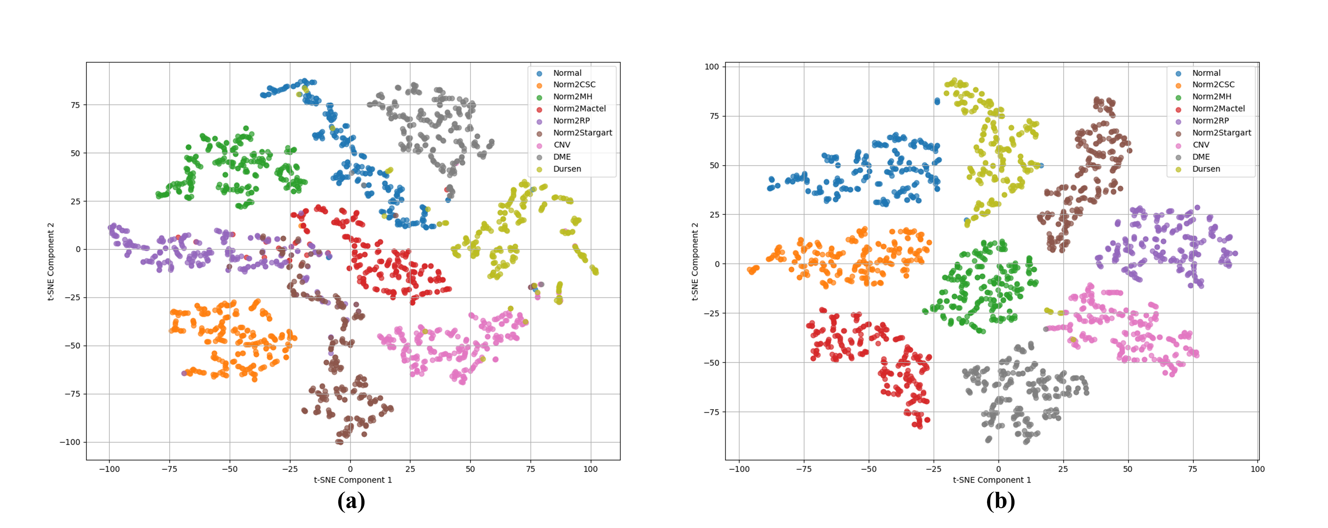
Without any augmentation or balancing, the baseline model (Imb + NoAug) achieved high overall accuracy but low balanced accuracy (0.550), revealing poor performance in detecting rare diseases in Table 1. Introducing CycleGAN-based generative augmentation improved balanced accuracy to 0.856 — an increase of approximately 30% — though this came at the cost of a slight decrease in overall accuracy by about 3.42%. In contrast, U-GAT-IT outperformed CycleGAN across all metrics; with 5,000 U-GAT-IT generated images (Imb + U5000), the model reached 95.66% accuracy—roughly 2% higher than the CycleGAN counterpart (Imb + C5000) in Table 1. The results of the t-SNE [9] algorithm show that the classification model trained with U-GAT-IT-generated images provides better visualization of rare groups compared to those generated by CycleGAN in Fig. 1. After rebalancing all classes to 5,000 images, the CycleGAN-based strategy (Bal + C5000) reduced the accuracy gap between major and average classes from 10.5% to 3.8%. In contrast, the U-GAT-IT-based strategy (Bal + U5000) further narrowed the gap from 6.5% to 4%, while outperforming the CycleGAN approach by approximately 4% across all key evaluation metrics. Under balanced conditions, training with 10,000 U-GAT-IT-generated images (Bal + U10000) the model achieved 96.18% accuracy, a Cohen’s of 0.949, and balanced accuracy of 0.959. Furthermore, incorporating attention mechanisms led to additional improvements. The CBAM[5]-enhanced model achieved accuracy, Cohen’s , RCI, Matthews correlation, and balanced accuracy of 97.85%, 0.972, 0.921, 0.972, and 0.972, respectively. Similarly, the SE[4]-enhanced model (SE) also demonstrated competitive performance, with values of 97.54%, 0.967, 0.914, 0.967, and 0.926, respectively.
IV Conclusion
This paper presents an enhanced framework for few-shot retinal disease classification using OCT images, addressing the limitations of prior generative approaches and feature extraction strategies. Results show that the proposed method achieved an overall accuracy of 97.85%, marking an approximate 4% improvement over the baseline model. These findings further confirm that lightweight attention modules, such as CBAM [5], can effectively improve classification robustness across both common and rare retinal disease categories.
| Method | Accuracy (%) | Cohen’s | RCI | MCC | BA |
|---|---|---|---|---|---|
| Imb + NoAug | 97.18 ± 0.25 | 0.963 ± 0.003 | 0.964 ± 0.005 | 0.963 ± 0.003 | 0.550 ± 0.001 |
| Imb + C5000 | 93.76 ± 1.44 | 0.922 ± 0.019 | 0.865 ± 0.039 | 0.924 ± 0.019 | 0.856 ± 0.062 |
| Imb + U5000 | 95.66 ± 1.13 | 0.943 ± 0.015 | 0.898 ± 0.019 | 0.944 ± 0.014 | 0.844 ± 0.062 |
| Bal + C5000 | 92.76 ± 1.06 | 0.904 ± 0.014 | 0.778 ± 0.025 | 0.904 ± 0.014 | 0.937 ± 0.010 |
| Bal + U5000 | 95.90 ± 0.38 | 0.946 ± 0.005 | 0.863 ± 0.011 | 0.946 ± 0.005 | 0.977 ± 0.009 |
| Bal + U10000 | 96.18 ± 0.58 | 0.949 ± 0.008 | 0.872 ± 0.016 | 0.949 ± 0.008 | 0.959 ± 0.027 |
| Bal + CBAM | 97.85 ± 0.31 | 0.972 ± 0.004 | 0.921 ± 0.011 | 0.972 ± 0.004 | 0.972 ± 0.020 |
| Bal + SE | 97.54 ± 0.30 | 0.967 ± 0.004 | 0.914 ± 0.008 | 0.967 ± 0.004 | 0.926 ± 0.023 |
| Yoo et al.[1] | 93.9 ± 4.5 | 0.910 ± 0.065 | 0.969 ± 0.028 | 0.911 ± 0.062 |
-
•
Note: Imb, imbalanced; NoAug, without augmentation; Bal, balanced; Cohen’s , Unweighted Cohen’s kappa; RCI, relative classifier information; MCC, Matthews correlation coefficient; BA, balanced accuracy; C5000, CycleGAN (2k real + 3k generated); U5000, U-GAT-IT (2k real + 3k generated); U10000, U-GAT-IT (2k real + 8k generated).
| Method | NORMAL | CNV | DME | Drusen | CSC | MH | MacTel | RP | Stargardt |
|---|---|---|---|---|---|---|---|---|---|
| Imb + NoAug | 100 | 100 | 99.60 | 96.80 | 0 | 100 | 0 | 0 | 0 |
| Imb + C5000 | 100 | 100 | 98.80 | 99.20 | 80.00 | 100 | 75.00 | 50.00 | 50.00 |
| Imb + U5000 | 100 | 100 | 99.20 | 99.60 | 100 | 100 | 100 | 75.00 | 75.00 |
| Bal + C5000 | 96.00 | 94.20 | 94.20 | 94.90 | 80.00 | 100 | 100 | 100 | 75.00 |
| Bal + U5000 | 98.00 | 96.59 | 96.59 | 97.76 | 100 | 100 | 100 | 100 | 100 |
| Bal + U10000 | 98.60 | 95.90 | 98.98 | 93.63 | 100 | 100 | 100 | 100 | 100 |
| Bal + CBAM | 98.40 | 97.80 | 99.39 | 97.54 | 100 | 100 | 100 | 100 | 100 |
| Bal + SE | 98.40 | 96.99 | 100 | 94.87 | 100 | 100 | 100 | 100 | 75.00 |
| Yoo et al.[1] | 91.2 | 94.8 | 93.2 | 88.8 | 100 | 100 | 100 | 75 | 75 |
-
•
Note: CNV, choroidal neovascularization; DME, diabetic macular edema; CSC, central serous chorioretinopathy; MacTel, macular telangiectasia;
References
- [1] T. K. Yoo, J. Y. Choi, and H. K. Kim, ”Feasibility study to improve deep learning in OCT diagnosis of rare retinal diseases with few-shot classification,” Med. Biol. Eng. Comput., vol. 59, pp. 401–415, Jan. 2021.
- [2] J.-Y. Zhu, T. Park, P. Isola, and A. A. Efros, ”Unpaired image-to-image translation using cycle-consistent adversarial networks,” in Proc. IEEE ICCV, Venice, Italy, Oct. 2017, pp. 2242–2251.
- [3] J. Kim, M. Kim, H. Kang, and K. Lee, ”U-GAT-IT: Unsupervised generative attentional networks with adaptive layer-instance normalization for image-to-image translation,” in Proc. ICLR, Addis Ababa, Ethiopia, 2020.
- [4] J. Hu, L. Shen, and G. Sun, ”Squeeze-and-excitation networks,” in Proc. IEEE CVPR, Salt Lake City, UT, USA, 2018, pp. 7132–7141. doi: 10.1109/CVPR.2018.00745
- [5] S. Woo, J. Park, J.-Y. Lee, and I. S. Kweon, ”CBAM: Convolutional block attention module,” in Proc. ECCV, Munich, Germany, Sep. 2018, pp. 3–19.
- [6] C. Szegedy, V. Vanhoucke, S. Ioffe, J. Shlens, and Z. Wojna, ”Rethinking the Inception architecture for computer vision,” in Proc. IEEE CVPR, Las Vegas, NV, USA, Jun. 2016, pp. 2818–2826.
- [7] T. Yoo, ”Data for: Improved accuracy in OCT diagnosis of rare retinal disease using few-shot learning with generative adversarial networks,” Mendeley Data, vol. 2, 2020.
- [8] D. Kermany, K. Zhang, and M. Goldbaum, ”Labeled optical coherence tomography (OCT) and chest X-ray images for classification,” Mendeley Data, vol. 2, 2018.
- [9] L. van der Maaten and G. Hinton, ”Visualizing data using t-SNE,” J. Mach. Learn. Res., vol. 9, pp. 2579–2605, 2008.