55email: xiaofan.zhang@sjtu.edu.cn, wangzhong@lzu.edu.cn††footnotetext: Contributed equally to this work.
MFM-DA: Instance-Aware Adaptor and Hierarchical Alignment for Efficient Domain Adaptation in Medical Foundation Models
Abstract
Medical Foundation Models (MFMs), trained on large-scale datasets, have demonstrated superior performance across various tasks. However, these models still struggle with domain gaps in practical applications. Specifically, even after fine-tuning on source-domain data, task-adapted foundation models often perform poorly in the target domain. To address this challenge, we propose a few-shot unsupervised domain adaptation (UDA) framework for MFMs, named MFM-DA, which only leverages a limited number of unlabeled target-domain images. Our approach begins by training a Denoising Diffusion Probabilistic Model (DDPM), which is then adapted to the target domain using a proposed dynamic instance-aware adaptor and a distribution direction loss, enabling the DDPM to translate source-domain images into the target domain style. The adapted images are subsequently processed through the MFM, where we introduce a designed channel-spatial alignment Low-Rank Adaptation (LoRA) to ensure effective feature alignment. Extensive experiments on optic cup and disc segmentation tasks demonstrate that MFM-DA outperforms state-of-the-art methods. Our work provides a practical solution to the domain gap issue in real-world MFM deployment. Code will be available at here.
Keywords:
Few-shot domain adaptation Few-shot image generation Foundation model Medical image segmentation.1 Introduction
Deep learning has made significant strides in medical image analysis [23, 8, 10]. However, these models often require large volumes of annotated data for training and tend to be task-specific, which limits their generalization across diverse clinical applications. To overcome this limitation, various MFMs have recently been proposed [25, 2, 26, 11, 9], which learn universal representations from vast medical datasets. These models are capable of performing a wide range of clinical tasks either directly or after fine-tuning.
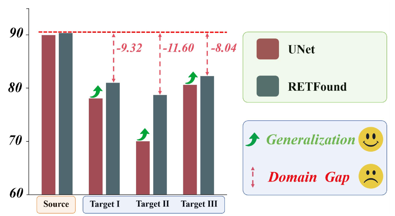
When fine-tuned on specific tasks within a source domain, these MFMs exhibit superior generalization performance in the target domain compared to models trained from scratch. However, they remain susceptible to domain shifts, often showing a noticeable decline in performance when applied to the target domain. For example, RETFound[26], a foundation model trained on 1.6 million unannotated retinal images using self-supervised learning, was evaluated on cup-to-disc segmentation tasks for fundus images. Our experiments reveal that RETFound demonstrates superior generalization compared to models trained from scratch, but its performance still suffers from domain gaps, as shown in Fig. 1. Despite outperforming UNet [18] trained from scratch in the target domain, RETFound’s results are significantly lower than its performance in the source domain, highlighting the impact of domain shifts.
To mitigate domain shifts, unsupervised domain adaptation (UDA) [4] has been widely explored to improve model performance on unannotated target data using labeled source domain data. UDA typically addresses domain shifts in two ways: image adaptation [21], which aligns the image appearances through pixel-level transformations, and feature adaptation [20]. However, UDA often requires large amounts of unannotated target data to reduce domain distribution differences, which is not always feasible in real-world medical scenarios. In contrast, Few-shot Domain Adaptation (FSDA) [13] offers a more practical solution, as it only requires a limited number of target samples during training.
To address the challenge of domain shifts in MFM with limited target data, we propose the MFM-DA framework, which requires only source domain data and a small number of target-domain images. As shown in Fig. 2, it mainly contains two stages. In Stage 1, we train a Denoising Diffusion Probabilistic Model (DDPM) [5] on source domain data, which is then adapted to the target domain using our proposed Dynamic Instance-Aware Adaptator and distribution consistency loss. In Stage 2, we fine-tune the foundation model with a combination of source and generated target-domain images, using LoRA [22] for adjusting attention mechanisms and a Pyramid Hierarchical Alignment method to align features across hierarchical levels. This approach facilitates domain adaptation by ensuring alignment in both channel-wise semantics and spatial structures, thereby improving model performance in the target domain.
We conducted extensive experiments on optic cup and disc segmentation tasks, demonstrating the effectiveness of our approach across source and target domains for MFMs. Our contributions include:
-
1.
To our best knowledge, we are the first to propose a specifically designed framework to address the few-shot unsupervised domain adaptation for medical foundation models;
-
2.
Introducing the Dynamic Instance-Aware Adaptator, which adapts the distribution of generated images to better match the target domain, even in few-shot scenarios.
-
3.
Proposing Pyramid Hierarchical Alignment, which aligns source and target-domain features at different levels to achieve robust domain adaptation.
2 Method
The framework we propose for few-shot unsupervised domain adaptation in medical image segmentation is illustrated in Fig. 2. We introduce a two-stage domain adaptation framework. In the first stage, DDPM is used to perform domain adaptation on 10 unannotated target domain images from the perspective of image generation distribution. In the second stage, we fine-tune the foundation model on the generated unannotated target domain images to perform domain adaptation at the feature level. This approach effectively narrows the performance gap caused by domain differences in the foundation model.
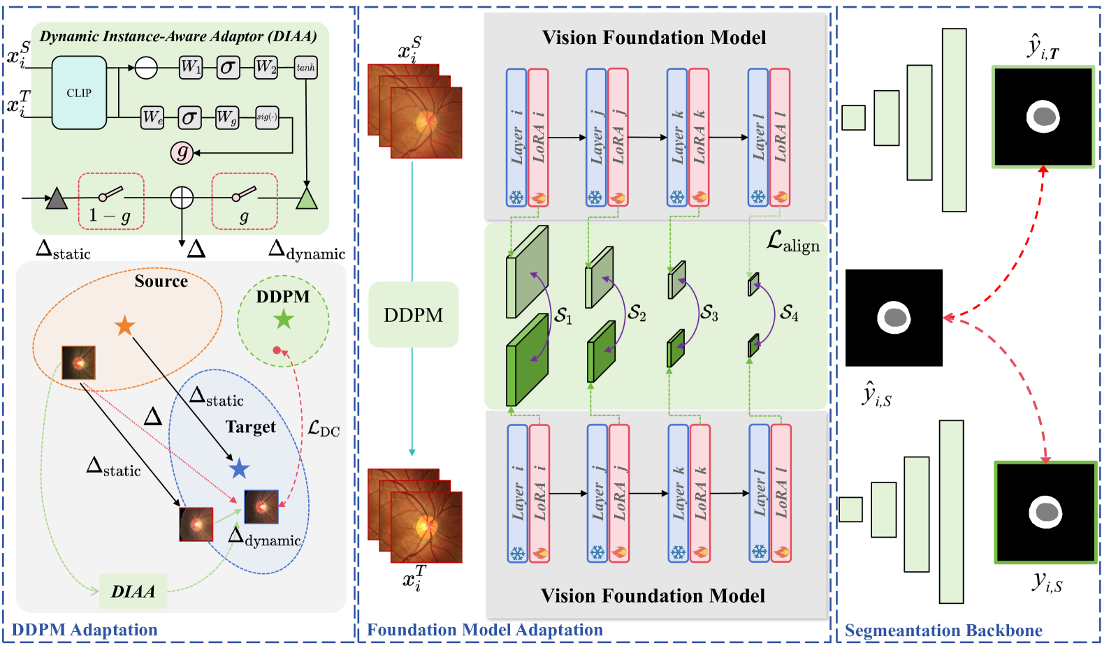
2.1 Dynamic Instance-Aware Adaptor
In the few-shot scenario, models are highly susceptible to overfitting. To address the overfitting issue, [7] proposes the Directional Distribution Consistency loss (DDC), which to extracts the feature distribution centers of all data from both the source domain and the target domain, and relies on the vector between these two centers to guide the DDPM for adaptation to the target domain. However, it suffer from rigid feature translation due to their reliance on fixed geometric directions ( in Eq. 1), which fails to capture instance-specific domain shifts. Our key innovation addresses this limitation through a learnable direction adapter that enables dynamic instance-aware adaptation while preserving global domain statistics.
Specifically, given the source dataset S = and target dataset T = , we extract the image features of each dataset using CLIP [17]. Then, we compute the static cross-domain direction vector from the center of the source domain to the center of the target domain in the feature space:
| (1) |
This captures global feature distribution differences but lacks instance awareness. Our proposed bottleneck network generates batch-specific adjustments conditioned on the source features and the global target domain center for each batch:
| (2) |
Where and , is the dimension of the features, and is the batch size. This allows the model to learn the directional changes for each batch, mapping to a wider range of regions in the target domain. For training stability, we start by using static vectors to guide the model’s learning and progressively introduce dynamic adjustments as the learning process advances, enabling broader coverage of the target domain. We employ a learnable gating network to dynamically fuse static and dynamic components:
| (3) |
| (4) |
Where and . We utilize the dynamic directional vector to constrain the structure of the generated distribution, ensuring it covers the original distribution as much as possible while aligning its center with that of the target distribution. This is achieved through the following distribution consistency loss.
| (5) |
where is the source image and is the output image in the target domain. Through this loss function, we explicitly enforce the spatial structural consistency between the generated and original distributions during domain adaptation.
2.2 Pyramid Hierarchical Alignment
Medical imaging domains often exhibit discrepancies in intensity distributions while sharing underlying anatomical structures. Traditional domain adaptation methods focusing solely on global feature alignment may fail to capture critical local geometric relationships. As illustrated in Fig.2, we propose a adaptation method with pyramid hierarchical feature alignment, addressing both channel-wise semantics and spatial structures. Given paired images from source and target domains, our medical foundation model produces multi-scale pyramid features:
| (7) | ||||
where denotes the -th level feature tensor, represent the number of features extracted, which we empirically set to 4.
Align features across four pyramid levels to capture organ structures at varying granularities. For each hierarchy level , flatten spatial dimensions while preserving channel correlations and compute cosine similarity between corresponding spatial locations across domains:
| (8) |
| (9) |
Finally, Combine losses across all pyramid levels, enforce position-wise similarity in both channel responses and spatial layouts:
| (10) |
We fine-tune the output of each layer using trainable low-rank matrices in the attention mechanism:
| (11) |
where and , represents the dimensionality of , is the sequence length of , and is the rank with , which reduces the number of parameters required for fine-tuning. During training, only 0.8% of the parameters (LoRA matrices) are updated.
The total loss combines segmentation supervision and alignment constraints:
| (12) | ||||
3 Experiments and Results
Materials and Evaluation Metrics. This study utilizes the joint optic cup / optic disc segmentation dataset RIGA+ [6, 3, 1] and the REFUGE dataset [16]. The RIGA+ dataset provides images from five different domains: BinRushed, Magrabia, BASE1, BASE2, and BASE3. In the REFUGE dataset, the training set and test set were captured using different devices, making them suitable for use as data from different domains. In our experiments, we used REFUGE (Train) as the source domain for model training and REFUGE (Test), BinRushed, and BASE2 as the target domains, which were labeled as I, II, and III, respectively. For image generation tasks, we used IC-LPIPS [14] to measure the diversity of generated images and FID [19] to evaluate the similarity between the generated images and the target domain. For segmentation tasks, we used the Dice similarity coefficient (D, %) to assess segmentation performance.
| Methods | Domain I | Domain II | Domain III | Average | ||||
|---|---|---|---|---|---|---|---|---|
| Syn | FID | IC | FID | IC | FID | IC | FID | IC |
| Finetune | 100.83 | 0.276 | 209.85 | 0.194 | 105.28 | 0.308 | 138.65 | 0.259 |
| FreezeD [12] | 84.12 | 0.263 | 177.49 | 0.196 | 108.15 | 0.268 | 123.25 | 0.242 |
| DDC [7] | 94.56 | 0.293 | 129.62 | 0.301 | 102.15 | 0.306 | 108.78 | 0.300 |
| ours | 62.79 | 0.371 | 88.27 | 0.347 | 91.52 | 0.345 | 80.86 | 0.354 |
| Seg | Dice | JI | Dice | JI | Dice | JI | Dice | JI |
| Unet | 78.079.14 | 65.2811.45 | 70.0317.61 | 57.5618.49 | 80.629.54 | 68.6211.54 | 76.24 | 63.82 |
| +Coral [20] | 82.278.34 | 70.910.91 | 72.3714.71 | 60.1716.24 | 84.129.37 | 73.7811.4 | 79.58 | 68.28 |
| +FDA [24] | 81.698.53 | 70.7311.31 | 78.3411.32 | 67.2013.67 | 85.416.24 | 75.308.99 | 81.81 | 71.07 |
| RETFound [26] | 80.9913.34 | 71.1114.66 | 78.7115.73 | 68.2516.87 | 82.2710.75 | 71.3813.12 | 80.66 | 70.24 |
| +Coral [20] | 82.1410.27 | 74.5312.16 | 78.8212.28 | 71.317.17 | 83.288.59 | 72.6311.11 | 81.41 | 72.82 |
| +FDA [24] | 84.1310.66 | 71.3412.94 | 81.3115.30 | 67.8717.37 | 83.519.61 | 72.5612.21 | 82.99 | 70.59 |
| +Reins [22] | 84.197.21 | 73.929.89 | 77.7512.88 | 66.4815.84 | 81.607.37 | 70.4110.09 | 81.18 | 70.27 |
| +ours | 84.838.09 | 74.8110.41 | 80.1610.87 | 68.9213.32 | 84.396.20 | 73.538.91 | 83.13 | 72.42 |
Implementation Details. To ensure consistent resolution between image generation and segmentation, all images are resized to 224×224 pixels. The initial learning rate is set to , and the maximum number of epochs is fixed at 100 to ensure convergence of all methods. All experiments are implemented using the PyTorch framework and run on five NVIDIA 4090 GPUs. Training DDPM requires 63 GPU hours.
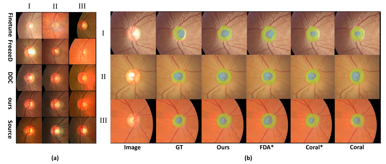
Comparative Experiments. We compared the two stages of MFM-DA with their respective baseline methods and state-of-the-art (SOTA) approaches. In each domain adaptation experiment for image generation models, only 10 images from the target domain were used for training to ensure that the model could access only unlabeled data from a single target domain during the training process. In the feature alignment domain adaptation experiments, the comparison methods were applied not only to the UNet architecture but also to MFMs to enable a more fair comparison. Table 1 presents the results of our method for generation tasks, showing significant improvements over competing approaches. This demonstrates that the Dynamic Instance-Aware Adaptor not only brings the model’s distribution closer to that of the target domain, but also enhances diversity, significantly improving the domain adaptation performance. Additionally, the segmentation results in Table 1 reveal that Pyramid Hierarchical Alignment achieves excellent feature alignment for the MFMs. The experimental results further indicate that the MFM-DA fine-tuned MFMs significantly reduces the domain gap with the target domain. Fig. 3 illustrates some representative results from various methods. Although “Finetune” generates images more similar in style by memorizing target images, our method preserves the structure of the source domain, captures the style of the target domain and produces more diverse images without memorization.
Ablation Analysis. To evaluate the effectiveness of the multi-level alignment loss used in the medical foundation model, we conducted ablation experiments on four features of the medical foundation model, each with a separate alignment loss. The average performance results are shown in Table 3. The results indicate that the use of multi-level alignment loss contributed to performance improvement.
Analysis of Few-shot Setting. As shown in Table 3, to evaluate the effectiveness of our method under different few-shot conditions, we tested the cases of 1-shot, 3-shot, 5-shot, 7-shot, and 10-shot with the target domain REFUGE (test). The results indicate that our method remains effective even under extremely few target domain scenarios.
Generalization to different foundation models. As shown in Table 3, we evaluate the generalizability of our method by applying MFM-DA to various foundation models [17, 15]. The baseline model used is U-Net. The experimental results demonstrate that MFM-DA effectively reduces the domain gap across different foundation models in the medical image segmentation task.
| Methods | Average |
|---|---|
| L1 | 84.17 |
| L2 | 84.09 |
| L3 | 84.04 |
| L4 | 83.39 |
| L-All | 84.83 |
| Setting | LPIPS | IC |
|---|---|---|
| 1-Shot | 90.17 | - |
| 3-Shot | 68.93 | 0.356 |
| 5-Shot | 100.47 | 0.315 |
| 7-Shot | 83.74 | 0.335 |
| 10-Shot | 62.79 | 0.371 |
| Methods | Target |
|---|---|
| Baseline | 78.07 |
| DINOv2 | 79.90 |
| +ours | 80.91 |
| CLIP | 82.52 |
| +ours | 83.46 |
4 Conclusion
In this paper, we propose a novel few-shot adaptation method for medical foundation models, named MFM-DA. In MFM-DA, the Dynamic Instance-Aware Adaptor generates feature transfer directions for each instance, requiring only 10 images to produce more diverse unlabeled target-domain images. Meanwhile, the Pyramid Hierarchical Alignment loss aligns source-domain and generated target-domain images in the feature space, enabling the medical foundation model to adapt to the target-domain distribution and improve performance on the target domain. Our experimental results on two datasets demonstrate the effectiveness of MFM-DA, showcasing its potential as a promising domain adaptation approach for MFMs. However, MFM-DA has so far only been experimented on fundus images and requires access to source domain data, which raises certain limitations in privacy protection. In our future work, we will extend MFM-DA to more modalities of medical imaging and develop techniques that do not require access to source domain data.
References
- [1] Almazroa, A., Alodhayb, S., Osman, E., et al.: Retinal fundus images for glaucoma analysis: the RIGA dataset. In: Medical Imaging 2018: Imaging Informatics for Healthcare, Research, and Applications. vol. 10579, pp. 55–62. SPIE (2018)
- [2] Chen, R.J., Ding, T., Lu, M.Y., et al.: Towards a general-purpose foundation model for computational pathology. Nature Medicine 30(3), 850–862 (2024)
- [3] Decencière, E., Zhang, X., Cazuguel, G., et al.: Feedback on a publicly distributed image database: the Messidor database. Image Analysis & Stereology pp. 231–234 (2014)
- [4] Ganin, Y., Lempitsky, V.: Unsupervised domain adaptation by backpropagation. In: International conference on machine learning. pp. 1180–1189 (2015)
- [5] Ho, J., Jain, A., Abbeel, P.: Denoising diffusion probabilistic models. Advances in Neural Information Processing Systems 33, 6840–6851 (2020)
- [6] Hu, S., Liao, Z., Xia, Y.: Domain specific convolution and high frequency reconstruction based unsupervised domain adaptation for medical image segmentation. In: International Conference on Medical Image Computing and Computer Assisted Intervention. pp. 650–659. Springer (2022)
- [7] Hu, T., Zhang, J., Liu, L., et al.: Phasic content fusing diffusion model with directional distribution consistency for few-shot model adaption. In: Proceedings of the IEEE/CVF International Conference on Computer Vision. pp. 2406–2415 (2023)
- [8] Jiang, J.X., Li, Y., Wang, Z.: Structure-aware single-source generalization with pixel-level disentanglement for joint optic disc and cup segmentation. Biomedical Signal Processing and Control 99, 106801 (2025)
- [9] Lei, W., Chen, H., Zhang, Z., et al.: A data-efficient pan-tumor foundation model for oncology ct interpretation. arXiv preprint arXiv:2502.06171 (2025)
- [10] Lei, W., Mei, H., Sun, Z., et al.: Automatic segmentation of organs-at-risk from head-and-neck ct using separable convolutional neural network with hard-region-weighted loss. Neurocomputing 442, 184–199 (2021)
- [11] Lei, W., Xu, W., Li, K., et al.: Medlsam: Localize and segment anything model for 3d ct images. Medical Image Analysis 99, 103370 (2025)
- [12] Mo, S., Cho, M., Shin, J.: Freeze the discriminator: a simple baseline for fine-tuning gans. arXiv preprint arXiv:2002.10964 (2020)
- [13] Motiian, S., Jones, Q., Iranmanesh, S., et al.: Few-shot adversarial domain adaptation. Advances in Neural Information Processing Systems 30 (2017)
- [14] Ojha, U., Li, Y., Lu, J., Efros, A.A., Lee, Y.J., Shechtman, E., Zhang, R.: Few-shot image generation via cross-domain correspondence. In: Proceedings of the IEEE/CVF Conference on Computer Vision and Pattern Recognition. pp. 10743–10752 (2021)
- [15] Oquab, M., Darcet, T., Moutakanni, T., et al.: DINOv2: Learning robust visual features without supervision. Transactions on Machine Learning Research Journal pp. 1–31 (2024)
- [16] Orlando, J.I., Fu, H., Breda, J.B., et al.: REFUGE challenge: A unified framework for evaluating automated methods for glaucoma assessment from fundus photographs. Medical Image Analysis 59, 101570 (2020)
- [17] Radford, A., Kim, J.W., Hallacy, C., et al.: Learning transferable visual models from natural language supervision. In: International Conference on Machine Learning. pp. 8748–8763 (2021)
- [18] Ronneberger, O., Fischer, P., Brox, T.: U-net: Convolutional networks for biomedical image segmentation. In: Medical Image Computing and Computer Assisted Intervention–MICCAI 2015: 18th international conference, Munich, Germany, October 5-9, 2015, proceedings, part III 18. pp. 234–241. Springer (2015)
- [19] Seitzer, M.: pytorch-fid: FID Score for PyTorch. https://github.com/mseitzer/pytorch-fid (August 2020), version 0.3.0
- [20] Sun, B., Saenko, K.: Deep coral: Correlation alignment for deep domain adaptation. In: Computer vision–ECCV 2016 workshops: Amsterdam, the Netherlands, October 8-10 and 15-16, 2016, proceedings, part III 14. pp. 443–450. Springer (2016)
- [21] Tsai, Y.H., Hung, W.C., Schulter, S., Sohn, K., Yang, M.H., Chandraker, M.: Learning to adapt structured output space for semantic segmentation. In: Proceedings of the IEEE Conference on Computer Vision and Pattern Recognition. pp. 7472–7481 (2018)
- [22] Wei, Z., Chen, L., Jin, Y., Ma, X., Liu, T., Ling, P., Wang, B., Chen, H., Zheng, J.: Stronger fewer & superior: Harnessing vision foundation models for domain generalized semantic segmentation. In: Proceedings of the IEEE/CVF Conference on Computer Vision and Pattern Recognition. pp. 28619–28630 (2024)
- [23] Wu, Y., Zhang, X., Zhang, H., et al.: Mamba-SAM: An adaption framework for accurate medical image segmentation. In: 2024 IEEE International Conference on Bioinformatics and Biomedicine (BIBM). pp. 3856–3859. IEEE (2024)
- [24] Yang, Y., Soatto, S.: FDA: Fourier domain adaptation for semantic segmentation. In: Proceedings of the IEEE/CVF Conference on Computer Vision and Pattern Recognition. pp. 4085–4095 (2020)
- [25] Zhang, S., Metaxas, D.: On the challenges and perspectives of foundation models for medical image analysis. Medical Image Analysis 91, 102996 (2024)
- [26] Zhou, Y., Chia, M.A., Wagner, S.K., et al.: A foundation model for generalizable disease detection from retinal images. Nature 622(7981), 156–163 (2023)