An anatomically-informed correspondence initialisation method to improve learning-based registration for radiotherapy
| Abstract We propose an anatomically-informed initialisation method for interpatient CT non-rigid registration (NRR), using a learning-based model to estimate correspondences between organ structures. A thin plate spline (TPS) deformation, set up using the correspondence predictions, is used to initialise the scans before a second NRR step. We compare two established NRR methods for the second step: a B-spline iterative optimisation-based algorithm and a deep learning-based approach. Registration performance is evaluated with and without the initialisation by assessing the similarity of propagated structures. Our proposed initialisation improved the registration performance of the learning-based method to more closely match the traditional iterative algorithm, with the mean distance-to-agreement reduced by 1.8mm for structures included in the TPS and 0.6mm for structures not included, while maintaining a substantial speed advantage (5 vs. 72 seconds). |
1 Introduction
Non-rigid registration (NRR) is widely used for many applications in radiotherapy to spatially align images, including fusion of complementary images, contour propagation, evaluating treatment response, dose mapping [1, 2]. Traditional image registration algorithms function by performing iterative updates to parameters of a chosen deformation model to optimise either an intensity- and/or feature-based metric [1]. Intensity-based metrics use the images greyscale intensity information directly to evaluate similarity. Feature-based metrics use features extracted from the images such as anatomical landmarks, structure boundaries, contours and fiducial markers to evaluate alignment.
Hybrid image registration techniques have been developed which aim to combine the strengths of intensity- and feature-based algorithms, leveraging greyscale information as well as anatomical structures or landmarks to guide the registration [3, 4]. However, hybrid methods tend to be slow and labour intensive, taking a long time to set up as they require landmark annotation or structure contouring.
Recently image registration methods using deep learning (DL) have garnered much attention as they can offer large time savings over iterative algorithms [5]. DL approaches are trained ahead of time on large datasets of examples; once trained, these methods can estimate a full deformation vector field in a single function evaluation, often taking less than a second on modern hardware [6]. However, current learning-based approaches are predominantly applied to easier tasks such as the registration of brain MRI scans where the inter-subject variability is relatively low and the soft-tissue contrast is high [5, 7]. There has been little evidence to show that learning-based registration methods are suitable for large deformations, such as inter-subject CT registration [8, 6].
Many learning-based registration approaches are trained to optimise intensity-based metrics. Some can leverage segmentations present in the images, but only at training time through semi-supervised learning [5]. In this study we incorporate ideas from hybrid registration methods to improve learning-based registration performance. To achieve this we introduce a new anatomical correspondence-informed non-rigid initialisation to align labelled anatomical structures prior to using established methods to register non-labelled tissue.
2 Materials and Methods
We used an open-access dataset of 31 head and neck CT scans with highly consistent organ-at-risk (OAR) segmentations [9].
2.1 Registration pipelines
We propose the use of a previously developed learning-based correspondence model [10] and a thin plate spline (TPS) to form an anatomically informed method to initialise inter-patient non-rigid registration. The impact of the proposed initialisation was compared for two established NRR methods: a) NiftyReg, a traditional B-spline iterative-optimisation based image registration method [11] and b) Voxelmorph, an unsupervised, DL-based approach [12]. From now on, the proposed initialisation method is referred to as CorrTPS.
2.1.1 Anatomically informed initialisation (CorrTPS)
Correspondence model
In a previous study, we showed an unsupervised geometric learning model could be trained to identify dense, point-to-point, correspondences for head and neck OARs [10]. The developed model can automatically identify corresponding anatomical points on the surface of 3D organ shapes and was shown to be effective for the brainstem, mandible, parotid glands, spinal cord and submandibular glands. The input for the correspondence model is two organ shapes which are converted to triangular meshes. The model predicts an assignment linking all vertices from the source to the target organ. Correspondence predictions are based on the organ geometry alone, however, CT imaging was utilised in the training process to improve prediction quality [10].
Mesh generation
To create triangular meshes of the OAR structures, we used the marching cubes algorithm followed by Taubin smoothing, mesh decimation, optimisation and simplification operations. The full details of this process have been described fully in our previous work [10].
A segmentation of the bony anatomy was acquired automatically from the CT scan by removing the table, thresholding at 400 Hounsfield units (HU), cropping at the base of the neck, and meshing using the marching cubes algorithm. A mesh of the external envelope comprising of the patient’s body was generated using a lower threshold level of -200 HU.
Correspondence inference
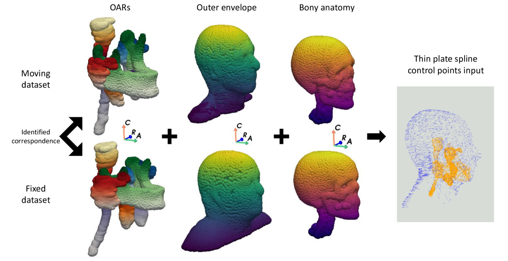
For non-rigid initialisation, dense correspondences were estimated for all segmented structures in the HN, as well as the bony anatomy and outer envelope of each patient (Figure 1).
Thin plate spline setup
Once estimated, the correspondences were used as the control points to set up a TPS deformation model [13]. The TPS is generalizable to 3D and has a regularisation parameter, TPS, which controls the allowed degree of local deformations.
Control points from each structure were gathered and a single TPS was created for each registration pair. In total, the TPS deformations were performed using about 14,000 control points (range: 10,500 - 16,000). Regions that were identified as null correspondences were excluded from the control point set driving the TPS. In this way, we prevent the creation of anatomically infeasible scenarios, e.g. inverting and folding around tissue mismatches.
For each fixed-moving image pair, we generated a displacement vector field by evaluating the TPS on the grid positions of the moving image. Then, we resampled the fixed image by inverse warping using the DisplacementFieldTransform from the SimpleITK package. We refer to these as non-rigidly initialised scans. A second NRR algorithm was then used to fine-tune the registration using the image intensities for the whole anatomy.
2.1.2 Non-rigid registration
To demonstrate the added value of CorrTPS, we compared the performance of two established intensity-based NRR methods with and without our proposed initialisation.
|
|
|
|||||||
|---|---|---|---|---|---|---|---|---|---|
| Voxelmorph | 1.6 | 0.3 | 1.9 | ||||||
| Voxelmorph + CorrTPS | (1.2 + 0.1 + 1.4 + 0.2) + 0.3 | 4.8 | |||||||
| NiftyReg | 70.7 | 72.3 | |||||||
| NiftyReg + CorrTPS | (1.2 + 0.1 + 1.4 + 0.2) + 70.7 | 75.2 |
Deep learning-based registration (Voxelmorph)
We trained multiple Voxelmorph models from scratch [12]. Image volumes were automatically cropped to dimensions of such that the whole HN anatomy was within the field-of-view while retaining the original voxel spacing. Additionally, the images were normalised from Hounsfield units onto the range [0,1] using contrast windowing with wide window settings (W 1600HU, L 0HU). The weight of the smoothing loss term which regularises the predicted deformations was set to 0.005.
When performing the five-fold cross-validation, each fold contains 7 testing patients. This leads to a testing pairs for each fold (trivial self-registration cases excluded). The same Voxelmorph models were applied to the non-rigidly initialised scans.
Iterative registration (NiftyReg)
NiftyReg was first used to non-rigidly register the CT scans after rigid pre-alignment alone [11]. Following an empirical parameter evaluation, the configuration settled upon was a GPU-accelerated cubic B-spline using normalised mutual information loss with a bending energy of 0.001. The same configuration of NiftyReg was then used to register the non-rigidly initialised CT scans.
2.2 Evaluation
For every pipeline, we report the execution time including all the necessary pre-processing steps and the results image- and segmentation-based metrics.
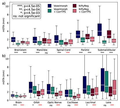
The mean distance-to-agreement (mDTA) was calculated for each of the structures included within CorrTPS as well as the brain, cochleae, optic nerves, orbits following non-rigid alignment of the moving anatomy to the fixed reference patient. Wilcoxon signed-rank hypothesis tests were used to compare each registration algorithm with and without the CorrTPS initialisation. A Bonferroni correction was applied to minimise the risk of type I errors from multiple hypothesis tests (corrected p value: 0.0045).
Implementation details
Model training and evaluation was performed on a workstation with an AMD Ryzen 9 3950X 16-Core Processor, 64GB of RAM and an NVidia GeForce RTX 4090 GPU.
3 Results
The average runtime for each approach is shown in Table 1.
Figure 2a shows boxplots of the mDTA results for the organ structures included in the CorrTPS, while 2b shows similar results for structures not included in CorrTPS. Significant improvements in registration performance were observed for most structures included in CorrTPS across both methods when using the proposed initialisation. For structures not included in CorrTPS, some improvements were observed for the learning-based registration method (the brain, orbits and lacrimal glands), however, the performance of the traditional registration algorithm was degraded.
4 Discussion
We proposed a novel method to non-rigidly pre-register an image pair prior to inter-subject registration of head and neck CT scans using correspondence of anatomical structures. The initialisation method is based on a geometric learning-based correspondence model which, when coupled with a TPS deformation, allowed the moving scans to be deformed to align segmented OARs before a final registration using established NRR methods. Our initialisation approach showed promise to improve the registration performance of the learning-based NRR method, Voxelmorph, improving the quality of the registrations in terms of the segmentation-based evaluation measure. The performance of our proposed method with the traditional iterative algorithm, NiftyReg, was less clearly defined, with some improvements for structures included in the initialisation and degradations for those not included. The use of CorrTPS required a limited increase in execution time.
In terms of the mDTA, the benefit of our proposed initialisation method is evident for structures included within CorrTPS. The one exception to this was for the spinal cord when using NiftyReg, in which the use of the initialisation method harmed registration performance.
Little improvement was observed when using our method for the structures not included in CorrTPS, which were predominantly in the superior region of the head. This is likely due to the TPS deformation in this region comprising of an interpolation between the envelope and bony anatomy and the controlling structures which were mainly further inferior in the head and neck region. This shows a sensitivity of the proposed method to the spatial distribution of controlling structures. For registrations with the iterative algorithm, the use of the proposed method degraded the performance for all structures not included in CorrTPS.
While we included some of the structures in this study and excluded others from CorrTPS, the proposed method is applicable to any of the OAR structures segmented for this study, i.e., the complete set of structures could also be included in CorrTPS, thereby fitting a wider distribution of control points within the head and neck anatomy. The implementation of TPS used is fast, estimating a deformation within a second for control points.
We deployed our proposed initialisation method with an intensity-based traditional NRR algorithm and a DL-based method. In future it would be useful to compare the performance of our hybrid-inspired registration framework against a hybrid registration methods such as ANACONDA [3] to further assess the efficacy of the proposed method.
In this study we focused solely on performing inter-patient registration, despite the task of intra-patient registration being more common within the radiotherapy treatment pathway. Inter-patient registration is important for tasks such as outcome modelling and can be challenging due to the increased variability in anatomy between subjects. We hypothesize that our initialisation method would be particularly effective in intra-patient cases such as in the re-irradiation setting, where large and complex anatomical changes are common, e.g. resulting from surgical interventions, which hinder the accuracy of currently available image registration algorithms [2, 14].
The segmentations used in this study were manually performed. Future introduction of auto-segmentation could mitigate the impact of inter- and intra-observer variation and improve correspondence reliability. Additionally, the use of auto-segmentations with the proposed framework would facilitate the implementation of a fully automated anatomically-informed registration pipeline.
5 Conclusion
The proposed method to initialise an image pair prior to NRR using anatomical correspondence shows particular promise to enhance the registration performance of DL-based registration methods to more closely match that of a traditional iterative NRR algorithm, whilst maintaining a substantial speed advantage. With further development and validation, the proposed approach could additionally raise the performance of traditional iterative NRR algorithms.
References
- [1] Kristy K. Brock et al. “Use of image registration and fusion algorithms and techniques in radiotherapy: Report of the AAPM Radiation Therapy Committee Task Group No. 132” In Medical Physics 44.7 Wiley, 2017, pp. e43–e76 DOI: 10.1002/mp.12256
- [2] Martina Murr et al. “Applicability and usage of dose mapping/accumulation in radiotherapy” In Radiotherapy and Oncology 182 Elsevier BV, 2023, pp. 109527 DOI: 10.1016/j.radonc.2023.109527
- [3] Ola Weistrand and Stina Svensson “The ANACONDA algorithm for deformable image registration in radiotherapy” In Medical Physics 42.1 Wiley, 2015, pp. 40–53 DOI: 10.1118/1.4894702
- [4] Jinkoo Kim et al. “A novel approach for establishing benchmark CBCT/CT deformable image registrations in prostate cancer radiotherapy” In Physics in Medicine & Biology 58.22 IOP Publishing, 2013 DOI: 10.1088/0031-9155/58/22/8077
- [5] Jing Zou, Bingchen Gao, Youyi Song and Jing Qin “A review of deep learning-based deformable medical image registration” In Frontiers in Oncology 12 Frontiers Media SA, 2022 DOI: 10.3389/fonc.2022.1047215
- [6] Jonas Teuwen, Zeno A.R. Gouw and Jan-Jakob Sonke “Artificial Intelligence for Image Registration in Radiation Oncology” In Seminars in Radiation Oncology 32.4 Elsevier BV, 2022, pp. 330–342 DOI: 10.1016/j.semradonc.2022.06.003
- [7] Mattias P. Heinrich and Lasse Hansen “Voxelmorph++” In Biomedical Image Registration Springer International Publishing, 2022, pp. 85–95 DOI: 10.1007/978-3-031-11203-4_10
- [8] R. Bhattacharjee et al. “Evaluation of a Learning-based Deformable Registration Method on Abdominal CT Images” In IRBM 42.2 Elsevier BV, 2021, pp. 94–105 DOI: 10.1016/j.irbm.2020.04.002
- [9] Stanislav Nikolov et al. “Deep learning to achieve clinically applicable segmentation of head and neck anatomy for radiotherapy” In ArXiv e-prints, 2018 DOI: 10.48550/arXiv.1809.04430
- [10] Edward G.. Henderson, Marcel van Herk, Andrew F. Green and Eliana M. Vasquez Osorio “Unsupervised Correspondence with Combined Geometric Learning and Imaging for Radiotherapy Applications” In Shape in Medical Imaging Springer Nature Switzerland, 2023, pp. 75–89 DOI: 10.1007/978-3-031-46914-5_7
- [11] Marc Modat et al. “Fast free-form deformation using graphics processing units” In Computer Methods and Programs in Biomedicine 98.3 Elsevier BV, 2010, pp. 278–284 DOI: 10.1016/j.cmpb.2009.09.002
- [12] Guha Balakrishnan et al. “VoxelMorph: A Learning Framework for Deformable Medical Image Registration” In IEEE Transactions on Medical Imaging 38.8 Institute of ElectricalElectronics Engineers (IEEE), 2019, pp. 1788–1800 DOI: 10.1109/tmi.2019.2897538
- [13] F.L. Bookstein “Principal warps: thin-plate splines and the decomposition of deformations” In IEEE Transactions on Pattern Analysis and Machine Intelligence 11.6 Institute of ElectricalElectronics Engineers (IEEE), 1989, pp. 567–585 DOI: 10.1109/34.24792
- [14] Lena Nenoff et al. “Review and recommendations on deformable image registration uncertainties for radiotherapy applications” In Physics in Medicine & Biology IOP Publishing, 2023 DOI: 10.1088/1361-6560/ad0d8a