Evaluating and Improving the Effectiveness of Synthetic Chest X-Rays for Medical Image Analysis
Abstract
-
•
Purpose: To explore best-practice approaches for generating synthetic chest X-ray images and augmenting medical imaging datasets to optimize the performance of deep learning models in downstream tasks like classification and segmentation.
-
•
Materials and Methods: We utilized a latent diffusion model to condition the generation of synthetic chest X-rays on text prompts and/or segmentation masks. We explored methods like using a proxy model and using radiologist feedback to improve the quality of synthetic data. These synthetic images were then generated from relevant disease information or geometrically-transformed segmentation masks and added to ground truth training set images from the CheXpert [7], CANDID-PTX[3], SIIM [14], and RSNA Pneumonia [12] datasets in order to measure improvements in classification and segmentation model performance on the test sets. F1 and Dice scores were used to evaluate classification and segmentation respectively. One-tailed t-tests with Bonferroni correction were used to assess the statistical significance of performance improvements with synthetic data.
-
•
Results: Across all experiments, the synthetic data we generated resulted in a maximum mean classification F1 score improvement of 0.150453 (CI: 0.099108, 0.201798, P=0.0031) compared to using only real data. For segmentation, the maximum Dice score improvement was 0.14575 (CI: 0.108267, 0.183233; P=0.0064).
-
•
Conclusion: We find best practices for generating synthetic chest X-ray images for downstream tasks include conditioning on single-disease labels or geometrically-transformed segmentation masks, as well as potentially using proxy modeling for fine-tuning such generations.
Introduction
Developing AI tools for medical image analysis is inherently challenging due to limited data availability and the need for carefully labeled medical datasets. Data scarcity in publicly available medical imaging datasets arises from constraints such as logistical challenges, patient privacy requirements, and the high cost of expert annotations. Synthetic data generation has emerged as a promising approach to address these limitations and can be utilized for applications such as augmenting datasets for disease segmentation and classification tasks. While previous studies have demonstrated the feasibility of generating synthetic data for chest X-rays using earlier methods like generative adversarial networks (GANs) [5], these models often struggle with producing clinically meaningful details, which limits their effectiveness in downstream applications [4, 11]. Recent diffusion-based approaches, such as vision-language foundation models for chest X-ray generation, have since advanced synthetic image quality by producing more realistic, high-resolution outputs [1]. However, these diffusion models often lack the ability to generate paired images and labels, limiting their utility for segmentation tasks where such pairs are essential [8]. Additionally, the effects of the quality and precision of synthetic medical images on downstream tasks, such as classification and segmentation, remain under-explored.
To address these challenges, this work uses a latent diffusion model [10] based on the ControlNet architecture [15] designed to generate synthetic chest X-rays conditioned on both text prompts and segmentation masks. Unlike prior models, this framework produces paired images and labels, enhancing its applicability for a broader range of downstream tasks, including segmentation and classification. This study involves extensive experimentation to examine several hypotheses about synthetic data generation for medical imaging. We hypothesize that synthetic data can enhance performance in data-scarce environments, especially when the dataset volume is increased. We also explore whether producing visually realistic, low-hallucination images leads to improved outcomes in classification and segmentation tasks. Finally, we investigate the impact of selecting synthetic images based on a pseudo-model mimicking the downstream task, positing that this approach can improve synthetic data selection and optimize performance.
Through these contributions, this work aims to deepen the understanding of synthetic data generation in medical imaging and establish effective strategies for leveraging these images in classification and segmentation tasks under data-limited conditions.
Results
Synthetic chest X-rays can be generated by conditioning on custom mask and reports to mimic diseases and conditions
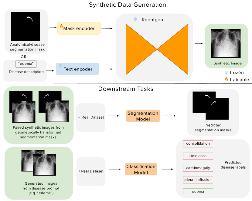
The Bluethgen et al. foundation model [1] adopts a pre-trained latent diffusion architecture to generate synthetic chest X-ray images conditioned on summarized radiology reports. In this work, we show that a latent diffusion model can be easily extended to generate synthetic chest X-rays condition on beyond just text. To do this, our latent diffusion model framework utilizes the ControlNet architecture [15], which integrates visual conditioning (e.g. segmentation masks) into pretrained, frozen text-to-image latent diffusion models like Stable Diffusion [10]. ControlNet integrates these conditions by cloning the frozen parameters to create trainable counterparts that accept conditioning vectors. These trainable counterparts are linked through zero-initialized convolution layers, enabling the model to learn new conditional patterns without affecting its original generative abilities. We reuse the model weights of Bluethgen et al., as it already is trained on a large-scale dataset of chest X-rays and reports. With this, we create a flexible framework as seen in Figure 1, which can take in both a mask and a text prompt to create accurate synthetic chest X-rays grounded by the input conditions we provide.
Synthetic chest X-rays help improve model performance on classification and segmentation tasks
We tested if synthetic data can improve the performance of both binary and multilabel disease classification tasks. For binary pneumonia classification, we trained a ResNet 50 [6] model to predict the presence or absence of pneumonia in the RSNA Pneumonia dataset. For multilabel classification, we trained the MGCA model [13] on the CheXpert dataset to classify five disease labels: atelectasis, edema, consolidation, cardiomegaly, and pleural effusion. We evaluated the F1 scores of both on the real test sets, and then synthetic data was added to augment the training datasets at 2x, 5x, 10x, and 25x the size of the original real training data and evaluate them on the same real test sets.
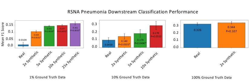
Binary classification for pneumonia showed substantial improvements with the inclusion of synthetic data (Figure 2, Plot 3). Synthetic pneumonia chest X-rays were generated using a single-disease prompt (i.e. ”pneumonia”). At 1% ground truth data availability, the mean F1 score improved from 0.0104 (real-only training) to 0.1541 with 25x synthetic augmentation. At 10% ground truth data, the F1 score increased from 0.0950 to 0.2760 with 10x synthetic data. The benefits of synthetic data diminished at 100% ground truth data, where the F1 score plateaued (0.326 with real data vs. 0.344 with 2x synthetic data).
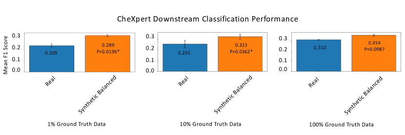
In multilabel classification on CheXpert dataset, we also found it was most effective to generate the synthetic data using single-disease prompts (e.g. ”cardiomegaly”), and the synthetic data was used to balance all classes to the count of the most frequent disease class. At 1% ground truth data, the mean F1 score increased from 0.209 with real data to 0.289 with 2x synthetic data. At 10% ground truth data, the F1 score improved from 0.255 to 0.323 with 2x synthetic data. At 100% ground truth data, synthetic data provided marginal improvements, with the F1 score increasing from 0.310 to 0.354 with balanced 2x synthetic augmentation (Figure 2, Plot 4). These results demonstrate that the strategic addition of synthetic data can address class imbalance and significantly improve multilabel classification performance, especially in low-resource scenarios.
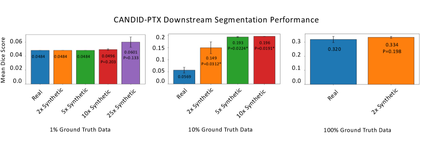
For segmentation, the UNet++ [16] architecture was employed to segment pneumothorax regions in the CANDID-PTX and SIIM datasets. Geometric transformations, including dilation, erosion, horizontal flipping, and translation, were applied to ground truth masks to generate diverse variations. These augmented masks served as input conditions to the diffusion model, which produced paired synthetic images and labels. Performance was evaluated on the real test sets, with synthetic data augmenting the training sets at 2x, 5x, 10x, and 25x the size of the real training data.
At 1% ground truth data availability, the mean Dice score improved from 0.0484 with real data to 0.0601 with 25x synthetic augmentation. At 10% ground truth data, synthetic data improved the Dice score from 0.0569 to 0.196 with 10× synthetic data. At 100% ground truth data, segmentation performance gains were marginal, with Dice scores increasing from 0.320 to 0.334 with 2x synthetic data (Figure 2, Plot 1).
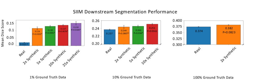
Similar trends were observed for the SIIM dataset. At 1% ground truth data, the Dice score increased from 0.2072 with real data to 0.2550 with 25x synthetic augmentation. At 10% ground truth data, the Dice score improved from 0.237 to 0.251 with 10x synthetic data. At 100% ground truth data, the improvements were minimal, with Dice scores increasing from 0.374 to 0.382 with 2x synthetic data (Figure 2, Plot 2). These findings show that synthetic data effectively improves segmentation performance, especially in data-scarce settings. However, the benefit diminishes as the availability of real data increases.
Overall, the addition of synthetic data improves the mean Dice score for segmentation tasks and mean F1 score for classification tasks, as demonstrated by the performance increase across almost all conditions when comparing the use of only ground truth data versus combinations of ground truth and synthetic data. This suggests that synthetic data can effectively augment existing datasets, particularly in settings where limited ground truth data is available.
More synthetic data results in better model performance
Increasing the proportion of synthetic data further boosts downstream segmentation and classification performance, particularly in cases with minimal ground truth data (e.g., 1% and 10% ground truth data). Models trained with progressively more synthetic data tend to achieve higher mean Dice and F1 scores compared to models trained with lower levels of synthetic augmentation. This progressive improvement underscores the value of synthetic data in augmenting small datasets, where limited real data may be insufficient for achieving optimal performance. However, this trend plateaus at higher data availability (e.g., 100% ground truth data), indicating that synthetic data is most beneficial when ground truth data is scarce and may offer diminishing returns when real data availability is robust.
Enhancing synthetic data with a proxy model boosts performance
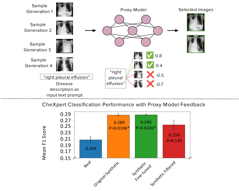
Chest X-rays exhibit fine-grained nuances and complexities that are challenging to capture in synthetic data, making fine-tuning helpful for improving representations. Radiologist feedback helps steer the model toward established medical knowledge, offering valuable guidance to refine the generation process. However, collecting such expert input at scale remains difficult. As a result, medical foundation models can serve as proxies for radiologists by efficiently offering metrics like cosine similarity between captions and images to evaluate the quality of synthetic generations, providing a scalable alternative to direct involvement from clinical experts.
We employed a proxy model approach using BioMedCLIP [2] to filter high-quality synthetic images based on cosine similarity between disease prompts and generated images. The filtered synthetic data was then used to fine-tune the diffusion model. In the CheXpert classification task, this approach yielded marginal improvements. For example, at 1% ground truth data, fine-tuned synthetic data improved the F1 score from 0.209 (real-only) to 0.290. However, using only the filtered high-quality synthetic images resulted in lower performance (F1 score = 0.258), suggesting that while filtering improves data quality, the volume and diversity of synthetic data remain critical for downstream performance.
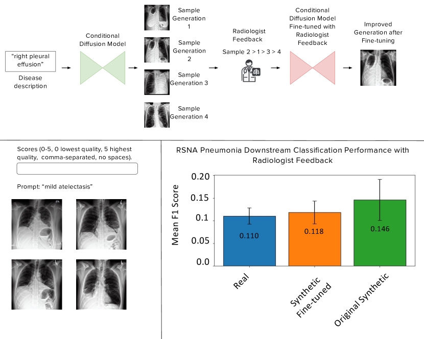
Aligning the diffusion model on radiologist feedback yields inconsistent results
To refine the synthetic image generation process, radiologist feedback was incorporated through a preference-ranking protocol. We found that the fine-tuned synthetic data showed minimal improvements in downstream tasks. For example, in the RSNA Pneumonia classification task, synthetic data fine-tuned with radiologist feedback improved the F1 score from 0.110 (real-only) to 0.118, while the original synthetic data (without fine-tuning) achieved a higher score of 0.146. These results suggest that while radiologist feedback can enhance image quality, its impact on task performance may be limited, particularly when compared to the original synthetic data pipeline.
Materials and Methods
| Dataset | Training/Test Size | Conditions |
|---|---|---|
| CheXpert [7] | 191,229 / 235 | Atelectasis, cardiomegaly, consolidation, edema, pleural effusion |
| CANDID-PTX [3] | 17,237 / 2,000 | Lungs, heart, pneumothorax, rib fractures, chest tubes |
| SIIM [14] | 1,903 / 476 | Pneumothorax |
| RSNA Pneumonia [12] | 21,885 / 4,801 | Pneumonia |
Datasets
Details regarding all datasets used in our experiments can be found in Table 1. We used the CANDID-PTX chest X-ray dataset for training our diffusion model. A total of 17,237 randomly selected images were used for training, while the remaining 2,000 images were reserved for evaluation. These 2,000 held-out images were then employed for downstream pneumothorax segmentation, with the data split randomly into an 80/20 ratio for training and testing. Following the MGCA [13] paper, we utilized the validation set of CheXpert as our test set. To more pointedly test segmentation performance, we only use the SIIM images with nonzero segmentation masks and randomly split these images 80/20 for training and testing. Following th MGCA [13] paper, we also randomly split the RSNA Pneumonia training set 80/20, given the public availability of only the training annotations.
Model Architecture and Training
Our latent diffusion model framework incorporates segmentation mask visual conditioning into the preinitialized Bluethgen et al. model [1] and its corresponding text encoder using the ControlNet framework. While training our model on segmentation masks, text conditions were set to “N/A,” prompting the model to convert text conditions to zero vectors. During sampling, if “N/A” was provided as a text condition or an all-black segmentation mask was used, the corresponding condition vectors were zeroed. This approach enabled the trained ControlNet model to generate synthetic data from any combination of text and mask conditions.
We train our model with the ControlNet loss function. It takes in an input image and progressively adds random noise to produce a noisy version , where is the given time step. Given the mask condition , text condition , and time step , the model trains by learning a noise prediction network to predict the noise added to , as shown in Equation 1:
| (1) |
We trained the model on eight NVIDIA A4000 GPUs. Gradient accumulation was set to 16, and the learning rate was . Other hyperparameters remained consistent with those in the original ControlNet implementation.
Improving the synthetic data
Proxy Model
As a proxy model, we employ BiomedCLIP [2] to identify and filter medical images depicting one of the five diseases outlined in the CheXpert classification tasks (i.e. ”cardiomegaly”, ”pleural effusion,” ”edema,” ”atelectasis”, and ”consolidation”). Our approach calculates cosine similarity scores between features extracted from each image and the corresponding disease text label. Images from a specified dataset are processed individually, and similarity scores are computed to assess how closely each image aligns with the target label. To ensure specificity, we apply a 90th-percentile threshold, selecting images with the highest similarity scores. This percentile-based filtering metric is instrumental in isolating images with the strongest relevance to the specified medical condition. The model was trained for an additional 9,000 steps on the filtered images.
Radiologist Feedback
For reinforcement learning fine-tuning, we collected preferences from radiologists to enhance the model’s outputs. We assembled approximately 200 sets of four sampled images each, with the distribution of condition types as follows: 75% text-only conditioning (T2I), 15% mask-only conditioning (M2I), and 10% combined text and mask conditioning (TM2I). The percentages were chosen based on the sampling diversity allowed by each condition type.
Text conditions for validation were generated from the impressions section of the MIMIC-CXR validation split and the held-out CANDID-PTX reports. These were summarized into concise five-word captions using GPT-4 with a standardized prompt to ensure consistency and avoid bias:
“Based on these chest X-ray reports, please write a five-word caption with the main finding. Don’t make comparisons with previous studies, so do not use words such as ’unchanged’, ’improved’, ’worsened’, ’no change’, ’increased’, ’decreased’, etc. in the caption. Don’t use commas or quotation marks in the caption. If it is normal or no problems are detected, just return ’Normal’ as the caption.”
The image sets were hosted on Gradio, and four radiologists independently scored each image set on a scale from 0 (low quality) to 5 (high quality). For images with combined conditions, separate scores were collected for text and mask condition quality. The data were anonymized, and the radiologists were blinded to the source of the images.
We employed Direct Preference Optimization (DPO) [9] to utilize these preferences in model finetuning. DPO operates directly on the preference scores, and the loss function is defined as:
| (2) |
where is a scaling factor, is the model policy parameterized by , is the reference model policy, represents an example in dataset where is a more preferred sample, is a less preferred sample, and is the corresponding condition. Separate losses are calculated for each condition type (text or mask) and zeroed or averaged based on the presence or absence of each condition. We use the parameters of the DPO paper, except for a learning rate of and a gradient accumulation of 1. The model was trained for an additional 4,000 steps on the collected preference data.
Downstream Evaluation
To assess the effectiveness of synthetic data in enhancing performance on out-of-distribution datasets, we conducted a series of downstream segmentation and classification experiments across multiple disease conditions. For segmentation, we utilized the UNet++ [16] architecture, an advanced encoder-decoder network with nested dense skip pathways, to segment pneumothorax regions. We augmented the CANDID-PTX and SIIM training datasets by applying geometric transformations—including dilation, erosion, horizontal flipping, and translation—to ground truth masks. These modified masks were then inputted into the model to generate new synthetic data paired with each mask, thus expanding the datasets with diversified synthetic instances.
In the classification domain, multi-class disease classification was performed using the MGCA [13] architecture to identify pleural effusion, atelectasis, edema, cardiomegaly, and consolidation on the CheXpert dataset. Single-class classification for pneumonia detection was carried out using a ResNet-50 [6] model. The training datasets for both tasks were augmented with synthetic images, with each synthetic instance corresponding to a single disease label to enhance model specificity. Model training parameters followed those outlined in the respective original architecture publications, ensuring consistency in evaluation.
We used F1 scores to evaluate classification and Dice scores for segmentation. This study posits three hypotheses: (1) synthetic data augmentation enhances performance in both segmentation and classification tasks, particularly in data-scarce settings; (2) increasing the volume of synthetic data correlates positively with model performance; (3) the generation of visually realistic, low-hallucination synthetic images, which can be generated using feedback from either experts or proxy models, contributes to improved task outcomes.
Statistical Analysis
One-tailed t-tests with Bonferroni correction of the performance differences between ground truth and synthetic-data-augmented downstream models were used to assess the statistical significance of performance improvements with synthetic data. We used two trials to calculate the mean values for each experimental setting and used a significance level of to determine general improvements with synthetic data.
Discussion and Conclusion
This study demonstrates that integrating synthetic chest X-ray images generated via a latent diffusion model can enhance downstream segmentation and classification performance, especially in data-scarce settings. By incorporating paired image-label generation and using feedback from radiologists and proxy models, the framework offers promise in providing a scalable solution for augmenting limited medical datasets. The results confirm that strategically adding synthetic data improves model performance and highlights its potential to address challenges related to dataset scarcity in medical imaging. Moreover, synthetic data can help improve long-tail disease classification by generating additional samples for rare conditions and enhancing the ability of the model to recognize underrepresented diseases.
This study validates that:
-
1.
Synthetic data augmentation can enhance performance in both segmentation and classification tasks, particularly in settings with limited ground truth data.
-
2.
Increasing the volume of synthetic data further correlates positively with improved model performance, demonstrating that larger synthetic datasets can mitigate the challenges of data scarcity.
-
3.
Generating visually realistic, low-hallucination synthetic images guided by a proxy model can contribute to improved outcomes in downstream tasks.
However, we found that limited radiologist feedback is minimally effective in refining model outputs due to the limited size of the expert cohort providing preferences.
Despite these advancements, the study has some limitations. Our evaluation focused exclusively on chest X-rays, which limits understanding of how well the approach works for other imaging modalities. The tasks we explored were also narrow, as segmentation experiments only targeted pneumothorax. Additionally, relying on a small group of radiologists to collect preference data for fine-tuning may have constrained the model’s optimization, reducing its generalizability. Expanding this work to include diverse imaging types, a broader range of tasks, and feedback from more radiologists could strengthen the effectiveness and applicability of this approach.
References
- [1] Christian Bluethgen, Aoxiao Liu, Mohammad Mehdipour, et al. A vision–language foundation model for the generation of realistic chest x-ray images. Nature Biomedical Engineering, 2024.
- [2] Harini Eswaran, Wenhao Zhang, Yifan Zhang, et al. Biomedclip: Visual-language models for biomedical applications. 2022. arXiv preprint arXiv:2210.12359.
- [3] Sijing Feng, Damian Azzollini, Ji Soo Kim, et al. Curation of the candid-ptx dataset with free-text reports. Radiology: Artificial Intelligence, 3(6):e210136, 2021.
- [4] Maayan Frid-Adar, Eyal Klang, Michal Amitai, et al. Gan-based synthetic medical image augmentation for increased cnn performance in liver lesion classification. Neurocomputing, 321:321–331, 2018.
- [5] Ian Goodfellow, Jean Pouget-Abadie, Mehdi Mirza, Bing Xu, David Warde-Farley, Sherjil Ozair, Aaron Courville, and Yoshua Bengio. Generative adversarial nets. In Advances in neural information processing systems, volume 27, 2014.
- [6] Kaiming He, Xiangyu Zhang, Shaoqing Ren, and Jian Sun. Deep residual learning for image recognition. 2015. arXiv preprint arXiv:1512.03385.
- [7] Jeremy Irvin, Pranav Rajpurkar, Michael Ko, et al. Chexpert: A large chest radiograph dataset with uncertainty labels and expert comparison. Proceedings of the AAAI Conference on Artificial Intelligence, 33:590–597, 2019.
- [8] Amir Kazerouni, Dwarikanath Mahapatra, Lin Chen, et al. Diffusion models for medical image analysis: A comprehensive survey. Medical Image Analysis, 82:102615, 2023.
- [9] Rumen Rafailov, Aditi Sharma, Eric Mitchell, et al. Direct preference optimization: Your language model is secretly a reward model. 2023. arXiv preprint arXiv:2305.18290.
- [10] Robin Rombach, Andreas Blattmann, Dominik Lorenz, Patrick Esser, and Björn Ommer. High-resolution image synthesis with latent diffusion models. arXiv preprint arXiv:2112.10752, 2022.
- [11] Hoo-Chang Shin, Nathan A Tenenholtz, John K Rogers, et al. Medical image synthesis for data augmentation and anonymization using generative adversarial networks. In Proceedings of MICCAI, pages 1–10, 2018.
- [12] Alex Stein, Chao Wu, Chris Carr, et al. Rsna pneumonia detection challenge, 2018.
- [13] Fei Wang, Yuyin Zhou, Shun Wang, et al. Multi-granularity cross-modal alignment for generalized medical visual representation learning. In Advances in Neural Information Processing Systems, volume 35, pages 33536–33549, 2022.
- [14] Andrew Zawacki, Chao Wu, George Shih, et al. Siim-acr pneumothorax segmentation, 2019.
- [15] Lianmin Zhang, Aohan Rao, and Maneesh Agrawala. Adding conditional control to text-to-image diffusion models. 2023. arXiv preprint arXiv:2302.05543.
- [16] Zongwei Zhou, Md Mahfuzur Rahman Siddiquee, Nima Tajbakhsh, and Jianming Liang. Unet++: A nested u-net architecture for medical image segmentation. 2018. arXiv preprint arXiv:1807.10165.