Molecular Dynamics and Machine Learning Unlock Possibilities in Beauty Design—A Perspective
Abstract
Computational molecular design—the endeavor to design molecules, with various missions, aided by machine learning and molecular dynamics approaches, has been widely applied to create valuable new molecular entities, from small molecule therapeutics to protein biologics. In the small data regime, physics-based approaches model the interaction between the molecule being designed and proteins of key physiological functions, providing structural insights into the mechanism. When abundant data has been collected, a quantitative structure-activity relationship (QSAR) can be more directly constructed from experimental data, from which machine learning can distill key insights to guide the design of the next round of experiment design. Machine learning methodologies can also facilitate physical modeling, from improving the accuracy of force fields and extending them to unseen chemical spaces, to more directly enhancing the sampling on the conformational spaces. We argue that these techniques are mature enough to be applied to not just extend the longevity of life, but the beauty it manifests. In this perspective, we review the current frontiers in the research & development of skin care products, as well as the statistical and physical toolbox applicable to addressing the challenges in this industry. Feasible interdisciplinary research projects are proposed to harness the power of machine learning tools to design innovative, effective, and inexpensive skin care products.
I Introduction: Beauty design and computer-aided molecular discovery.
The human race has been using cosmetics and skin-care products for more than 8000 years bri —the history of which arguably predates that of medical practice. Through these 8 millenniums, while medical and medicinal sciences have been transformed into quantitative, mechanistic, rigorous, and digitalized disciplines, the design of cosmetics and skin-care products, referred to as beauty design henceforth, remains largely empirical, experimental, and trial-and-error-based.
The in silico approaches to design new molecule entities, namely therapeutics, using computer simulation and modeling are collectively termed computer-aided molecular discovery. Roughly, they can be characterize into two categories, physics-inspired and data-driven, which correspond to ligand-based and structure-based drug discovery in medicinal chemistry, respectively. The former models the conformational landscape of a biomolecular system , which adopts a Boltzmann-type distribution:
| (1) |
based on which ensemble properties of the system can be estimated using the samples drawn from this distribution :
| (2) |
And molecular dynamics (MD) simulations can be employed to generate such samples. If one only wishes to draw insights from the minima of Equation 1, they can further simplify Equation 2 to be:
| (3) |
where the optima are found by structural methods such as docking Meng et al. (2011) or protein folded pose prediction Jumper et al. (2021); Abramson et al. (2024), although this can be a very crude approximation neglecting the dynamic nature of biomolecular systems.
On the other hand, data-driven methods seek insights directly from data, most typically the molecule-measurement pairs. Quantitative structure-activity relationship (QSAR) models are constructed to model the mapping from the molecular topological graphs to scalar physical, chemical, or physiological properties. This is where machine learning has become an indispensable workhorse for designing new molecular entities Retchin et al. (2024); Dara et al. (2022); Wang et al. (2023). In principle, this school of molecular modeling characterizes the posterior predictive distribution
| (4) |
where the property of a molecule given a model can be written as that dependent upon the parameters to the model , marginalized across the parameter distribution conditioned on the existing data . This principled Bayesian approach is usually replaced with a single point maximum-likelihood estimate (MLE) on the parameter space.
While traditional fingerprint-based Wigh, Goodman, and Lapkin (2022) methods are still used, small molecules are now more ubiquitously modeled as graphs, and processed through a graph neural network (GNN) Kipf and Welling (2016a); Xu et al. (2018); Gilmer et al. (2017); Hamilton, Ying, and Leskovec (2017); Battaglia et al. (2018); Wang and Cho (2024) to form useful representations for downstream prediction tasks. Once the backbone of this mapping is constructed, it can also be used to quantify the uncertainty of its predictions in a Bayesian setting Wang and Karaletsos (2021); Wang et al. , for example with variational inference Blei, Kucukelbir, and McAuliffe (2017), or in an active learning setting to guide the design of the next round of experiments Kristiadi et al. (2024).
Additionally, machine learning techniques are also applied in a structure-based molecular discovery setting, where the accuracy or efficiency of traditional physical models are revolutionized by machine learning methodologies. On the one hand, machine learning provides new functional forms Smith, Isayev, and Roitberg (2017); Abramson et al. (2024); Schütt, Unke, and Gastegger (2021); Thölke and Fabritiis (2022); Unke and Meuwly (2019); Eastman et al. (2023); Wang and Chodera (2023) and parametrization methods Wang et al. (2022a); Takaba et al. (2024); Wang et al. (2024a, b, c) for force fields to be used in molecular dynamics (MD) simulations. On the other hand, it has also been applied to directly predict the structure or conformational distributions Noé et al. (2019). When it comes to proteins, in particular, the determination of the folded conformation thereof has been dramatically accelerated using machine learning-based models Jumper et al. (2021); Abramson et al. (2024); Ahdritz et al. (2024), and with the predicted structures one can readily design Kuhlman et al. (2003) new proteins to fulfill desired properties or functions.
In this perspective, we first review the overarching goals and specific challenges in beauty design. Next, we identify the physical (MD-based) and machine learning toolboxes useful in modeling and optimizing the properties of interest in beauty design. Finally, we also envision beauty design areas where MD simulation and ML modeling can be of immediate utility, and propose feasible projects.
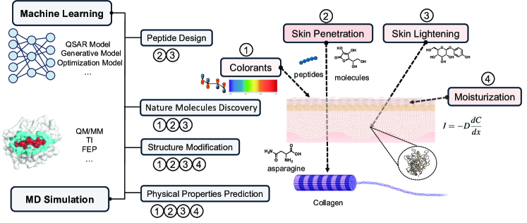
II Objectives in skincare and cosmetics
In this section, we review the primary goals in beauty design, the general taxonomy, and the physical laws to be considered in each axis.
II.1 Coloration.
Cosmetic colorants can be classified into two major categories based on their chemical composition: inorganic and organic colorants Hefford (2011). The global annual production of inorganic colorants is approximately 10 million tons, accounting for 80% of the total colorant production Lewis (1998). Key inorganic colorants include titanium dioxide, iron oxides, carbon black, zinc oxide, ultramarine pigments, and mica-based pearlescent pigments, most of which are derived from natural minerals Giancola and Schlossman (2015); Neytal, Maji, and Maji (2024)—these pigments are widely used in facial, eye, lip, and nail products. Due to their stable crystalline structure, inorganic pigments exhibit high stability, excellent opacity from their high refractive index, and natural coloration Lam (2021). Nevertheless, they also have certain limitations, such as a narrow range of light absorption, making it difficult to cover a broader visible spectrum, high surface energy leading to aggregation, and large particle size affecting skin permeability Gürses et al. (2016).
As to organic colorants, they can be further divided into natural and synthetic colorants Zollinger (2003). Natural colorants are typically derived from plant and insect pigments, such as carmine, carotenoids, anthraquinones, betalains, and flavonoids. These colorants are popular among consumers due to their natural origin Sigurdson, Tang, and Giusti (2017), although they also face challenges such as color instability and susceptibility to environmental degradation. In contrast, synthetic colorants generally offer excellent color stability, a wide range of spectral options, and superior light and heat resistance Lanzendörfer-Yu (2021). This is mainly due to their molecular structures, which often contain a large number of conjugated double bonds (e.g., azo and bonds, triphenylmethane structures). These conjugated systems extend electron delocalization, enabling absorption over a broader wavelength range of visible light and producing vivid colors Lewis (1998). The highly conjugated structures not only enhance molecular stability and reduce structural breakdown under light or heat exposure but also enable optimal electronic transitions, effectively absorbing specific wavelengths of light to improve color vibrancy and stability Kiernan (2001). Synthetic colorants encompass a wide variety of types, including azo dyes, triphenylmethane dyes, flavonoid dyes, and anthraquinone dyes. Through optimized molecular design, these different types of dyes ensure color performance and stability across various application environments. Moreover, the consistency achieved through batch synthesis results in highly uniform molecular structures and purity, further enhancing the clarity and stability of the colors. However, due to environmental and health concerns, the design of synthetic colorants requires more stringent molecular design criteria to minimize potential risks Renita et al. (2023).
II.2 Moisturization.
The moisturizing efficacy of skincare products is typically assessed through Transepidermal Water Loss (TEWL), which measures the rate at which water diffuses from the stratum corneum into the atmosphere Rogiers (2001), where an evaporimeter is used to quantify the amount of water lost per square meter of skin surface per hour, providing insights into the skin’s water kinetics. Since TEWL primarily reflects the process of water diffusing from the deeper layers of the skin through the epidermis to the external environment, this process can be approximately described by Fick’s First Law of Diffusion Rodrigues et al. (1999):
| (5) |
where is the water flux is the diffusion coefficient of water in the stratum corneum and is the concentration gradient of water .
Based upon this relationship, different types of moisturizing products regulate TEWL by adjusting the diffusion coefficient or the concentration gradient , thus achieving a moisturizing effect. Typically, moisturizing ingredients in skincare products fall into one or more of the following three categories: humectants, occlusives, and emollients Neytal, Maji, and Maji (2024). Humectants are generally compounds containing multiple hydroxyl groups (–OH), such as glycerin, hyaluronic acid, Hydroxy Acids, and propylene glycol. These molecules form stable hydrogen-bond networks with water, increasing the water content in the stratum corneum and lowering the chemical potential of water. By increasing the water concentration within the stratum corneum, humectants reduce the concentration gradient . According to Fick’s first law, when humectants increase the water concentration in the stratum corneum, the concentration gradient decreases, leading to a reduction in the water flux and consequently reducing the rate of water evaporation. Occlusives, such as petroleum jelly, mineral oil, and silicone oils, are typically hydrophobic compounds that form a physical barrier on the skin surface de Clermont-Gallerande (2020). This barrier prevents water evaporation and thereby reduces the rate of water flux from the skin, effectively minimizing transepidermal water loss. Emollients work by softening and smoothing the skin, filling the gaps between skin cells, many of which possess both occlusive and emollient properties.
II.3 Skin Lightening
To achieve skin lightening typically requires intervention in the processes of melanin production, transport, and metabolism in the skin Smit, Vicanova, and Pavel (2009). Inhibiting melanin production is primarily achieved by targeting tyrosinase, a copper-containing metalloenzyme with two copper ions (CuA and CuB) in its active site, which are responsible for substrate binding and catalysis Lai et al. (2018); Chang (2009). There are several strategies for inhibiting tyrosinase activity. The most direct approach involves inhibiting its active site. For example, arbutin effectively reduces tyrosinase activity through competitive inhibition, thereby decreasing melanin production Boo (2021). Another approach is to chelate the copper ions (CuA and CuB) at the active site, preventing the enzyme from functioning properly. Kojic acid, for instance, can form chelates with the copper ions in tyrosinase, significantly inhibiting its catalytic function Chang (2009). Additionally, retinoic acid can suppress TYR gene promoter activity, reducing tyrosinase transcription and consequently lowering melanin synthesis at the source Zaidi et al. (2019). Tyrosinase activity is also influenced by glycosylation modifications, and inhibitors like mannose can reduce tyrosinase glycosylation, thereby decreasing its enzymatic activity Petrescu et al. (1997). Moreover, linoleic acid and -sitosterol promote tyrosinase ubiquitination and proteasomal degradation, reducing the amount of active enzyme in the cell, and further inhibiting melanin production Logesh et al. (2023); Shi et al. (2022).
Blocking melanin transport is also an essential strategy for achieving skin whitening. By interfering with the transfer of melanosomes from melanocytes to surrounding keratinocytes, the distribution of melanin in the epidermis can be reduced. Niacinamide, for example, can inhibit protease-activated receptor-2 (PAR-2) on keratinocytes, thereby reducing melanosome transfer Smit, Vicanova, and Pavel (2009). Similarly, licorice extract interferes with the melanosome transport process by inhibiting signaling pathways such as PI3K/Akt and MAPK, further reducing melanin deposition in the epidermis Wang et al. (2022b).
II.4 Skin Penetration.
An important function in cosmetics is to activate collagen production and enhance antioxidant effects at the deeper layers of the skin Kusumawati and Indrayanto (2013); Costa and Santos (2017). The skin serves as a protective barrier against external environmental factors. Therefore, for cosmetic molecules to be effective, they must penetrate this barrier to reach the underlying layers. Common skin-penetrating ingredients in cosmetic products can be broadly categorized into small molecules, peptides and liposomes.
In particular, small molecules in cosmetic products, such as Vitamin C, Vitamin A, Vitamin E, salicylic acid, and niacinamide, play crucial roles in processes such as amino acid hydroxylation in collagen, activation of signaling pathways, and antioxidation Murad et al. (1981); Mohiuddin (2019); Talakoub, Neuhaus, and Yu (2016); Sajna et al. (2015); Traber and Atkinson (2007); Dinarello (2010). Due to their molecular weights ranging from 122 to 430, these small molecules can penetrate the deeper layers of the skin through aqueous channels or the lipid bilayer. Despite their favorable permeability, the rate of penetration is influenced by several factors, including formulation, concentration, temperature, and pH. For example, Vitamin C requires a water-based formulation with a pH lower than its pKa (pKa 4.2) to reduce the charge density on the molecule Stamford (2012). Similarly, the skin permeability of salicylates exhibits a parabolic relationship with the partition coefficient, with optimal permeability typically occurring at a log P value of around 2.5. At this pH, salicylates have sufficient solubility in the lipid layer to facilitate diffusion through the stratum corneum while maintaining enough hydrophilicity to penetrate the viable epidermal tissue Benson (2005). To enhance the skin penetration of these small molecules while preserving their efficacy, various optimization strategies can be employed. These include molecular group modification, chemical potential optimization, eutectic systems, and coordination compounds.
In addition to their direct effects on the skin, small molecules also play a role in facilitating the penetration of peptides and proteins into the body. Peptides and larger protein molecules are particularly effective in wrinkle reduction, skin vitality maintenance, and collagen repair, as well as in sensitive factor attachment protein receptor (SNARE) regulation Rostkowska et al. (2023); Lim et al. (2018). However, due to their larger molecular weight and hydrophilic nature, these molecules face significant challenges in penetrating the hydrophobic lipid matrix of the stratum corneum Mortazavi and Moghimi (2022). To address these permeability issues, various strategies are being developed, such as the use of small-molecule chemical penetration enhancers (CPEs) Ahad et al. (2009). These include azone derivatives, fatty acids, alcohols, esters, sulfoxides, pyrrolidones, glycols, surfactants, and terpenes Kumar and Ghirnikar (2016); Karande et al. (2005). These chemicals can enhance the permeability of peptides and proteins by increasing the diffusion rate within the skin, inducing lipid fluidization, optimizing thermodynamic activity, influencing partition coefficients, and enhancing the release of drugs from formulations into the upper layers of the skin. However, while CPEs significantly improve permeability, they may also cause skin irritation and inflammation, and so far, no ideal chemical penetration enhancer has been identified Zeng et al. (2022).
In the way of searching for more effective, non-invasive, non-toxic, and non-irritating skin penetration enhancers, short-sequence peptides have been identified as safer alternatives to small molecule CPEs Kumar et al. (2015); Hmingthansanga et al. (2022). By designing and synthesizing specific short-chain peptides, it is possible to facilitate the delivery of other substances to the deeper layers of the skin. These short peptides have small molecular weights, making them more likely to penetrate the skin. Among them, cell-penetrating peptides (CPPs) and skin-penetrating peptides (SPPs) are recognized as valuable delivery tools due to their high transduction efficiency and low cytotoxicity Copolovici et al. (2014); Guidotti, Brambilla, and Rossi (2017); Gennari et al. (2016); Hsu and Mitragotri (2011). A notable example is the TAT peptide (Trans-Activator of Transcription), a short peptide sequence (amino acids 47-57) that has been extensively studied and applied in cell penetration and drug delivery. When combined with the tripeptide GKH, the resulting TAT-GKH complex significantly enhances GKH’s skin penetration efficiency and is utilized in lipolytic cosmetic products Lim et al. (2003). Additionally, Chen et al. reported the use of the skin-penetrating peptide SPACE to enhance the topical delivery of high molecular weight hyaluronic acid (HA, molecular weight: 200-325 kDa) Chen et al. (2014). Carmichael and colleagues demonstrated that botulinum toxin type A (BoNT-A) could be administered subcutaneously or topically with a novel transdermal delivery peptide to reduce inflammation by inhibiting the activation of nociceptors in the skin Carmichael, Dostrovsky, and Charlton (2010). Similar to small molecules, modifying the side chains of these peptides can also enhance their permeability. For example, the carrier peptide GHK can bind to Cu(II) and Mn ions in the skin Hur et al. (2020); Hussain and Goldberg (2007); Errante et al. (2020). Through chemical modification, GHK can be linked to a palmitoyl group (a long-chain fatty acid), increasing its hydrophobicity and enhancing its interaction with the lipid bilayer, thereby improving its ability to penetrate the skin Arul et al. (2005); Jeong et al. (2019).
Besides, liposomes are utilized to encapsulate peptides and proteins, enhancing their ability to penetrate the skin barrier more effectively El Maghraby, Barry, and Williams (2008); Peralta et al. (2018). Liposomes offer several advantages for the transdermal delivery of peptides and proteins, including improved permeability, protection of active ingredients, and controlled release. With the advent of novel liposome technologies, such as elastic liposomes, cationic liposomes, and pH-sensitive liposomes, delivery efficiency has been further enhanced Rai, Pandey, and Rai (2017); Bouwstra and Honeywell-Nguyen (2002); liu2013formati
III Frontiers in molecular discovery for beauty design
In §II, we have outlined the general guidelines of beauty design. In this section, we focus on two directions particularly popular in beauty design—collagen formation and microhyaluronic acid application.
III.1 The odyssey towards collagen
Collagen, the most common type of protein found in skin as well as tendons and bones, assists fibroblasts forming in the dermis, promoting the growth of new cells and replacing dead skin cells. Oral supplement of collagen has been linked to improved firmness, suppleness, moisture, smoothness, and elasticity of skin Pu et al. (2023); Inoue, Sugihara, and Wang (2016); Sugihara, Inoue, and Wang (2015), although larger-scale study is needed to determine whether commercially available products are useful for long-term use. Used topically, certain mixtures of amino acids, such as arginine and ornithine, have been proven to promote the disposition of collagen in wounds Williams, Abumrad, and Barbul (2002); Arribas-López et al. (2021); Albaugh, Mukherjee, and Barbul (2017). Topically, the application of amino acids have also shown to be useful to boost collagen formation and skin moisture Trookman et al. (2009).
III.2 Micro hyaluronic acids: potency and risks
Micro hyaluronic acids with low molecular weight are well known for their ability to penetrate the skin effectively. These tiny molecules can go through the skin’s protective barrier and provide moisture directly to the deeper layers of the skin improving its elasticity and minimizing noticeable aging signs. This property is crucial in cosmetic dermatology, where hydration and skin suppleness are key to a youthful appearance. The effectiveness of micro hyaluronic acids in promoting skin hydration and reducing wrinkles has been substantiated by various studies, which highlight its efficacy as a humectant, with the ability to hold water up to a thousand times their weight and facilitate better absorption compared to their higher molecular counterparts Bravo et al. (2022); Juhaščik, Kováčik, and Huerta-Ángeles (2022). Advancements in skincare technology have leveraged the properties of micro hyaluronic acids to develop sophisticated delivery systems that maximize skin benefits. Techniques such as encapsulation in liposomes or binding to nanoparticles have been explored to further enhance the penetration and longevity of hyaluronic acids in skin layers. These innovations not only improve the immediate hydrating effects but also contribute to the long-term anti-aging benefits by maintaining higher levels of skin hydration over extended periods Juhaščik, Kováčik, and Huerta-Ángeles (2022).
Despite their benefits, microhyaluronic acids come with potential risks that must be managed carefully. Topical applications are generally safe but can occasionally cause skin irritation or mild allergic reactions in sensitive individuals. More invasive applications, such as injections, carry greater risks including infection, tissue necrosis, or adverse reactions at the injection site. It is vital that these treatments are administered by qualified healthcare professionals to minimize risks and ensure safety. Additionally, Bravo et al. (2022) conducted a comprehensive review and noted that extremely low molecular weights of hyaluronic acid may trigger inflammatory responses in the skin. It is necessary to have precise control over the molecular size used in skin care formulations.
Because of the growing usage of hyaluronic acid in the field of aesthetics, notably in injectable skin rejuvenation treatments, it has become essential to build a solid regulatory framework in order to protect the efficacy and safety of this substance. Regulatory scrutiny ensures that hyaluronic acid products, classified as medical devices, meet stringent safety standards and are marketed accurately regarding their benefits and potential side effects. The framework also highlights the importance of the administration by licensed practitioners and the need for ongoing research to support the claims made by manufacturers regarding the cellular effects of these injections Allen and Dodou (2024). Hyaluronic acid usage regulations are made to protect the general public’s health as well as satisfy the increasing market for anti-aging goods. This involves guaranteeing the chemicals’ moral provenance and scientific backing. Strict regulatory measures are required as demand for these therapies rises in order to guard against abuse and guarantee that the goods live up to expectations without sacrificing safety.
IV In silico beauty design: recent advances.
Efforts using machine learning to design peptides for skin care.
Current trends in cosmetic molecule development closely align with those in medicinal chemistry David, Wolfender, and Dias (2015); Pillaiyar, Manickam, and Namasivayam (2017). In recent years, machine learning has played a significant role in peptide design and optimization, greatly improving the development efficiency of cosmetic peptides. Specifically, Yue et al. (2024) reported using machine learning to predict the antimicrobial activity and hemolytic properties of peptides, leading to the design of a novel -defensin-based antimicrobial peptide, IK-16-1. Additionally, Hsueh et al. design a multifunctional peptides which can efficiently penetrate the cells and combined with melanin Hsueh et al. (2023). In the area of skin penetration, De Oliveira et al. reported the development of BChemRF-CPPred, which extracts descriptors from peptide structures and sequences to distinguish cell-penetrating peptides (CPPs) from non-CPPs de Oliveira et al. (2021). The design of short peptides has become a prominent strategy in molecular discovery, primarily due to their strong targeting capabilities, high efficacy, stability, and low allergenicity Apostolopoulos et al. (2021). Although substantial progress has been made utilizing scientists’ domain knowledge, the de novo design of short peptides remains relatively limited Aruan et al. (2023); Ganceviciene et al. (2012).
Natural molecular discovery for skin care.
Natural molecular discovery is emerging as a new trend in molecular design. This shift is largely driven by the perception that natural molecules are inherently safer and exhibit lower toxicity, aligning with consumer preferences and regulatory standards for safer cosmetic and therapeutic products. Consequently, most animal and plant extracts are widely recognized as safe, leading to the screening of many cosmetic molecules using chemical libraries derived from natural sources Martins et al. (2014). For example, Bournez et al. employed homology modeling and molecular docking to screen 4,534 natural products for potential SIK2 inhibitors, aiming to enhance melanin production and achieve tanning effects without UV-induced damage Bournez et al. (2024). Besides, Su et al. compiled a database of 5,616 natural compounds and, through XP docking and molecular dynamics simulations, identified five compounds with promising dynamic binding properties. Subsequent in vivo experiments on zebrafish revealed that two components, Sennoside B (SB) and Sennoside C (SC), exhibited inhibitory effects on melanin production Su et al. (2024).
Structural modification for cosmetic molecules.
Structure modification is a primary method for developing new cosmetic molecules Ai et al. (2014). Typically, chemical structures are altered to enhance their biological activity, stability, and Absorption, Distribution, Metabolism, Excretion, and Toxicity (ADMET) properties. In addition to these direct approaches, derivatives can also be generated based on known targets and established cosmetic molecules. Specifically, a typical example is the molecular modification of vitamins, to address the inherent instability and challenges with the topical application of vitamin C, formulation chemists have engineered derivatives such as ascorbyl 2-phosphate (SAP and MAP) and ascorbyl 6-palmitate (AA-Pal) Stamford (2012). These derivatives stabilize vitamin C within cosmetic formulations and enhance skin penetration by maintaining stability at a neutral pH and protecting the molecule from oxidation, facilitating their inclusion in various skincare products. Similarly, many researchers have modified natural products to enhance their bioactivity. Kumari et al. demonstrated that isoliquiritigenin and its derivatives, such as 5’-formylisoliquiritigenin and isoliquiritigenin dimers, exhibit higher binding affinity and more potent tyrosinase inhibition compared to isoliquiritigenin alone Kumari et al. (2023). Molecular docking studies revealed that these derivatives have lower binding energies with tyrosinase, indicating their potential as more effective tyrosinase inhibitors for the prevention and treatment of melanogenesis and related hyperpigmentation disorders.
In addition to these methods, cosmetic formulations following molecular editing are further evaluated through their physical properties. Our article focuses on the use of chemical computational tools for prediction and combination. Therefore, in the subsequent Physical Toolbox section, we briefly introduce the relevant prediction tools, which will not be reiterated here.
V The physical toolbox: molecular dynamics (MD) simulation.
Characterizing Equation 1 by drawing samples while integrating Newton’s second motion of law, MD simulation finds use in all schools of molecular modeling. There have been a wide variety of mature and widely used pipelines for modeling the energy landscape of biomolecular systems of therapeutical interest, which can be seamlessly adopted to model those of cosmeceutical significance.
Modeling of cosmeceutical molecule-target binding.
To design effective cosmetic molecules, molecular dynamics (MD) simulations Case et al. (2021); Lee et al. (2016); Abraham et al. (2015); Eastman et al. (2023) can be employed to model the interactions between cosmeceutical molecules and their biological targets. To be more specific, MD simulations enable the accurate prediction of binding free energy, a key metric in assessing the strength and stability of the interaction between a small molecule, such as a cosmetic agent, and a target protein Mobley and Gilson (2017). One of these methods is commonly categorized as endpoint techniques, such as molecular mechanics generalized Born surface area (MM-GBSA) and molecular mechanics Poisson-Boltzmann surface area (MM-PBSA), to estimate free energy changes by evaluating the energy differences between the bound and unbound states with data sampled from the same molecular dynamics trajectory Wang et al. (2019); Genheden and Ryde (2015). In both approaches, the binding free energy is divided into several components, including enthalpic and entropic contributions. The former component is calculated as the sum of the gas-phase interaction energy between the ligand and protein and the solvation free energy. At the same time, the latter is computed as conformational entropy change related to ligand binding Tuccinardi (2021). To enhance computational efficiency, these endpoint methods employ an implicit continuum solvent model, which significantly reduces the complexity of the calculations by omitting explicit water molecules from the MD simulations. While this approach accelerates calculations, it may also compromise the accuracy, as it overlooks the potentially significant role of specific water-mediated interactions. Besides, a key limitation of MM-PBSA and MM-GBSA is their treatment of entropy. These methods often neglect entropic contributions or handle them in a less rigorous manner. For instance, the configurational entropy is either approximated using simplified models or ignored altogether Wang et al. (2016). Therefore, MM-GBSA/PBSA exhibit moderate accuracy while being computationally efficient, which makes them well-suited for the rapid screening of potential small molecules in the early stages of drug discovery, such as anti-tumor and anti-virus drugs Hu and Contini (2019); Forouzesh and Mishra (2021); Shridhar Deshpande et al. (2021). For the calculation of complex interactions and highly flexible ligands, we developed the alanine scanning with generalized Born and interaction entropy (ASGBIE) method Liu et al. (2018); Yan et al. (2017). Compared to MM-GBSA, ASGBIE provides more accurate binding free energy estimations derived from improved solvation energy calculations and a more rigorous consideration of entropic contributions, which are often neglected in MM-GBSA. Additionally, the method employs variable dielectric coefficients ranging from 1 to 10 for different categories of amino acids, including non-polar, polar, and charged groups, allowing for more nuanced electrostatic calculations. In a comparison of ASGBIE and MM-GBSA on 24 complex systems with 433 pairs of mutation, ASGBIE achieved a mean unsigned error (MUE) of 0.2 kcal/mol, outperformed the MUE of 1.0 kcal/mol for MM-GBSA Yan et al. (2017). Additionally, ASGBIE has been applied to predict hotspots with the highest contributions in binding free energy on a protein-peptide system, aiding peptide modifications to enhance binding affinity, and estimating relative binding free energy changes between wild-type and mutated peptides with reported accuracy Zhou et al. (2024).
More sophisticated methods can also be considered for estimating binding free energies. One important category is alchemical methods, including thermodynamic integration (TI) Kirkwood (1935) and free energy perturbation (FEP) Zwanzig (1954). These methods estimate free energy changes by sampling intermediate states between initial and final configurations based on sampling configurations guided by the potential energy of the initial state. Two more extended functions were then developed based on the FEP equation. Bennett derived a Bennett acceptance ratio (BAR) Bennett (1976) function requiring sampling configuration at both states to minimize the expected square error of the free energy difference. Meanwhile, a multistate Bennett acceptance ratio (MBAR) Shirts and Chodera (2008) function takes into account simulation data from multiple states, involving a more comprehensive sampling. These methods involve the calculation of free energy difference between two states by introducing a coupling parameter () that varies from 0 to 1 to sample configurations for both states, either based on the ensemble average of the state 0 or that of both states, creating a series of intermediate states that improve phase space overlap and facilitate a smoother transformation. The utilization of soft-core potential further avoids the dispersed energy distribution on endpoints of 0 and 1. Furthermore, alchemical methods have been extensively applied in estimating binding free energy between target proteins and small molecules, such as large-scale screenings or docking poses evaluation Bhati and Coveney (2022); Goldfeld et al. (2015); Kaus et al. (2015). Some approaches combine alchemical methods with replica exchange solute tempering (FEP/REST), further improving accuracy by overcoming energy barriers associated with local minima Bourion-Bédès et al. (2021); Lockhart et al. (2023); Ngo et al. (2020). Several software platforms support alchemical methods. For instance, AMBER18 enables GPU-accelerated TI calculations, and AMBER20 package was evaluated for its accuracy on a dataset of around 200 ligand mutations spanning 8 protein targets, achieving a mean unsigned error (MUE) of 0.84 kcal/mol Lee et al. (2020). Similarly, a commercial software FEP+ exploited FEP/REST approach on the same dataset and achieved a comparable MUE of 0.86 kcal/mol Wang et al. (2015). These alchemical approaches offer more efficient sampling of the protein-ligand phase space and have demonstrated strong correlations with experimental data, making them reliable for accurately predicting the binding free energy of small cosmeceutical molecules interacting with their targets. Another category of methods includes enhanced sampling techniques like umbrella sampling and metadynamics, which are useful for large-scale molecular systems Williams-Noonan, Yuriev, and Chalmers (2018). However, these methods significantly increase computational demand due to the exponential growth in the number of molecules and interactions, necessitating the use of high-performance computing (HPC) resources and substantial storage capacity.
Physical property prediction.
Usually, Cosmetic compounds necessitate a thorough evaluation of ADMET profiles to guarantee their suitability for topical application Martins et al. (2014). This comprehensive assessment ensures that active ingredients are effectively absorbed through the skin barrier, distributed appropriately within the skin layers, metabolized without generating harmful by-products, and excreted without accumulation that could lead to adverse effects. Traditionally, ADMET evaluation has relied primarily on experimental methods to determine these key pharmacokinetic and toxicological properties of drugs or active molecules. Recently, MD simulations have emerged as an important tool for aiding in the computational analysis of ADMET Srivastava et al. (2025); Balkrishna et al. (2024). By simulating the behavior of molecules within cell membranes and skin tissues, researchers can predict how these molecules penetrate the skin barrier, their distribution within the skin, and their interactions with proteins. Additionally, MD simulations can be combined with other computational methods, such as machine learning and computational fluid dynamics, to predict the fluid properties of products under various conditions, including viscosity, surface tension, and diffusion coefficients. These predictions can effectively guide the design of cosmetic formulations, reduce development costs, and improve the application performance and stability of products.
VI The machine learning toolbox
Graph-based machine learning.
Graph neural networks Kipf and Welling (2016a); Xu et al. (2018); Gilmer et al. (2017); Hamilton, Ying, and Leskovec (2017); Battaglia et al. (2018); Wang and Cho (2024) have be come the de facto workhorse of all schools of molecular modeling. A graph neural network can be most generally defined as one adopting a layer-wise updating scheme that aggregates representations from a node’s neighborhood and updates its embedding:
| (6) |
where are the update and aggregate function, repsectively. Omitting the nonlinear transformation step ubiquitous in all neural models, and assuming a convolutional aggregate function, or , a graph neural network layer is characterized by the aggregation/convolution operation that pools representations from neighboring nodes, forming an intermediary representation , which on a global level, with activation function and weights , can be written as: . The primary difference among architectures amounts to distinct effective adjacency matrix . The most classical examples include: graph convolutional networks (GCN) Kipf and Welling (2016a), which normalizes by the in-degree of nodes and graph attention networks (GAT) Veličković et al. (2017), which takes as the attention score;
| (7) |
The resulting representation is rich and naturally permutation-invariant, providing a strong starting point for downstream tasks.
Generative modeling.
Once we have a reliable model for the prediction of activities given the structure of the graph, we can couple it with a generative model for the true de novo design of molecules and peptides. This can be achieved through a principled, probabilistic manner, where the conditional distribution in Equation 4 is replaced by the distribution of the chemical graphs without measurements:
| (8) |
and the model is typically trained by maximizing the (lower bound of) the likelihood of data . For the generation of molecular structures, common approaches include variational autoencoder (VAE)-based models, where molecules are generated as strings Gómez-Bombarelli et al. (2018), graphs Kipf and Welling (2016b), or a Markov-decision process to construct from fragments Jin, Barzilay, and Jaakkola (2019); flow-based models Zang and Wang (2020); and diffusion-based models Liu et al. (2023). The conformations of molecules can also be simultaneously or conditionally generated in the 3-dimensional space Hoogeboom et al. (2022).
Active learning.
With some over-simplification, molecular discovery can be abstracted as a sequential active learning process. Given a finite pool of candidate chemical compounds, represented by chemical graphs , each associated with a potency , of which a noisy observation is expensive to acquire, we are interested in finding the compound with the highest potency with fewest function evaluations. This can be achieved via active learning, starting with an empty portfolio , and in each round, choosing a compound from the candidate pool, , based on a decision informed by the current portfolio, . Subsequently, we subtract this compound from the candidate pool to add it to the portfolio, .
Bayesian optimization (BO) is a powerful approach in active learning where the decision is constructed in a modular way. Firstly, we extract fixed -dimensional features from the compounds to form a representation: ; secondly, this representation is used to construct a predictive posterior distribution (Eqaution 4) with uncertainty quantification: , whose parameters is is mapped from the graph representations. Lastly, an acquisition function, , is applied on this predictive posterior to form a score, on the basis of which a decision is made: . This process can be executed iteratively until a satisfactory compound has been identified.
Large-language models.
Large language models (LLMs)—neural networks composed primarily of transformer Vaswani et al. (2023)—have shown surprising promise Touvron et al. (2023); OpenAI (2023); Brown et al. (2020); Bender et al. (2021); Ni et al. (2024) in representing and generating text-structured data and gained popularity very quickly in recent years. There have been a plethora of LLMs for chemical tasks Jablonka et al. (2023), the most popular ones include Yu et al. (2024); Zhang et al. (2024a), which have been fine-tuned on instruction datasets Zhang et al. (2024b). LLMs can be integrated into various workflows discussed here, from planning experiments to be carried out in an active learning setting, to writing code to prepare biomolecular systems for simulations.
VII Roadmap: Potential use cases
Having reviewed the goals and challenges in the beauty design industry and the powerful methodologies in our physical and statistical toolboxes, in this section, we propose three projects that are immediately feasible, to showcase the potential power of combining MD and ML approaches in beauty design.
The molecular modification of arbutin via multiobjective active learning.
Inhibiting the activity of tyrosinase, an essential enzyme for melanin synthesis in skin, arbutin has been ubiquitously used as skin-lightening agents targeting hyperpigmentation issues. Usually extracted from the leaves of plants, such as the bearberry, the efficacy of this natural product is bottlenecked by its inability to penetrate the skin to reach the melanocytes Masyita et al. (2021). We propose to develop arbutin derivatives displaying similar efficacy while showing significantly greater lipophilicity for better skin penetration.
First, docking and molecular dynamics simulations will be carried out to better understand the binding mechanism of arbutin. With these structural insights, we will carry out molecular modification, for example using fragment-based tools such as Zhang et al. (2024c), to replace functional groups on arbutin. This can be done in an ultra-high throughput manner, generating a large library of arbutin-derivatives. Multitask Bayesian optimization algorithms can then design experiments to be carried out sequentially, to optimize for both lipophilicity and melanocytes-inhibiting activity, which can be accessed either experimentally or using free energy calculations.
Optimizing the ADMET profile of hyluronic acid.
The extremely small molecular weight of hyaluronic acid is both a blessing and a curse—it enables the easy penetration of the skin while also triggering inflammatory reactions Bravo et al. (2022). The chemical modification of hyaluronic acid Bokatyi, Dubashynskaya, and Skorik (2024) has been proven to be an effective avenue towards efficient drug delivery. Using the modification strategies outlined in Bokatyi, Dubashynskaya, and Skorik (2024), we can chemically decorate the backbone with inactive functional groups to moderately increase the molecular weight of hyaluronic acid, while not altering its ability to hold water. These modifications can be proposed either by generative models or fragment-based structure enumeration and library generation tools such as Polishchuk (2020), while the physical properties and the ADMET profile can be predicted using a machine learning-enabled QSAR model.
Cosmetic peptide design.
Peptides are currently predominantly composed of oligopeptides (containing fewer than 10 amino acids), forming a vast polypeptide space. The design of synthetic peptides still relies heavily on traditional, resource-intensive methods that are both time-consuming and laborious. These conventional approaches often result in peptides that lack the necessary modifications to enhance their skin penetration and bioactivity. However, advancements in technology have made peptide sequence prediction and optimization more efficient and precise. In terms of de novo peptide design, researchers can now leverage diffusion models trained on medicinal peptide datasets to capture the complex patterns of peptide sequence distribution and functional properties, enabling the generation of novel peptides with desired characteristics. Additionally, protein function prediction tools, such as DeepFRI, can be employed to predict the corresponding protein functions of these peptides Gligorijević et al. (2021). From a peptide modification perspective, targeted chemical modifications can be introduced to enhance the permeability and stability of peptides. For instance, acetyl tetrapeptide-15, a chemically modified version of Tyr-Pro-Phe-Phe, demonstrates how modifications like cyclization, lipid chain conjugation, or terminal glycosylation can be applied. These modified peptides can then be evaluated using molecular dynamics (MD) simulations. Through iterative cycles of model evaluation and screening, the efficacy and safety of the generated peptides can be validated.
Towards precision beauty design: personalized mixture for cosmetics and skin care.
Drawing inspirations from the rich literature of drug combinations Jaaks et al. (2022), which characterizes the synergistic effect among multiple small molecule drugs on multi-factorial diseases, we propose to design a system to design formulae of skin-care and cosmetic mixtures that cater to personal conditions and requirements. For instance, such a system can take a picture of a person’s face, and propose various solutions for lipstick mixtures, which contain waxes, oils, pigments, moisturizers, and antioxidants, and simulate their effects based upon techniques such as spectra prediction.
VIII Conclusions
From this perspective, we discuss the contributions of computational tools, including molecular dynamics and machine learning, to the discovery and development of cosmetic molecules. Cosmetic molecules exhibit a high degree of similarity to drug molecules, and the development of corresponding cosmetic molecules is still in its early stages, offering significant potential for further growth. At the end of this paper, we present a roadmap outlining several feasible approaches. These approaches represent the future of small molecules, proteins, and physics-based comprehensive product development, respectively.
Disclosures.
YW has limited financial interests in Flagship Pioneering, Inc. and its subsidiaries.
Funding.
YW acknowledges support from the Schmidt Science Fellowship, in partnership with the Rhodes Trust, and the Simons Center for Computational Physical Chemistry at New York University.
References
- (1) “Why Did We Start Wearing Makeup? | Britannica — britannica.com,” [Accessed 07-10-2024].
- Meng et al. (2011) X.-Y. Meng, H.-X. Zhang, M. Mezei, and M. Cui, “Molecular docking: a powerful approach for structure-based drug discovery,” Current computer-aided drug design 7, 146–157 (2011).
- Jumper et al. (2021) J. Jumper, R. Evans, A. Pritzel, T. Green, M. Figurnov, O. Ronneberger, K. Tunyasuvunakool, R. Bates, A. Žídek, A. Potapenko, et al., “Highly accurate protein structure prediction with alphafold,” nature 596, 583–589 (2021).
- Abramson et al. (2024) J. Abramson, J. Adler, J. Dunger, R. Evans, T. Green, A. Pritzel, O. Ronneberger, L. Willmore, A. J. Ballard, J. Bambrick, et al., “Accurate structure prediction of biomolecular interactions with alphafold 3,” Nature , 1–3 (2024).
- Retchin et al. (2024) M. Retchin, Y. Wang, K. Takaba, and J. D. Chodera, “Druggym: A testbed for the economics of autonomous drug discovery,” bioRxiv , 2024–05 (2024).
- Dara et al. (2022) S. Dara, S. Dhamercherla, S. S. Jadav, C. M. Babu, and M. J. Ahsan, “Machine learning in drug discovery: a review,” Artif. Intell. Rev. 55, 1947–1999 (2022).
- Wang et al. (2023) H. Wang, T. Fu, Y. Du, W. Gao, K. Huang, Z. Liu, P. Chandak, S. Liu, P. Van Katwyk, A. Deac, et al., “Scientific discovery in the age of artificial intelligence,” Nature 620, 47–60 (2023).
- Wigh, Goodman, and Lapkin (2022) D. S. Wigh, J. M. Goodman, and A. A. Lapkin, “A review of molecular representation in the age of machine learning,” WIREs Comput. Mol. Sci. 12, e1603 (2022).
- Kipf and Welling (2016a) T. N. Kipf and M. Welling, “Semi-supervised classification with graph convolutional networks,” CoRR abs/1609.02907 (2016a), arXiv:1609.02907 .
- Xu et al. (2018) K. Xu, W. Hu, J. Leskovec, and S. Jegelka, “How powerful are graph neural networks?” arXiv preprint arXiv:1810.00826 (2018).
- Gilmer et al. (2017) J. Gilmer, S. S. Schoenholz, P. F. Riley, O. Vinyals, and G. E. Dahl, “Neural message passing for quantum chemistry,” arXiv preprint arXiv:1704.01212 (2017).
- Hamilton, Ying, and Leskovec (2017) W. Hamilton, Z. Ying, and J. Leskovec, “Inductive representation learning on large graphs,” in Advances in neural information processing systems (2017) pp. 1024–1034.
- Battaglia et al. (2018) P. W. Battaglia, J. B. Hamrick, V. Bapst, A. Sanchez-Gonzalez, V. Zambaldi, M. Malinowski, A. Tacchetti, D. Raposo, A. Santoro, R. Faulkner, et al., “Relational inductive biases, deep learning, and graph networks,” arXiv preprint arXiv:1806.01261 (2018).
- Wang and Cho (2024) Y. Wang and K. Cho, “Non-convolutional graph neural networks,” (2024), arXiv:2408.00165 [cs.LG] .
- Wang and Karaletsos (2021) Y. Wang and T. Karaletsos, “Stochastic aggregation in graph neural networks,” (2021), arXiv:2102.12648 [stat.ML] .
- (16) Y. Wang, M. Nguyen, M. Retchin, J. D. Chodera, and T. Karaletsos, “Bayesian active drug discovery,” in https://realworldml.github.io/files/cr/48_BADD_paper_workshop.pdf.
- Blei, Kucukelbir, and McAuliffe (2017) D. M. Blei, A. Kucukelbir, and J. D. McAuliffe, “Variational inference: A review for statisticians,” J. Am. Stat. Assoc. 112, 859–877 (2017).
- Kristiadi et al. (2024) A. Kristiadi, F. Strieth-Kalthoff, M. Skreta, P. Poupart, A. Aspuru-Guzik, and G. Pleiss, “A sober look at llms for material discovery: Are they actually good for bayesian optimization over molecules?” (2024), arXiv:2402.05015 [cs.LG] .
- Smith, Isayev, and Roitberg (2017) J. S. Smith, O. Isayev, and A. E. Roitberg, “Ani-1: an extensible neural network potential with dft accuracy at force field computational cost,” Chem. Sci. 8, 3192–3203 (2017).
- Schütt, Unke, and Gastegger (2021) K. T. Schütt, O. T. Unke, and M. Gastegger, “Equivariant message passing for the prediction of tensorial properties and molecular spectra,” (2021), arXiv:2102.03150 [cs.LG] .
- Thölke and Fabritiis (2022) P. Thölke and G. D. Fabritiis, “Torchmd-net: Equivariant transformers for neural network based molecular potentials,” (2022), arXiv:2202.02541 [cs.LG] .
- Unke and Meuwly (2019) O. T. Unke and M. Meuwly, “Physnet: A neural network for predicting energies, forces, dipole moments, and partial charges,” J. Chem. Theory Comput. 15, 3678–3693 (2019).
- Eastman et al. (2023) P. Eastman, R. Galvelis, R. P. Peláez, C. R. Abreu, S. E. Farr, E. Gallicchio, A. Gorenko, M. M. Henry, F. Hu, J. Huang, et al., “Openmm 8: molecular dynamics simulation with machine learning potentials,” J. Phys. Chem. B 128, 109–116 (2023).
- Wang and Chodera (2023) Y. Wang and J. D. Chodera, “Spatial attention kinetic networks with e(n)-equivariance,” in ICLR 2023 (2023).
- Wang et al. (2022a) Y. Wang, J. Fass, B. Kaminow, J. E. Herr, D. Rufa, I. Zhang, I. Pulido, M. Henry, H. E. B. Macdonald, K. Takaba, et al., “End-to-end differentiable construction of molecular mechanics force fields,” Chem. Sci. 13, 12016–12033 (2022a).
- Takaba et al. (2024) K. Takaba, A. J. Friedman, C. E. Cavender, P. K. Behara, I. Pulido, M. M. Henry, H. MacDermott-Opeskin, C. R. Iacovella, A. M. Nagle, A. M. Payne, et al., “Machine-learned molecular mechanics force fields from large-scale quantum chemical data,” Chem. Sci. (2024).
- Wang et al. (2024a) L. Wang, P. K. Behara, M. W. Thompson, T. Gokey, Y. Wang, J. R. Wagner, D. J. Cole, M. K. Gilson, M. R. Shirts, and D. L. Mobley, “The open force field initiative: Open software and open science for molecular modeling,” J. Phys. Chem. B (2024a).
- Wang et al. (2024b) Y. Wang, I. Pulido, K. Takaba, B. Kaminow, J. Scheen, L. Wang, and J. D. Chodera, “Espalomacharge: Machine learning-enabled ultrafast partial charge assignment,” J. Phys. Chem. A 128, 4160–4167 (2024b).
- Wang et al. (2024c) Y. Wang, K. Takaba, M. S. Chen, M. Wieder, Y. Xu, T. Zhu, J. Z. H. Zhang, A. Nagle, K. Yu, X. Wang, D. J. Cole, J. A. Rackers, K. Cho, J. G. Greener, P. Eastman, S. Martiniani, and M. E. Tuckerman, “On the design space between molecular mechanics and machine learning force fields,” (2024c), arXiv:2409.01931 [physics.chem-ph] .
- Noé et al. (2019) F. Noé, S. Olsson, J. Köhler, and H. Wu, “Boltzmann generators – sampling equilibrium states of many-body systems with deep learning,” (2019), arXiv:1812.01729 [stat.ML] .
- Ahdritz et al. (2024) G. Ahdritz, N. Bouatta, C. Floristean, S. Kadyan, Q. Xia, W. Gerecke, T. J. O’Donnell, D. Berenberg, I. Fisk, N. Zanichelli, et al., “Openfold: Retraining alphafold2 yields new insights into its learning mechanisms and capacity for generalization,” Nat. Methods , 1–11 (2024).
- Kuhlman et al. (2003) B. Kuhlman, G. Dantas, G. C. Ireton, G. Varani, B. L. Stoddard, and D. Baker, “Design of a novel globular protein fold with atomic-level accuracy,” science 302, 1364–1368 (2003).
- Hefford (2011) R. Hefford, “Colourants and dyes for the cosmetics industry,” in Handbook of textile and industrial dyeing (Elsevier, 2011) pp. 175–203.
- Lewis (1998) P. A. Lewis, “Colorants: organic and inorganic pigments,” in AZimuth, Vol. 1 (Elsevier, 1998) pp. 283–312.
- Giancola and Schlossman (2015) G. Giancola and M. L. Schlossman, “Decorative cosmetics,” Cosmeceuticals and Active Cosmetics 191 (2015).
- Neytal, Maji, and Maji (2024) P. Neytal, N. Maji, and S. Maji, “Advances in cosmetic products towards a new future,” in Preserving Health, Preserving Earth: The Path to Sustainable Healthcare (Springer, 2024) pp. 193–214.
- Lam (2021) H. Lam, “Factors enhancing adhesion of color cosmetic products to skin: the role of pigments and fillers,” Surf. Sci. Adhes. Cosmet. , 487–541 (2021).
- Gürses et al. (2016) A. Gürses, M. Açıkyıldız, K. Güneş, M. S. Gürses, A. Gürses, M. Açıkyıldız, K. Güneş, and M. S. Gürses, “Colorants in health and environmental aspects,” Dyes Pigments , 69–83 (2016).
- Zollinger (2003) H. Zollinger, Color chemistry: syntheses, properties, and applications of organic dyes and pigments (John Wiley & Sons, 2003).
- Sigurdson, Tang, and Giusti (2017) G. T. Sigurdson, P. Tang, and M. M. Giusti, “Natural colorants: Food colorants from natural sources,” Annu. Rev. Food Sci. Technol. 8, 261–280 (2017).
- Lanzendörfer-Yu (2021) G. Lanzendörfer-Yu, “Colorants in cosmetic applications,” Phys. Sci. Rev. 6, 835–877 (2021).
- Kiernan (2001) J. A. Kiernan, “Classification and naming of dyes, stains and fluorochromes,” Biotech. Histochem. 76, 261–278 (2001).
- Renita et al. (2023) A. A. Renita, T. K. Gajaria, S. Sathish, J. A. Kumar, D. S. Lakshmi, J. Kujawa, and W. Kujawski, “Progress and prospective of the industrial development and applications of eco-friendly colorants: an insight into environmental impact and sustainability issues,” Foods 12, 1521 (2023).
- Rogiers (2001) V. Rogiers, “Eemco guidance for the assessment of transepidermal water loss in cosmetic sciences,” Skin Pharmacol. Physiol. 14, 117–128 (2001).
- Rodrigues et al. (1999) L. Rodrigues, P. Pinto, N. Galego, P. A. Da Silva, and L. M. Pereira, “Transepidermal water loss kinetic modeling approach for the parameterization of skin water dynamics,” Skin Res. Technol. 5, 72–82 (1999).
- de Clermont-Gallerande (2020) H. de Clermont-Gallerande, “Functional roles of lipids in make-up products,” OCL 27, 33 (2020).
- Smit, Vicanova, and Pavel (2009) N. Smit, J. Vicanova, and S. Pavel, “The hunt for natural skin whitening agents,” Int. J. Mol. Sci 10, 5326–5349 (2009).
- Lai et al. (2018) X. Lai, H. J. Wichers, M. Soler-Lopez, and B. W. Dijkstra, “Structure and function of human tyrosinase and tyrosinase-related proteins,” Chem. Eur. J. 24, 47–55 (2018).
- Chang (2009) T.-S. Chang, “An updated review of tyrosinase inhibitors,” Int. J. Mol. Sci. 10, 2440–2475 (2009).
- Boo (2021) Y. C. Boo, “Arbutin as a skin depigmenting agent with antimelanogenic and antioxidant properties,” Antioxidants 10, 1129 (2021).
- Zaidi et al. (2019) K. U. Zaidi, S. A. Ali, A. Ali, and I. Naaz, “Natural tyrosinase inhibitors: Role of herbals in the treatment of hyperpigmentary disorders,” Mini Rev. Med. Chem. 19, 796–808 (2019).
- Petrescu et al. (1997) S. M. Petrescu, A.-J. Petrescu, H. N. Titu, R. A. Dwek, and F. M. Platt, “Inhibition of n-glycan processing in b16 melanoma cells results in inactivation of tyrosinase but does not prevent its transport to the melanosome,” J. Biol. Chem. 272, 15796–15803 (1997).
- Logesh et al. (2023) R. Logesh, S. R. Prasad, S. Chipurupalli, N. Robinson, and S. K. Mohankumar, “Natural tyrosinase enzyme inhibitors: A path from melanin to melanoma and its reported pharmacological activities,” Biochim. Biophys. Acta Rev. Cancer , 188968 (2023).
- Shi et al. (2022) J. Shi, Y. Guo, H. Wang, Y. Xiao, W. Liu, and L. Lyu, “The ubiquitin-proteasome system in melanin metabolism,” J. Cosmet. Dermatol. 21, 6661–6668 (2022).
- Wang et al. (2022b) J.-y. Wang, X.-y. Xie, Y. Deng, H.-q. Yang, X.-s. Du, P. Liu, and Y. Du, “Licorice zinc suppresses melanogenesis via inhibiting the activation of p38mapk and jnk signaling pathway in balb/c mice skin,” Acta Cir. Bras. 37, e371002 (2022b).
- Kusumawati and Indrayanto (2013) I. Kusumawati and G. Indrayanto, “Natural antioxidants in cosmetics,” Stud. Nat. Prod. Chem. 40, 485–505 (2013).
- Costa and Santos (2017) R. Costa and L. Santos, “Delivery systems for cosmetics-from manufacturing to the skin of natural antioxidants,” Powder Technol. 322, 402–416 (2017).
- Murad et al. (1981) S. Murad, D. Grove, K. Lindberg, G. Reynolds, A. Sivarajah, and S. Pinnell, “Regulation of collagen synthesis by ascorbic acid.” Proc. Natl. Acad. Sci. 78, 2879–2882 (1981).
- Mohiuddin (2019) A. K. Mohiuddin, “Skin aging & modern age anti-aging strategies,” Int. J. Clin. Dermatol. Res. 7, 209–240 (2019).
- Talakoub, Neuhaus, and Yu (2016) L. Talakoub, I. M. Neuhaus, and S. S. Yu, “Antiaging cosmeceuticals,” Int. Textb. Aesthet. Surg. , 1183–1209 (2016).
- Sajna et al. (2015) K. V. Sajna, L. D. Gottumukkala, R. K. Sukumaran, and A. Pandey, “White biotechnology in cosmetics,” in Industrial biorefineries & white biotechnology (Elsevier, 2015) pp. 607–652.
- Traber and Atkinson (2007) M. G. Traber and J. Atkinson, “Vitamin e, antioxidant and nothing more,” Free radical biology and medicine 43, 4–15 (2007).
- Dinarello (2010) C. A. Dinarello, “Anti-inflammatory agents: present and future,” Cell 140, 935–950 (2010).
- Stamford (2012) N. P. Stamford, “Stability, transdermal penetration, and cutaneous effects of ascorbic acid and its derivatives,” J. Cosmet. Dermatol. 11, 310–317 (2012).
- Benson (2005) H. A. Benson, “Transdermal drug delivery: penetration enhancement techniques,” Curr. Drug Deliv. 2, 23–33 (2005).
- Rostkowska et al. (2023) E. Rostkowska, E. Poleszak, K. Wojciechowska, and K. Dos Santos Szewczyk, “Dermatological management of aged skin,” Cosmetics 10, 55 (2023).
- Lim et al. (2018) S. H. Lim, Y. Sun, T. Thiruvallur Madanagopal, V. Rosa, and L. Kang, “Enhanced skin permeation of anti-wrinkle peptides via molecular modification,” Sci. Rep. 8, 1596 (2018).
- Mortazavi and Moghimi (2022) S. M. Mortazavi and H. R. Moghimi, “Skin permeability, a dismissed necessity for anti-wrinkle peptide performance,” Int. J. Cosmet. Sci. 44, 232–248 (2022).
- Ahad et al. (2009) A. Ahad, M. Aqil, K. Kohli, H. Chaudhary, Y. Sultana, M. Mujeeb, and S. Talegaonkar, “Chemical penetration enhancers: a patent review,” Expert Opin. Ther. Pat. 19, 969–988 (2009).
- Kumar and Ghirnikar (2016) M. Kumar and R. Ghirnikar, “Proteins and peptides in personal care,” in Biotechnology in Personal Care (CRC Press, 2016) pp. 81–110.
- Karande et al. (2005) P. Karande, A. Jain, K. Ergun, V. Kispersky, and S. Mitragotri, “Design principles of chemical penetration enhancers for transdermal drug delivery,” Proc. Natl. Acad. Sci. 102, 4688–4693 (2005).
- Zeng et al. (2022) L. Zeng, F. Huang, Q. Zhang, J. Liu, D. Quan, and W. Song, “Molecular perspective of efficiency and safety problems of chemical enhancers: bottlenecks and recent advances,” Drug Deliv. Transl. Res. , 1–19 (2022).
- Kumar et al. (2015) S. Kumar, M. Zakrewsky, M. Chen, S. Menegatti, J. A. Muraski, and S. Mitragotri, “Peptides as skin penetration enhancers: mechanisms of action,” J. Control. Release 199, 168–178 (2015).
- Hmingthansanga et al. (2022) V. Hmingthansanga, N. Singh, S. Banerjee, S. Manickam, R. Velayutham, and S. Natesan, “Improved topical drug delivery: Role of permeation enhancers and advanced approaches,” Pharmaceutics 14, 2818 (2022).
- Copolovici et al. (2014) D. M. Copolovici, K. Langel, E. Eriste, and U. Langel, “Cell-penetrating peptides: design, synthesis, and applications,” ACS Nano 8, 1972–1994 (2014).
- Guidotti, Brambilla, and Rossi (2017) G. Guidotti, L. Brambilla, and D. Rossi, “Cell-penetrating peptides: from basic research to clinics,” Trends Pharmacol. Sci. 38, 406–424 (2017).
- Gennari et al. (2016) C. G. Gennari, S. Franzè, S. Pellegrino, E. Corsini, G. Vistoli, L. Montanari, P. Minghetti, and F. Cilurzo, “Skin penetrating peptide as a tool to enhance the permeation of heparin through human epidermis,” Biomacromolecules 17, 46–55 (2016).
- Hsu and Mitragotri (2011) T. Hsu and S. Mitragotri, “Delivery of sirna and other macromolecules into skin and cells using a peptide enhancer,” Proc. Natl. Acad. Sci. 108, 15816–15821 (2011).
- Lim et al. (2003) J. M. Lim, M. Y. Chang, S. G. Park, N. G. Kang, Y. S. Song, Y. H. Lee, Y. C. Yoo, W. G. Cho, S. Y. Choi, and S. H. Kang, “Penetration enhancement in mouse skin and lipolysis in adipocytes by tat-gkh, a new cosmetic ingredient,” J. Cosmet. Sci. 54, 483–492 (2003).
- Chen et al. (2014) M. Chen, V. Gupta, A. C. Anselmo, J. A. Muraski, and S. Mitragotri, “Topical delivery of hyaluronic acid into skin using space-peptide carriers,” J. Control. Release 173, 67–74 (2014).
- Carmichael, Dostrovsky, and Charlton (2010) N. M. Carmichael, J. O. Dostrovsky, and M. P. Charlton, “Peptide-mediated transdermal delivery of botulinum neurotoxin type a reduces neurogenic inflammation in the skin,” Pain 149, 316–324 (2010).
- Hur et al. (2020) G.-H. Hur, S.-C. Han, A.-R. Ryu, Y. Eom, J.-W. Kim, and M.-Y. Lee, “Effect of oligoarginine conjugation on the antiwrinkle activity and transdermal delivery of ghk peptide,” J. Pept. Sci. 26, e3234 (2020).
- Hussain and Goldberg (2007) M. Hussain and D. J. Goldberg, “Topical manganese peptide in the treatment of photodamaged skin,” J. Cosmet. Laser Ther. 9, 232–236 (2007).
- Errante et al. (2020) F. Errante, P. Ledwoń, R. Latajka, P. Rovero, and A. M. Papini, “Cosmeceutical peptides in the framework of sustainable wellness economy,” Front. Chem. 8, 572923 (2020).
- Arul et al. (2005) V. Arul, D. Gopinath, K. Gomathi, and R. Jayakumar, “Biotinylated ghk peptide incorporated collagenous matrix: A novel biomaterial for dermal wound healing in rats,” J. Biomed. Mater. Res. B Appl. Biomater. 73, 383–391 (2005).
- Jeong et al. (2019) S. Jeong, S. Yoon, S. Kim, J. Jung, M. Kor, K. Shin, C. Lim, H. S. Han, H. Lee, K.-Y. Park, et al., “Anti-wrinkle benefits of peptides complex stimulating skin basement membrane proteins expression,” Int. J. Mol. Sci. 21, 73 (2019).
- El Maghraby, Barry, and Williams (2008) G. M. El Maghraby, B. W. Barry, and A. Williams, “Liposomes and skin: from drug delivery to model membranes,” Eur. J. Pharm. Sci. 34, 203–222 (2008).
- Peralta et al. (2018) M. F. Peralta, M. L. Guzmán, A. Pérez, G. A. Apezteguia, M. L. Fórmica, E. L. Romero, M. E. Olivera, and D. C. Carrer, “Liposomes can both enhance or reduce drugs penetration through the skin,” Sci. Rep. 8, 13253 (2018).
- Rai, Pandey, and Rai (2017) S. Rai, V. Pandey, and G. Rai, “Transfersomes as versatile and flexible nano-vesicular carriers in skin cancer therapy: The state of the art,” Nano Rev. Exp. 8, 1325708 (2017).
- Bouwstra and Honeywell-Nguyen (2002) J. Bouwstra and P. Honeywell-Nguyen, “Skin structure and mode of action of vesicles,” Adv. Drug Deliv. Rev. 54, S41–S55 (2002).
- Pu et al. (2023) S.-Y. Pu, Y.-L. Huang, C.-M. Pu, Y.-N. Kang, K. D. Hoang, K.-H. Chen, and C. Chen, “Effects of oral collagen for skin anti-aging: A systematic review and meta-analysis,” Nutrients 15, 2080 (2023).
- Inoue, Sugihara, and Wang (2016) N. Inoue, F. Sugihara, and X. Wang, “Ingestion of bioactive collagen hydrolysates enhance facial skin moisture and elasticity and reduce facial ageing signs in a randomised double-blind placebo-controlled clinical study,” Journal of the Science of Food and Agriculture 96, 4077–4081 (2016).
- Sugihara, Inoue, and Wang (2015) F. Sugihara, N. Inoue, and X. Wang, “Clinical effects of ingesting collagen hydrolysate on facial skin properties,” Jpn Pharmacol Ther 43, 67–70 (2015).
- Williams, Abumrad, and Barbul (2002) J. Z. Williams, N. Abumrad, and A. Barbul, “Effect of a specialized amino acid mixture on human collagen deposition,” Annals of surgery 236, 369–375 (2002).
- Arribas-López et al. (2021) E. Arribas-López, N. Zand, O. Ojo, M. J. Snowden, and T. Kochhar, “The effect of amino acids on wound healing: a systematic review and meta-analysis on arginine and glutamine,” Nutrients 13, 2498 (2021).
- Albaugh, Mukherjee, and Barbul (2017) V. L. Albaugh, K. Mukherjee, and A. Barbul, “Proline precursors and collagen synthesis: biochemical challenges of nutrient supplementation and wound healing,” The Journal of nutrition 147, 2011–2017 (2017).
- Trookman et al. (2009) N. S. Trookman, R. L. Rizer, R. Ford, E. Ho, and V. Gotz, “Immediate and long-term clinical benefits of a topical treatment for facial lines and wrinkles,” The Journal of clinical and aesthetic dermatology 2, 38 (2009).
- Bravo et al. (2022) B. Bravo, P. Correia, J. Gonçalves Junior, B. Sant’Anna, and D. Kerob, “Benefits of topical hyaluronic acid for skin quality and signs of skin aging: From literature review to clinical evidence,” Dermatol. Ther. 35, e15903 (2022).
- Juhaščik, Kováčik, and Huerta-Ángeles (2022) M. Juhaščik, A. Kováčik, and G. Huerta-Ángeles, “Recent advances of hyaluronan for skin delivery: From structure to fabrication strategies and applications,” Polymers 14 (2022), 10.3390/polym14224833.
- Allen and Dodou (2024) J. Allen and K. Dodou, “Current knowledge and regulatory framework on the use of hyaluronic acid for aesthetic injectable skin rejuvenation treatments,” Cosmetics 11 (2024), 10.3390/cosmetics11020054.
- David, Wolfender, and Dias (2015) B. David, J.-L. Wolfender, and D. A. Dias, “The pharmaceutical industry and natural products: historical status and new trends,” Phytochem. Rev. 14, 299–315 (2015).
- Pillaiyar, Manickam, and Namasivayam (2017) T. Pillaiyar, M. Manickam, and V. Namasivayam, “Skin whitening agents: Medicinal chemistry perspective of tyrosinase inhibitors,” J. Enzyme Inhib. Med. Chem. 32, 403–425 (2017).
- Yue et al. (2024) L. Yue, L. Song, S. Zhu, X. Fu, X. Li, C. He, and J. Li, “Machine learning assisted rational design of antimicrobial peptides based on human endogenous proteins and their applications for cosmetic preservative system optimization,” Sci. Rep. 14, 947 (2024).
- Hsueh et al. (2023) H. T. Hsueh, R. T. Chou, U. Rai, W. Liyanage, Y. C. Kim, M. B. Appell, J. Pejavar, K. T. Leo, C. Davison, P. Kolodziejski, et al., “Machine learning-driven multifunctional peptide engineering for sustained ocular drug delivery,” Nat. Commun. 14, 2509 (2023).
- de Oliveira et al. (2021) E. C. L. de Oliveira, K. Santana, L. Josino, A. H. Lima e Lima, and C. de Souza de Sales Júnior, “Predicting cell-penetrating peptides using machine learning algorithms and navigating in their chemical space,” Sci. Rep. 11, 7628 (2021).
- Apostolopoulos et al. (2021) V. Apostolopoulos, J. Bojarska, T.-T. Chai, S. Elnagdy, K. Kaczmarek, J. Matsoukas, R. New, K. Parang, O. P. Lopez, H. Parhiz, et al., “A global review on short peptides: frontiers and perspectives,” Molecules 26, 430 (2021).
- Aruan et al. (2023) R. R. Aruan, H. Hutabarat, A. A. Widodo, M. T. C. C. Firdiyono, C. Wirawanty, and L. Fransiska, “Double-blind, randomized trial on the effectiveness of acetylhexapeptide-3 cream and palmitoyl pentapeptide-4 cream for crow’s feet,” J. Clin. Aesthet. Dermatol. 16, 37 (2023).
- Ganceviciene et al. (2012) R. Ganceviciene, A. I. Liakou, A. Theodoridis, E. Makrantonaki, and C. C. Zouboulis, “Skin anti-aging strategies,” Dermato-endocrinology 4, 308–319 (2012).
- Martins et al. (2014) A. Martins, H. Vieira, H. Gaspar, and S. Santos, “Marketed marine natural products in the pharmaceutical and cosmeceutical industries: Tips for success,” Mar. Drugs 12, 1066–1101 (2014).
- Bournez et al. (2024) C. Bournez, J.-M. Gally, S. Aci-Sèche, P. Bernard, and P. Bonnet, “Virtual screening of natural products to enhance melanogenosis,” Mol. Inform. , e202300335 (2024).
- Su et al. (2024) Y. Su, S. Xu, X. Hu, R. Wang, M. Dong, Y. Wang, S. Wang, Y. Zhang, Q. Tian, and L. Han, “Rapid discovery of natural skin-lightening ingredients based on an integrated screening strategy based on molecular docking and zebrafish model,” J. Cosmet. Dermatol. (2024).
- Ai et al. (2014) N. Ai, W. J. Welsh, U. Santhanam, H. Hu, and J. Lyga, “Novel virtual screening approach for the discovery of human tyrosinase inhibitors,” PloS one 9, e112788 (2014).
- Kumari et al. (2023) A. Kumari, R. kumar, G. Sulabh, P. Singh, J. Kumar, V. K. Singh, and K. K. Ojha, “In silico admet, molecular docking and molecular simulation-based study of glabridin’s natural and semisynthetic derivatives as potential tyrosinase inhibitors,” Advances in Traditional Medicine 23, 733–751 (2023).
- Case et al. (2021) D. A. Case, H. M. Aktulga, K. Belfon, I. Ben-Shalom, S. R. Brozell, D. S. Cerutti, T. E. Cheatham III, V. W. D. Cruzeiro, T. A. Darden, R. E. Duke, et al., Amber 2021 (University of California, San Francisco, 2021).
- Lee et al. (2016) J. Lee, X. Cheng, S. Jo, A. D. MacKerell, J. B. Klauda, and W. Im, “Charmm-gui input generator for namd, gromacs, amber, openmm, and charmm/openmm simulations using the charmm36 additive force field,” Biophys. J. 110, 641a (2016).
- Abraham et al. (2015) M. J. Abraham, T. Murtola, R. Schulz, S. Páll, J. C. Smith, B. Hess, and E. Lindahl, “Gromacs: High performance molecular simulations through multi-level parallelism from laptops to supercomputers,” SoftwareX 1, 19–25 (2015).
- Mobley and Gilson (2017) D. L. Mobley and M. K. Gilson, “Predicting binding free energies: frontiers and benchmarks,” Annual review of biophysics 46, 531–558 (2017).
- Wang et al. (2019) E. Wang, H. Sun, J. Wang, Z. Wang, H. Liu, J. Z. Zhang, and T. Hou, “End-point binding free energy calculation with mm/pbsa and mm/gbsa: strategies and applications in drug design,” Chem. Rev. 119, 9478–9508 (2019).
- Genheden and Ryde (2015) S. Genheden and U. Ryde, “The mm/pbsa and mm/gbsa methods to estimate ligand-binding affinities,” Expert Opin. Drug Discov. 10, 449–461 (2015).
- Tuccinardi (2021) T. Tuccinardi, “What is the current value of mm/pbsa and mm/gbsa methods in drug discovery?” Expert Opinion on Drug Discovery 16, 1233–1237 (2021).
- Wang et al. (2016) C. Wang, P. H. Nguyen, K. Pham, D. Huynh, T.-B. N. Le, H. Wang, P. Ren, and R. Luo, “Calculating protein–ligand binding affinities with mmpbsa: Method and error analysis,” J. Comput. Chem. 37, 2436–2446 (2016).
- Hu and Contini (2019) X. Hu and A. Contini, “Rescoring virtual screening results with the mm-pbsa methods: beware of internal dielectric constants,” J. Chem. Inf. Model. 59, 2714–2728 (2019).
- Forouzesh and Mishra (2021) N. Forouzesh and N. Mishra, “An effective mm/gbsa protocol for absolute binding free energy calculations: A case study on sars-cov-2 spike protein and the human ace2 receptor,” Molecules 26, 2383 (2021).
- Shridhar Deshpande et al. (2021) N. Shridhar Deshpande, G. S. Mahendra, N. N. Aggarwal, B. F. D. Gatphoh, and B. C. Revanasiddappa, “Insilico design, admet screening, mm-gbsa binding free energy of novel 1, 3, 4 oxadiazoles linked schiff bases as parp-1 inhibitors targeting breast cancer,” Futur. J. Pharm. Sci. 7, 174 (2021).
- Liu et al. (2018) X. Liu, L. Peng, Y. Zhou, Y. Zhang, and J. Z. Zhang, “Computational alanine scanning with interaction entropy for protein–ligand binding free energies,” J. Chem. Theory Comput. 14, 1772–1780 (2018).
- Yan et al. (2017) Y. Yan, M. Yang, C. G. Ji, and J. Z. Zhang, “Interaction entropy for computational alanine scanning,” J. Chem. Inf. Model. 57, 1112–1122 (2017).
- Zhou et al. (2024) M. Zhou, F. Zhao, L. Yu, J. Liu, J. Wang, and J. Z. Zhang, “An efficient approach to the accurate prediction of mutational effects in antigen binding to the mhc1,” Molecules 29, 881 (2024).
- Kirkwood (1935) J. G. Kirkwood, “Statistical mechanics of fluid mixtures,” The Journal of chemical physics 3, 300–313 (1935).
- Zwanzig (1954) R. W. Zwanzig, “High-temperature equation of state by a perturbation method. i. nonpolar gases,” The Journal of Chemical Physics 22, 1420–1426 (1954).
- Bennett (1976) C. H. Bennett, “Efficient estimation of free energy differences from monte carlo data,” Journal of Computational Physics 22, 245–268 (1976).
- Shirts and Chodera (2008) M. R. Shirts and J. D. Chodera, “Statistically optimal analysis of samples from multiple equilibrium states,” The Journal of chemical physics 129 (2008).
- Bhati and Coveney (2022) A. P. Bhati and P. V. Coveney, “Large scale study of ligand–protein relative binding free energy calculations: actionable predictions from statistically robust protocols,” J. Chem. Theory Comput. 18, 2687–2702 (2022).
- Goldfeld et al. (2015) D. A. Goldfeld, R. Murphy, B. Kim, L. Wang, T. Beuming, R. Abel, and R. A. Friesner, “Docking and free energy perturbation studies of ligand binding in the kappa opioid receptor,” J. Phys. Chem. B 119, 824–835 (2015).
- Kaus et al. (2015) J. W. Kaus, E. Harder, T. Lin, R. Abel, J. A. McCammon, and L. Wang, “How to deal with multiple binding poses in alchemical relative protein–ligand binding free energy calculations,” J. Chem. Theory Comput. 11, 2670–2679 (2015).
- Bourion-Bédès et al. (2021) S. Bourion-Bédès, C. Tarquinio, M. Batt, P. Tarquinio, R. Lebreuilly, C. Sorsana, K. Legrand, H. Rousseau, and C. Baumann, “Psychological impact of the covid-19 outbreak on students in a french region severely affected by the disease: Results of the pims-cov 19 study,” Psychiatry Res. 295, 113559 (2021).
- Lockhart et al. (2023) C. Lockhart, X. Luo, A. Olson, B. M. Delfing, X. E. Laracuente, K. W. Foreman, M. Paige, K. Kehn-Hall, and D. K. Klimov, “Can free energy perturbation simulations coupled with replica-exchange molecular dynamics study ligands with distributed binding sites?” J. Chem. Inf. Model. 63, 4791–4802 (2023).
- Ngo et al. (2020) S. T. Ngo, T. H. Nguyen, N. T. Tung, P. C. Nam, K. B. Vu, and V. V. Vu, “Oversampling free energy perturbation simulation in determination of the ligand-binding free energy,” J. Comput. Chem. 41, 611–618 (2020).
- Lee et al. (2020) T.-S. Lee, B. K. Allen, T. J. Giese, Z. Guo, P. Li, C. Lin, T. D. McGee Jr, D. A. Pearlman, B. K. Radak, Y. Tao, et al., “Alchemical binding free energy calculations in amber20: Advances and best practices for drug discovery,” J. Chem. Inf. Model. 60, 5595–5623 (2020).
- Wang et al. (2015) L. Wang, Y. Wu, Y. Deng, B. Kim, L. Pierce, G. Krilov, D. Lupyan, S. Robinson, M. K. Dahlgren, J. Greenwood, et al., “Accurate and reliable prediction of relative ligand binding potency in prospective drug discovery by way of a modern free-energy calculation protocol and force field,” J. Am. Chem. Soc. 137, 2695–2703 (2015).
- Williams-Noonan, Yuriev, and Chalmers (2018) B. J. Williams-Noonan, E. Yuriev, and D. K. Chalmers, “Free energy methods in drug design: prospects of “alchemical perturbation” in medicinal chemistry: miniperspective,” J. Med. Chem. 61, 638–649 (2018).
- Srivastava et al. (2025) S. Srivastava, S. Pandey, A. Kumar, S. A. Siddiqui, and K. D. Dubey, “Synthesis admet molecular docking, md simulation and mm-pbsa investigations of a novel hetero-steroid with anti-proliferative and anti-inflammatory properties,” J. Mol. Struct. 1321, 139783 (2025).
- Balkrishna et al. (2024) A. Balkrishna, A. Dabas, N. Singh, P. Katiyar, V. Arya, and D. Sharma, “In-silico evaluation of phytochemicals for vitiligo: Admet, molecular docking, and md simulation approaches,” Nat. Prod. Res. , 1–13 (2024).
- Veličković et al. (2017) P. Veličković, G. Cucurull, A. Casanova, A. Romero, P. Lio, and Y. Bengio, “Graph attention networks,” arXiv preprint arXiv:1710.10903 (2017).
- Gómez-Bombarelli et al. (2018) R. Gómez-Bombarelli, J. N. Wei, D. Duvenaud, J. M. Hernández-Lobato, B. Sánchez-Lengeling, D. Sheberla, J. Aguilera-Iparraguirre, T. D. Hirzel, R. P. Adams, and A. Aspuru-Guzik, “Automatic chemical design using a data-driven continuous representation of molecules,” ACS central science 4, 268–276 (2018).
- Kipf and Welling (2016b) T. N. Kipf and M. Welling, “Variational graph auto-encoders,” (2016b), arXiv:1611.07308 [stat.ML] .
- Jin, Barzilay, and Jaakkola (2019) W. Jin, R. Barzilay, and T. Jaakkola, “Junction tree variational autoencoder for molecular graph generation,” (2019), arXiv:1802.04364 [cs.LG] .
- Zang and Wang (2020) C. Zang and F. Wang, “Moflow: An invertible flow model for generating molecular graphs,” in Proceedings of the 26th ACM SIGKDD International Conference on Knowledge Discovery & Data Mining, KDD ’20 (ACM, 2020).
- Liu et al. (2023) C. Liu, W. Fan, Y. Liu, J. Li, H. Li, H. Liu, J. Tang, and Q. Li, “Generative diffusion models on graphs: Methods and applications,” (2023), arXiv:2302.02591 [cs.LG] .
- Hoogeboom et al. (2022) E. Hoogeboom, V. G. Satorras, C. Vignac, and M. Welling, “Equivariant diffusion for molecule generation in 3d,” (2022), arXiv:2203.17003 [cs.LG] .
- Vaswani et al. (2023) A. Vaswani, N. Shazeer, N. Parmar, J. Uszkoreit, L. Jones, A. N. Gomez, L. Kaiser, and I. Polosukhin, “Attention is all you need,” (2023), arXiv:1706.03762 [cs.CL] .
- Touvron et al. (2023) H. Touvron, T. Lavril, G. Izacard, X. Martinet, M.-A. Lachaux, T. Lacroix, B. Rozière, N. Goyal, E. Hambro, F. Azhar, A. Rodriguez, A. Joulin, E. Grave, and G. Lample, “Llama: Open and efficient foundation language models,” (2023), arXiv:2302.13971 [cs.CL] .
- OpenAI (2023) OpenAI, “Chatgpt,” https://chat.openai.com (2023), accessed: 2024-09-25.
- Brown et al. (2020) T. B. Brown, B. Mann, N. Ryder, M. Subbiah, J. Kaplan, P. Dhariwal, A. Neelakantan, P. Shyam, G. Sastry, A. Askell, S. Agarwal, A. Herbert-Voss, G. Krueger, T. Henighan, R. Child, A. Ramesh, D. M. Ziegler, J. Wu, C. Winter, C. Hesse, M. Chen, E. Sigler, M. Litwin, S. Gray, B. Chess, J. Clark, C. Berner, S. McCandlish, A. Radford, I. Sutskever, and D. Amodei, “Language models are few-shot learners,” (2020), arXiv:2005.14165 [cs.CL] .
- Bender et al. (2021) E. M. Bender, T. Gebru, A. McMillan-Major, and S. Shmitchell, “On the dangers of stochastic parrots: Can language models be too big?” in Proceedings of the 2021 ACM conference on fairness, accountability, and transparency (2021) pp. 610–623.
- Ni et al. (2024) H. Ni, S. Meng, X. Chen, Z. Zhao, A. Chen, P. Li, S. Zhang, Q. Yin, Y. Wang, and Y. Chan, “Harnessing earnings reports for stock predictions: A qlora-enhanced llm approach,” (2024), arXiv:2408.06634 [q-fin.CP] .
- Jablonka et al. (2023) K. M. Jablonka, P. Schwaller, A. Ortega-Guerrero, and B. Smit, “Is gpt-3 all you need for low-data discovery in chemistry?” (2023).
- Yu et al. (2024) B. Yu, F. N. Baker, Z. Chen, X. Ning, and H. Sun, “Llasmol: Advancing large language models for chemistry with a large-scale, comprehensive, high-quality instruction tuning dataset,” (2024), arXiv:2402.09391 [cs.AI] .
- Zhang et al. (2024a) D. Zhang, W. Liu, Q. Tan, J. Chen, H. Yan, Y. Yan, J. Li, W. Huang, X. Yue, W. Ouyang, D. Zhou, S. Zhang, M. Su, H.-S. Zhong, and Y. Li, “Chemllm: A chemical large language model,” (2024a), arXiv:2402.06852 [cs.AI] .
- Zhang et al. (2024b) S. Zhang, L. Dong, X. Li, S. Zhang, X. Sun, S. Wang, J. Li, R. Hu, T. Zhang, F. Wu, and G. Wang, “Instruction tuning for large language models: A survey,” (2024b), arXiv:2308.10792 [cs.CL] .
- Masyita et al. (2021) A. Masyita, E. Salim, R. M. Asri, F. Nainu, A. Hori, R. Yulianty, M. Hatta, Y. Rifai, and T. Kuraishi, “Molecular modeling and phenoloxidase inhibitory activity of arbutin and arbutin undecylenic acid ester,” Biochem. Biophys. Res. Commun. 547, 75–81 (2021).
- Zhang et al. (2024c) Y. Zhang, Z. Zhang, D. Ke, X. Pan, X. Wang, X. Xiao, and C. Ji, “Fraggrow: A web server for structure-based drug design by fragment growing within constraints,” J. Chem. Inf. Model (2024c).
- Bokatyi, Dubashynskaya, and Skorik (2024) A. N. Bokatyi, N. V. Dubashynskaya, and Y. A. Skorik, “Chemical modification of hyaluronic acid as a strategy for the development of advanced drug delivery systems,” Carbohydrate Polymers , 122145 (2024).
- Polishchuk (2020) P. Polishchuk, “Crem: chemically reasonable mutations framework for structure generation,” Journal of Cheminformatics 12, 28 (2020).
- Gligorijević et al. (2021) V. Gligorijević, P. D. Renfrew, T. Kosciolek, J. K. Leman, D. Berenberg, T. Vatanen, C. Chandler, B. C. Taylor, I. M. Fisk, H. Vlamakis, et al., “Structure-based protein function prediction using graph convolutional networks,” Nat. Commun. 12, 3168 (2021).
- Jaaks et al. (2022) P. Jaaks, E. A. Coker, D. J. Vis, O. Edwards, E. F. Carpenter, S. M. Leto, L. Dwane, F. Sassi, H. Lightfoot, S. Barthorpe, et al., “Effective drug combinations in breast, colon and pancreatic cancer cells,” Nature 603, 166–173 (2022).