\ul \svgsetup inkscapeexe = your_path/inkscape.exe, inkscapelatex = false
These authors contributed equally to this work.
These authors contributed equally to this work.
[1,5]\fnmYuting \surHe
[1]\fnmYang \surChen
1]School of Computer Science and Engineering, Southeast University, China
2]School of Instrument Science and Engineering, Southeast University, China
3]Department of Computer and Data Sciences, Case Western Reserve University, USA
4]Department of Biomedical Engineering, Case Western Reserve University, USA
5]Department of Computer Science and Engineering, Hong Kong University of Science and Technology, Hong Kong SAR
6]Department of Electrical & Computer Engineering, Yale University, USA
Imaging foundation model for universal enhancement of non-ideal measurement CT
Abstract
Non-ideal measurement computed tomography (NICT), which sacrifices optimal imaging standards for new advantages in CT imaging, is expanding the clinical application scope of CT images. However, with the reduction of imaging standards, the image quality has also been reduced, extremely limiting the clinical acceptability. Although numerous studies have demonstrated the feasibility of deep learning for the NICT enhancement in specific scenarios, their high data cost and limited generalizability have become large obstacles. The recent research on the foundation model has brought new opportunities for building a universal NICT enhancement model - bridging the image quality degradation with minimal data cost. However, owing to the challenges in the collection of large pre-training datasets and the compatibility of data variation, no success has been reported. In this paper, we propose a multi-scale integrated Transformer AMPlifier (TAMP), the first imaging foundation model for universal NICT enhancement. It has been pre-trained on a large-scale physical-driven simulation dataset with 3.6 million NICT-ICT image pairs, and is able to directly generalize to the NICT enhancement tasks with various non-ideal settings and body regions. Via the adaptation with few data, it can further achieve professional performance in real-world specific scenarios. Our extensive experiments have demonstrated that the proposed TAMP has significant potential for promoting the exploration and application of NICT and serving a wider range of medical scenarios.
keywords:
Foundation model, Non-ideal measurement CT imaging, Universal enhancement1 Introduction
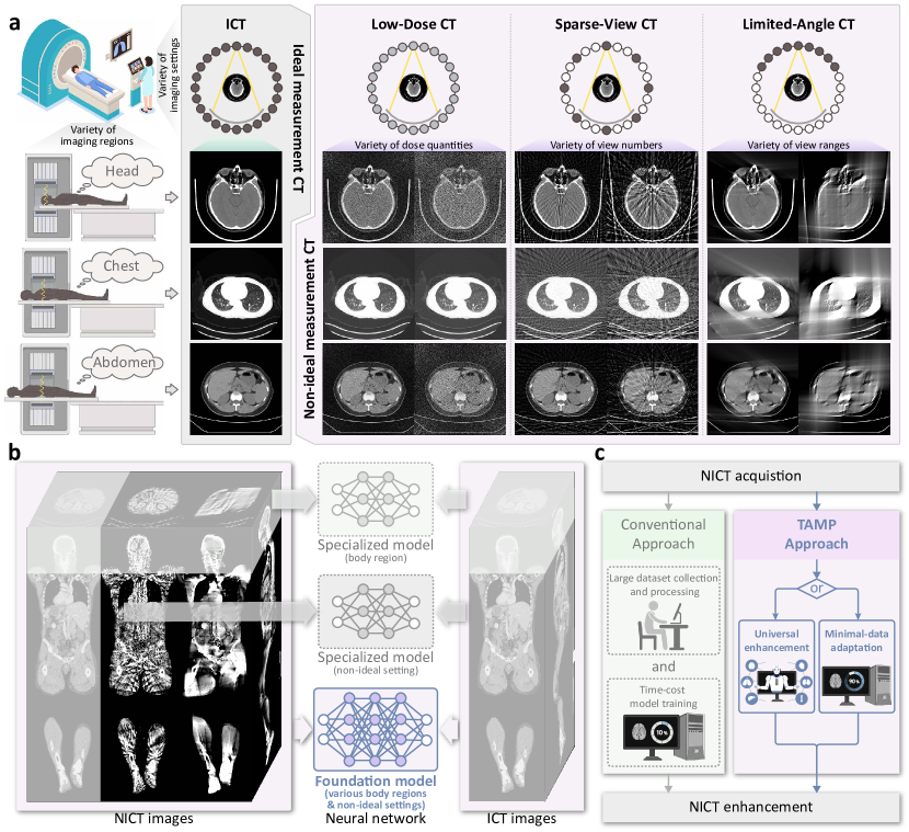
Non-ideal measurement computed tomography (NICT), whose imaging conditions do not conform to optimal standards [1], e.g., low-dose CT (LDCT) [2], sparse-view CT (SVCT), and limited-angle CT (LACT) [3], expands the scope of CT applications with the advantages of the radiation dose reduction, scanning acceleration, and adaptation of restricted scanning posture. However, with the reduction of imaging standards, the image quality has also been reduced, limiting the clinical acceptability [4]. As shown in Fig.1a, the LDCT reduces the tube current or tube voltage of the CT device [5], reducing damage from the X-ray radiation dose. The SVCT implements sparse angle sampling [6], accelerating the CT scanning speed. The LACT captures projections within a restricted range of angles [7, 8], enabling CT scanning in scenarios requiring restricted posture. These NICT settings have been used in wide clinical practices, e.g., lung cancer screening, breast cancer diagnosis, etc. [9, 10, 11], and medical device development, e.g., slow kVp switching dual energy CT [12], C-arm CT [13], etc. However, compared with the standard CT (we named ideal measurement CT (ICT) in this paper), NICT exhibits inferior imaging quality owing to the incomplete scanned information from the human body, losing the details of the tissues and structures, and aggravating the noise and artifacts [14]. Consequently, radiologists will be challenged to accurately identify clinical-concerned features on NICT images, hindering their clinical significance.
Although numerous studies [15, 16, 17, 18, 19] have demonstrated the feasibility of using deep learning models to enhance the quality of NICT images in specific scenarios, their high data cost and limited generalizability have become large obstacles. As shown in Fig.1c, the development of these specialized models has to take the large dataset collection and processing and time-cost model training, costing a lot of money and time. Therefore, it will extremely extend the development cycle and enlarge the cost of intelligent NICT imaging devices. Moreover, they also focus on specific body regions (head, chest, abdomen, etc.) and NICT settings (LDCT [16], SVCT [17], LACT [15]), so that it makes them only perform well in the NICT images with the same distribution as the training dataset (Fig.1b). Once the device is updated or the scanning protocol changes, the model with a large upfront cost will be unable to be applicable [20, 21, 22].
Foundation models (FMs) have shown impressive generalizability across diverse scenarios [23], highlighting their potential for universal NICT enhancement. However, owing to the challenges in the collection of large pre-training datasets and the compatibility of data variation, no success has been reported. a) Data quantity. Ethical concerns [24] restrict the creation of large datasets for NICT FM training. The radiation risks associated with CT scanning make it unethical to repeatedly scan individuals solely for data collection purposes [25]. This limitation hinders the direct collection of large NICT datasets for FM training, affecting generalizability in universal scenarios [26]. b) Data variation. Different physical processes in NICT settings lead to highly varied defect patterns in images. For instance, LDCT images exhibit detailed noise, while LACT images show significant angular defects (Fig. 1a). This variability poses a challenge for universal NICT enhancement models, which struggle to accommodate the diverse defect patterns. Additionally, existing specialized NICT enhancement models focus on specific defects in their targeted NICT settings, lacking compatibility for varied defect patterns, and limiting their universal representation and learning capabilities in FM training.
In this paper, we propose a multi-scale integrated Transformer AMPlifier (TAMP), an imaging FM for universal enhancement of NICT images. It constructs a physical-driven pre-training and parameter-efficient adaptation process that trains an imaging FM on more than 3.6 million simulated NICT-ICT image pairs for universal NICT enhancement ability and optimizes few parameters of the model to adapt to real-world NICT scenarios, minimizing the amount of additional real-world training data. The contributions of this work are summarized as follows:
-
•
To the best of our knowledge, TAMP is the first imaging FM for universal NICT enhancement. It has a powerful generalization ability that is beneficial to the enhancement of diverse NICT images including the LDCT, SVCT, and LACT across the large body regions including the head, chest, abdomen, and lower-limbs. It will minimize the data cost in the device development and improve the subjective quality and clinical acceptability of diverse NICT images, demonstrating strong potential for clinical application. Therefore, as shown in Fig.1c, it has two advanced properties, i.e., universal enhancement and minimal-data adaptation.
-
•
Universal enhancement: We propose a physical-driven pre-training paradigm for large-scale training of the NICT enhancement FM (Fig.1b). By simulating the defects that meet the physical process of non-ideal measurement in the CT’s projection domain [2, 3], NICT images are synthesized from ICT images for a large-scale NICT-ICT paired dataset. Then, a multi-scale integrated transformer network (MITNet) is designed to represent the multi-granularity defect features in the varied NICT data. Finally, a dual-domain enhancement learning (DDEL) is constructed to learn the universal NICT enhancement both in image and projection domains. Based on the above methods, our TAMP will be able to be generalized to multiple NICT settings across different body regions.
-
•
Minimal-data adaptation: We utilize a parameter-efficient fine-tuning strategy for the professional performance of our TAMP in specific scenarios with low data cost (Fig.1c). This incorporates low-rank adaptation (LoRA) [27], so that the TAMP can only tune a small number of parameters in the whole network, thus greatly reducing the risk of over-fitting caused by training too many parameters. With just a few additional training iterations and data slices, TAMP will quickly acquire knowledge for specific NICT settings and body regions, facilitating rapid development in NICT enhancement models, especially where the real-world training data is limited.
-
•
We construct and publicly released a large-scale simulated NICT dataset (SimNICT), offering researchers a valuable resource for exploring deep learning methods on NICT enhancement. It consists of 3.6 million NICT-ICT image pairs simulated from 9,639 ICT volumes, featuring LDCT, SVCT, and LACT settings across various defect degrees in the head, chest, abdomen, and lower-limbs. This dataset enables quick data acquisition for developing NICT enhancement models and establishes a standard for performance evaluation.
2 Results
2.1 SimNICT dataset with large NICT quantity and diversity
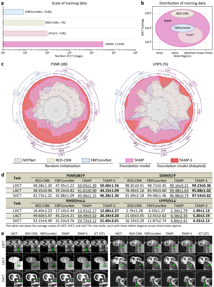
As shown in Fig.2a,2b, our SimNICT is a large-scale NICT dataset that contains 3.6 million images featuring various NICT settings and body regions. It starts from the ICT images from ten publicly CT datasets (Sec.5) with a total of 9,639 volumes. Three NICT settings (LDCT, SVCT, and LACT) across four body regions (head, chest, abdomen, and lower-limbs) have been enrolled into the SimNICT dataset (Fig.2b), having much larger diversity than the existing works. We compared the quantity and diversity of the NICT data used in the recent or typical NICT enhancement works, i.e., FBPConvNet [17], RED-CNN [16], and ProCT [18]. It has more than 360 times data quality compared with the existing works so our SimNICT is the currently largest NICT enhancement dataset. It will pre-train the model for the enhancement of extensive NICT images, supporting the model’s learning for universal enhancement ability. Although our SimNICT is a simulation dataset, it is simulated to meet the physical process of NICT imaging (illustrated in Sec.4.1), achieving realistic defects in the simulated NICT images. Our real-world validation, Sec.2.4, has demonstrated the generalizability of the model trained by the SimNICT on real-world data.
2.2 TAMP achieves universal NICT enhancement with powerful generalizability
Our TAMP, trained on the SimNICT, has powerful generalizability and effectiveness and is able to directly enhance diverse NICT images without additional training. After the parameter-efficient fine-tuning with LoRA, TAMP can be further specialized to specific NICT settings and body regions (TAMP-S), achieving professional performance (Fig.2b).
Experimental Setting: The powerful generalizability and effectiveness of our TAMP are evaluated in 27 NICT enhancement tasks. Specifically, the tasks cover the NICT settings of LDCT, SVCT, and LACT, with defect degrees of high, mid, and low, across body regions of the chest (from COVID-19 [28]), abdomen (from AMOS [29]), and whole-body (from AutoPET [30]). According to the data amount in the related studies [17, 16, 18], we allocate 2,089, 8,669, and 19,613 CT images from three datasets as training (80%) and testing (20%) sets, which are independent of TAMP pre-training. We compare the enhancement performance with two typical specialized NICT enhancement models, i.e., RED-CNN [16] (direct prediction architecture) and FBPConvNet [17] (residual-based architecture), and the proposed MITNet. For each task, RED-CNN, FBPConvNet, and MITNet are trained from scratch, our TAMP is used for enhancement without additional training, and TAMP-S is fine-tuned beginning with our TAMP’s pre-trained parameters. We utilize the peak signal-to-noise ratio (PSNR) and root mean square error (RMSE) to evaluate the pixel-level accuracy of enhancement, and the structural similarity index measure (SSIM) and learned perceptual image patch similarity (LPIPS) [31] to evaluate visual performance. The units for PSNR and RMSE are dB and Hu, and SSIM and LPIPS scores are multiplied by 100 and expressed as percentages to display finer details. More specific settings are described in our supplementary materials.
Observations: As shown in Fig.2, TAMP has universal NICT enhancement ability with powerful generalizability and effectiveness. There are two observations in Fig.2:
1) Our TAMP achieves superior performance in diverse NICT enhancement tasks across various NICT settings, body regions, and defect degrees, as shown in Fig.2c. Without adaptation, our TAMP outperforms the compared methods in PSNR across all 27 NICT enhancement tasks and in LPIPS on 24 (88.89%) tasks. After adaptation, TAMP-S improves the PSNR score on all tasks and the LPIPS score on 23 (85.19%) tasks, surpassing the comparison methods in both PSNR and LPIPS on all tasks. Specifically, the outstanding universal NICT enhancement capability of TAMP is demonstrated as follows:
NICT settings: Artifacts of varying scales and shapes appear in images under different NICT settings. Compared to FBPConvNet, RED-CNN achieves better PSNR and LPIPS scores on 6 out of 9 LDCT tasks. However, for the 9 LACT tasks, it outperforms in only 1 task for PSNR and 2 tasks for LPIPS. RED-CNN’s preference for LDCT tasks stems from its network structure, which is specifically designed for small-scale artifacts. However, this design limits its ability to generalize to NICT enhancement tasks involving other types of artifacts. In contrast, MITNet’s network design is well-suited for multi-scale artifacts across various NICT settings. After training with random initialization, MITNet outperforms RED-CNN on 8 out of 9 LACT tasks. Following large-scale pretraining, its significant potential enables TAMP to achieve superior performance across LDCT (50.05 PSNR), LACT (41.61 PSNR), and SVCT (45.06 PSNR) tasks, with respective improvements over the compared methods of 4.38%, 6.01%, and 5.08% (Fig.2d).
Body regions: CT images from different body regions show distinct characteristics, with the abdomen having higher values, the head showing concentrated high values, and the chest having lower values in the lungs and moderate values in soft tissues, posing greater challenges for model generalization. MITNet’s Transformer architecture expands network capacity, demonstrating initial generalization across body regions even during the random initialization training stage, surpassing RED-CNN on 5 tasks with the AutoPET whole-body dataset. After extensive large-scale training, this generalization is fully realized, with TAMP and TAMP-S outperforming comparison methods across all body region tasks.
Defect degrees: The defect degrees of NICT images directly influence the difficulty of enhancement, yet TAMP consistently improves image quality across all defect degrees. Compared to other methods, TAMP shows an average PSNR improvement of 4.89%, 5.75%, and 5.81% across nine tasks for high, medium, and low defect degrees, respectively. This indicates that TAMP not only excels in challenging tasks but also achieves even more significant improvements in simpler tasks. After adaptation, these improvements with TAMP-S increased to 7.61%, 9.24%, and 10.05%, meeting the more stringent demands of clinical applications.
2) Our TAMP effectively removes artifacts of various scales and shapes from NICT images (Fig.2e), significantly enhancing their image quality. The different physical processes in NICT settings lead to artifacts of varying scales and shapes in NICT images, placing higher demands on the model’s comprehensive enhancement capabilities. While RED-CNN’s convolutional network structure, designed for high-resolution features, performs well in addressing small-scale artifacts in LDCT and SVCT images, its limited receptive field hampers its ability to handle large-scale artifacts in SVCT and LACT. In contrast, our TAMP, with its convolutional and Transformer-based architecture across multiple resolution channels, excels in addressing multi-scale artifacts with different shapes in various NICT images. As shown in the first row of Fig.2e, small-scale speckle artifacts with discrete distributions in LDCT are effectively removed by all methods. However, the slightly larger-scale linear noise formed by continuous speckles is misinterpreted as artifacts by RED-CNN and FBPConvNet, whereas TAMP and TAMP-S effectively avoid this issue. Similarly, linearly distributed noise is also present in the SVCT images in the third row of Fig.2e, where TAMP and TAMP-S demonstrate more thorough artifact removal. More severely, in the LACT images in the second row of Fig.2e, large-scale wedge-shaped artifacts significantly distort the original CT image structures, making them difficult to infer based on adjacent image information. However, due to the expansive receptive fields and the universal NICT representations learned through large-scale pretraining, both TAMP and TAMP-S effectively reconstruct the disrupted image structures, making them easier to interpret. Consequently, TAMP’s ability to universally suppress various types of artifacts makes NICT images clearer in detail and more accurate in structure, providing greater value for clinical diagnosis.
2.3 TAMP effectively reduces the cost for specialized NICT enhancement
Our TAMP, as an imaging FM, offers a prepared initialization that enables the efficient development of specialized models with only a few data. We conducted data validation experiments to explore the performance of specialized models developed using TAMP across varying data quantities.
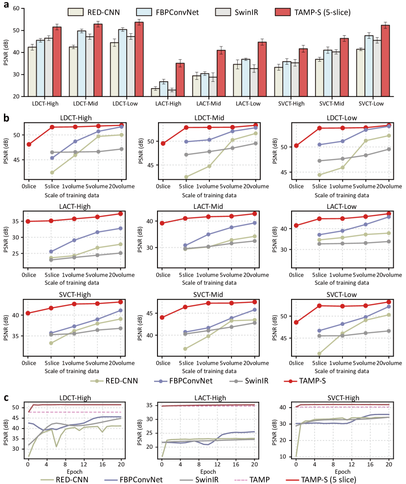
Experimental Setting: We evaluated the TAMP-S on nine NICT enhancement tasks by fine-tuning TAMP across varying data quantities. Two specialized NICT enhancement models (RED-CNN, FBPConvNet), and a pre-trained foundation model from natural images (SwinIR [32]) are used for comparison. These tasks contain three NICT settings (LDCT, SVCT, LACT) with three defect degrees (high, mid, low). On each task, we fine-tune TAMP and SwinIR, and train RED-CNN and FBPConvNet from scratch on five CT slices, one, five, and twenty subjects (abdomen regions simulated from AMOS dataset) to evaluate the influence of data amount on enhancement performance.
Observations: As shown in Fig.3, our TAMP only requires very small data amount and training iterations in the adaptation of specialized NICT enhancement models, effectively reducing the cost in NICT imaging application development. There are three observations in this experiment:
1) As shown in Fig.3a, TAMP-S achieves state-of-the-art performance across all tasks using five slices of training data. For the relatively simple tasks of LDCT-High, LDCT-Mid, and LDCT-Low, TAMP-S achieved PSNR scores of 51.57, 52.88, and 53.75 with just five slices of training data, respectively, surpassing the best comparison methods by 10.9%, 5.93%, and 6.14%, demonstrating its remarkably low data requirements for simpler tasks. For the most challenging task, LACT-High, TAMP-S still achieved a PSNR of 35.12, surpassing the highest score (27.11) of the comparison method by 29.55%, demonstrating its robust performance even in difficult scenarios. This excellent performance is due to its universal NICT enhancement capability as a foundation model that has undergone large-scale pre-training tailored for NICT enhancement. Although SwinIR was also pre-trained on large-scale data, the gap between its upstream task of grayscale image denoising and the downstream task of NICT image enhancement limits its performance. As a result, it is outperformed by FBPConvNet, which was trained with random initialization, in 88.89% of tasks.
2) As shown in Fig.3b, TAMP demonstrates efficient adaptation capacity in diverse NICT enhancement tasks with lower data cost. TAMP-S, fine-tuned with only 5 slices of data, achieves at least 94.11% of the PSNR performance obtained when fine-tuned with 20 volumes of data across 9 tasks and surpasses all comparison methods trained with 5 volumes, indicating that it has effectively adapted to each specific task. Moreover, as the amount of fine-tuning data increases, TAMP-S shows a stable improvement trend and consistently superior performance across varying quantities of training data in all NICT enhancement tasks. The pre-trained SwinIR outperforms RED-CNN on the three LDCT tasks, which are most similar to its upstream task, with 5 slices of training data, but is overtaken once the data volume exceeds 1 volume. This is because SwinIR’s network architecture and pre-trained representations are not well-suited for adapting to the NICT enhancement tasks, limiting its adaptation capability to this domain.
3) As shown in Fig.3c, the rapid and stable convergence performance of TAMP demonstrates its potential to reduce the consumption of computational time and resources. TAMP achieves at least 98.89% of its final convergence performance in just one epoch using five slices of data and maintains stability as training epochs increase. The effective convergence capability of TAMP is due to its LoRA-based fine-tuning strategy, which allows quick adaptation to specific tasks while retaining the complete pre-trained representations, thereby preventing significant performance fluctuations. In contrast, the compared methods require more epochs and exhibit performance fluctuations during the convergence process, necessitating additional time and resources to ensure optimal results.
2.4 Real-world validation: TAMP enhances real-world NICT images
Due to the difference between the real-world CT imaging process and the simulated process in this work, there will be a domain gap between the simulation-based NICT enhancement training and real-world NICT enhancement. Therefore, this experiment further evaluates the enhancement capability of our TAMP on real-world NICT images and shows our applicability in real-world clinical practices.
Experimental Setting: The real-world data is collected from Nanjing Drum Tower Hospital. It has three NICT settings, i.e., LDCT, LACT, and SVCT. The LDCT, which has 1496 image pairs from five volumes, is obtained by scanning the patient both under low and high tube current and voltage settings. 1,224 of them are used for training and 252 of them are used for testing. The SVCT and LACT are all reconstructed from the raw ICT projection data that has 750 images from 5 volumes. They are reconstructed by adjusting the raw projection data at sparse views and limited angles and mapping to the image domain. 625 of them are used for training and 125 of them are used for testing. We perform the TAMP, TAMP-S, RED-CNN, and FBPConvNet on these real-world data following Sec.2.2 to evaluate our TAMP’s enhancement ability in real-world situations.
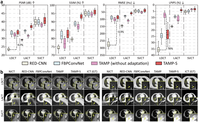
Observations: As shown in Fig.4, our TAMP and TAMP-S effectively enhance the quality of real-world NICT images, based on two observations:
1) Our TAMP, pre-trained on simulated NICT images, can directly enhance the quality of real-world NICT images without additional training, demonstrating its potential to accelerate imaging equipment development with a reliable enhancement model. Quantitatively, TAMP outperforms RED-CNN across four metrics in LDCT and LACT tasks, particularly achieving the best LPIPS score (7.83%) in the LDCT task, surpassing RED-CNN by 78.12% (Fig.4a). Qualitatively, TAMP reduces small-scale artifacts with clear structures reconstructed, and it also moderately reconstructs severely damaged areas (Fig.4b). Enhanced by it, both LDCT and SVCT images with small-scale light spot artifacts appear smoother, with clearly distinguishable edges and lines of structures such as blood vessels, muscles, and the heart. In contrast, the predictions of RED-CNN and FBPConvNet exhibit blurred edges and lines, as their mere reliance on mean squared error in the loss calculation promotes conservative predictions and hinders accurate detail inference. Moreover, severely damaged areas, such as the chest edges and blood vessels in LACT images, were also reconstructed by TAMP. However, RED-CNN failed to reconstruct them due to its pure convolutional network structure, which is solely designed for high-resolution features and cannot adapt to such larger-scale distortions in LACT images.
2) After adaptation, our TAMP-S exhibits the best metrics and visual performance, providing more precise and reliable support for clinical diagnosis. Quantitatively, TAMP-S outperforms two compared methods in all tasks across four metrics (Fig.4a). Qualitatively, TAMP-S retains the visual advantages of TAMP while demonstrating more thorough denoising and more accurate reconstruction of real-world NICT images (Fig.4b). It efficiently enhances the smoothness of homogenous regions in LDCT and SVCT images while maintaining the edge clarity advantage of TAMP, making structures easier to observe. The severely damaged chest edges and blood vessels in LACT images also achieve more precise reconstruction, featuring clearer and more accurate shapes. Integrating these features, TAMP-S’s visual performance closely resembles that of real CT images, which is due to its pre-training from large-scale CT data, allowing for a deeper understanding of CT features and reliable real-world noise reduction.
2.5 Radiologist validation: TAMP improves the clinical acceptability of NICT images
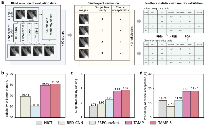
To evaluate TAMP’s ability to enhance the clinical acceptability of NICT images, we conducted a radiologist validation study. Specifically, we invite three radiologists (one with over 10 years of experience and two with more than 5 years each) to blindly rank these images according to subjective quality and score them based on clinical acceptability. We then statistically analyzed the scoring data to evaluate the performance of each model.
Experimental Setting: The experimental process of radiologist validation includes blind selection of evaluation data, blind expert evaluation, and feedback statistics with metrics calculation. One proficient (1P) and two competent radiologists (2C and 3C) from the Department of Radiology in the affiliated hospital of the medical school of Ningbo University were invited to score these images blindly. As shown in Fig.5a, four images were randomly selected from NICT, ICT, FBPConvNet enhanced, RED-CNN enhanced, TAMP enhanced, and TAMP-S enhanced NICT, to construct a validation group. Ninety groups are shuffled and randomly selected for evaluation. To calculate the final score, the scoring data from the three radiologists are weighted according to their years of experience (weights of 0.5 for 1P, and 0.25 for 2C and 3C) and analyzed using three designed metrics: probability of better than NICT (PBN), subjective quality ranking (SQR), and probability of clinical acceptability (PCA). The PBN represents the enhancement degree for the enhanced NICT images via the methods. The SQR reflects the subjective ranking of the enhanced images’ quality among the methods. The PCA represents the clinical acceptability of the various CT images. The detailed calculation process for these metrics is described in the supplementary materials.
Observations: Our results presented in Fig.5 demonstrate that NICT images enhanced by TAMP have higher subjective quality and are more likely to be clinically accepted, based on three observations:
1) Enhanced by TAMP and TAMP-S, the subjective quality of NICT images is considered significantly improved by radiologists. As shown in Fig.5b, the PBN for TAMP (79.49%) significantly exceeds 50% (the threshold indicating insufficient enhancement in NICT quality), surpassing RED-CNN by 14.47% and FBPConvNet by 32.48%, indicating that the majority of NICT images enhanced by TAMP are recognized by radiologists for their improved subjective quality. After fine-tuning, the PBN for TAMP-S further improved to 81.00%, reflecting its potential to achieve better subjective quality enhancements for NICT images in various specific scenarios. This robust subjective quality enhancement capability is due to TAMP’s universal NICT representation achieved through large-scale pre-training, enabling consistent performance across various types of NICT images.
2) Enhanced by TAMP and TAMP-S, the subjective quality of NICT images is considered efficiently improved by radiologists. As shown in Fig.5c, the SQR of TAMP (2.83) surpasses RED-CNN (1.92) by 47.40% and FBPConvNet by 26.91%, reflecting a 58.99% improvement in NICT images. After adaptation, the SQR of TAMP-S increased by 3.18%, achieving the best value among all methods and demonstrating its effectiveness in enhancing the quality of NICT images. Compared to the significance reflected by PBN, SQR focuses on the extent of image quality enhancement performance. For instance, FBPConvNet has a lower PBN than RED-CNN (60.00% vs. 69.44%) but a higher SQR (2.23 vs. 1.92), indicating that FBPConvNet is very effective for enhancing certain NICT images. However, our method surpasses the comparison methods in both PBN and SQR, reflecting its effective enhancement of subjective quality across various NICT images.
3) NICT images enhanced by TAMP and TAMP-S receive greater clinical acceptance from radiologists. As shown in (Fig.5d), TAMP and TAMP-S efficiently improve the PCA of NICT by 54.60% and 56.73% respectively, demonstrating their improvements in clinical acceptability are recognized by radiologists. An interesting observation is that although the SQR scores in Fig.5c demonstrate that the compared methods improve the subjective quality of NICT images, RED-CNN actually decreases the PCA of NICT images by a substantial 34.15%, while FBPConvNet achieves only a slight improvement of 1.45%. This is because, for clinical acceptance, radiologists are more concerned with factors beyond subjective quality, such as whether the regions of interest in the enhanced NICT images conform to the structural features of real-world CT images to provide reliable clinical diagnostic support. Thus, the improvement of NICT images in both SQR and PCA reflects the ability of TAMP and TAMP-S to enhance image subjective quality based on the features of real-world CT images, demonstrating significant potential for clinical application.
3 Discussion
In this paper, we have proposed and validated TAMP, the first imaging foundation model for the universal enhancement of NICT images. Through pre-training on the large-scale dataset SimNICT, our TAMP is able to directly enhance diverse NICT images, and effectively adapts to specific NICT scenarios with few training data.
Our work pioneers the application of foundation models in the NICT enhancement domain, utilizing the pre-training and adaptation paradigm to advance research in universal NICT enhancement technologies and enable efficient model deployment for specific NICT enhancement scenarios. Firstly, TAMP’s universal capability is showcased by its direct enhancement of diverse LDCT, LACT, and SVCT images with varying defects across body regions, achieved through pre-training on 10.8 million simulated NICT images. Among 27 NICT enhancement tasks (Sec.2.2), our TAMP outperforms compared methods in 19 tasks on the PSNR and in 22 tasks on the LPIPS, all without additional training. Secondly, TAMP can be rapidly adapted for specific NICT enhancement scenarios by fine-tuning with the LoRA method, requiring only five slices of training data to achieve excellent performance, outperforming compared methods trained with 20 subjects in most tasks (Sec.2.3). This efficient generalization capability also highlights TAMP’s advantage when using real-world NICT data for fine-tuning (Sec.2.4).
Clinically, TAMP’s suitability for a wide range of non-ideal imaging conditions and subjects facilitates the development of new CT imaging devices. Moreover, its efficient generalization capability reduces data and computing costs in specialized model development, promoting its application in broader and more demanding clinical scenarios. In addition, TAMP predictions closely align with the morphological features of real-world CT images, as demonstrated in Fig.2e and Fig.4b. This alignment facilitates clinical acceptance, as experimentally validated in Sec.2.5, aiding radiologists in using it more effectively and accurately for clinical diagnoses.
This study has two limitations. First, TAMP was pre-trained using simulated NICT images, which have a gap with real-world NICT images. However, since both our NICT image simulation and DDEL strategy are physical-driven, adhering to the physical processes of NICT imaging, TAMP is generalizable to real-world NICT images with few data, as has been experimentally demonstrated in Sec.2.4. Second, the multi-scale Transformer structure of TAMP provides a universal NICT representation but requires increased memory consumption during operation. Fortunately, techniques such as model pruning, knowledge distillation, and mixed precision training [33] have been applied to reduce memory usage, which will also be a focus of our future research.
4 Methods
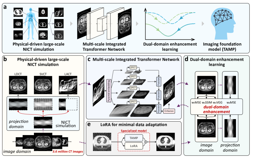
As shown in Fig.6a, our proposed imaging foundational model, TAMP, consists of a physical-driven large-scale NICT simulation, multi-scale integrated transformer network, and dual-domain enhancement learning, for universal enhancement of NICT. In this section, we illustrate the methods of our TAMP, and more specific details are described in our supplementary materials.
4.1 SimNICT: Physical-driven simulation for large-scale NICT dataset
SimNICT is the first dataset constructed for universal NICT enhancement model training. It starts from the ICT images from ten publicly CT datasets (Sec.5) that encompass whole-body regions, including the head, chest, abdomen, and lower-limbs, and are simulated into LDCT, SVCT, and LACT under different defect degrees. By removing the volumes with low quality, our SimNICT dataset finally obtains 3,633,465 images from 9,639 ICT volumes. It simulates the NICT images with three non-ideal settings (LDCT, SVCT, and LACT) and different defect degrees, thus finally achieving over 10.9 million NICT-ICT image pairs.
As shown in Fig.6b, we simulate the NICT images according to the physical processes of the non-ideal measurements. It projects the ICT images to the projection domain to simulate the maps of the CT raw signal. Then, according to the physical processes of LDCT, SVCT, and LACT, we simulate the defective copies of these CT raw signal maps. For the LDCT, according to the low anti-interference of low-dose radiation in the environment, we produce Gaussian noise on the maps to simulate the interfered measurement. For the SVCT, according to the sparse angle sampling, we reduce the views at equal intervals on the maps to simulate the sparse measurement. For the LACT, according to the restricted scanning angle range, we reduce the range of views on the maps to simulate the angular-defected measurement. Finally, the defective CT raw signal maps are back-projected to the image domain, thus achieving the NICT images with real-world defects. We utilize the ASTRA Toolbox111ASTRA: https://astra-toolbox.com/index.html to achieve this physical-driven simulation process.
4.2 MITNet: Multi-scale integrated transformer network for the representation of varied defect patterns
Our MITNet constructs a transformer [34] architecture that is compatible with multi-scale features, thus representing the varied defect patterns in different NICT images. As shown in Fig.6c, it has two important aspects: 1) Pyramid modelling extracts the NICT images’ features in different scales to adapt to the varied defect patterns for different NICT settings. The NICT images are put into a pyramid embedding module that adopts different patch scales and takes the linear layers to map the patches to the embedding space, thus achieving the embedding features in multiple pyramid levels. These features will be further represented in multiple Deep Feature Extraction Transformer (DFET) blocks [32] which is a stack of Swin Transformer Layers to learn to repair the defect information in each level. 2) Progressive multi-scale integration gradually fuses the features from low resolution to high resolution, thus achieving the features that are represented as compatible with varied defect patterns. In each fusion stage, the feature maps from different pyramid levels are concatenated and put into a convolution-ReLU layer for multi-scale feature integration. Finally, these features are input into several convolution layers for to predict the final enhanced NICT images.
4.3 Dual-domain enhancement learning for imaging pre-training
We design a dual-domain enhancement learning (DDEL) that trains our MITNet both in the projection domain and the image domain to learn to perceive the physical property and defect patterns of the NICT images, thus effectively extracting information [3] (Fig.6d). In the image domain, three kinds of losses are used to learn the enhancement of various defect patterns, including the mean square error (MSE) loss (pixel-level) , SSIM loss (region-level) [35], and VGG loss (image-level) [36]. These losses measure the similarity between the enhanced NICT images and the ICT images in different granularities, training the MITNet to represent the varied defect patterns in different NICT settings. In the projection domain, the enhanced NICT and ICT images are mapped to the projection domain for sinogram maps, and the MSE loss is calculated between these maps. Since NICT quality degradation occurs not only in the image domain but also in CT detector sampling projections and the process of reconstructing projection data into image data, the features of NICT are reflected within its projection space. Hence, the loss in the projection domain will encourage the model to perceive on the physical property of CT images. We utilize the Operator Discretization Library 222ODL: https://github.com/odlgroup/odl to implement this projection, which is capable of backpropagation during training. Totally, these four losses are weighted and summed for the training loss, i.e., value for model training.
4.4 Parameter-efficient fine-tuning for minimal-data adaptation
We employ the LoRA which is a parameter-efficient fine-tuning method to adapt our TAMP to specialized NICT enhancement tasks for a professional performance with very few additional training data (Fig.6e). It only tunes a small number of parameters in the whole network, thus greatly reducing the risk of over-fitting caused by training too many parameters. Therefore, it enables only very little data to stimulate the professional performance of our TAMP in specific scenarios achieving minimal data adaptation. Specifically, following the implementation of LoRA [27], we add the bypasses of low-rank matrix on the linear layers and convolutional layers in the whole network to adapt their representation to target scenarios. During adaptation, the parameters in the LoRA bypasses are tuned and the original parameters in the TAMP are fixed. The additional parameters from the LoRA bypasses will be fused back to the main path by the re-parameterization method so that no additional parameters will be added after the adaptation. We utilize the same training loss as the pre-training stage for adaptation.
4.5 Details of pre-training and adaptation
Our TAMP is implemented by PyTorch333PyTorch: https://pytorch.org/ for its pre-training and adaptation. To reduce the time consumption of inputting and outputting (IO) large-scale data on disk during training, a queued training process is designed. It loads (we set ) NICT volumes into memory as a queue and shuffles the slices for learning. To avoid over-fitting for a fixed defect pattern in the training process, we ensure that all NICT settings exist in the queue. Once all the slices have been iterated, the oldest volume is removed from the queue, and a new NICT volume is loaded, thus effectively reducing the IO cost. We take the Adan [37] as our optimizer, which is an outstanding optimizer targetedly designed for foundation model training. It accelerates the convergence speed and reduces the loss fluctuation in the learning process of transformer networks. For pre-training, we set the learning rate , b1, and b2 as 5e-4, 0.5, and 0.999, and the learning rate becomes 0.95 times, i.e., after the training of every 100 queues for finer fitting. For adaptation, we set the same learning rate , b1, and b2 as the pre-training, and the learning rate becomes 0.5 times, i.e., after the training of every 10 queues. We set the batch size as 5 and the input size as to train the NICT images in a high resolution. In our experiment, considering the scale difference of loss values, we set the weights of losses in the training loss as 1, 5e-3, 1e-4, and 5e-4.
5 Data Availability
This work has enrolled ten publicly available datasets to construct our SimNICT dataset, including COVID-19-NY-SBU dataset444COVID-19-NY-SBU: https://wiki.cancerimagingarchive.net/pages/viewpage.action?pageId=89096912 [38], STOIC dataset555STOIC: https://stoic2021.grand-challenge.org/ [39], MELA dataset666MELA: https://mela.grand-challenge.org/ [40], LUNA dataset777LUNA: https://luna16.grand-challenge.org/ [41], LNDb dataset888LNDb: https://lndb.grand-challenge.org/ [42], HECKTOR22 dataset999HECKTOR22: https://hecktor.grand-challenge.org [43], CT_COLONOGRAPHY dataset101010CT_COLONOGRAPHY: https://wiki.cancerimagingarchive.net/pages/viewpage.action?pageId=3539213 [44], AutoPET dataset111111AutoPET: https://autopet.grand-challenge.org/ [30], AMOS dataset121212AMOS: https://amos22.grand-challenge.org/ [29], and the CT Images in COVID-19131313CT Images in COVID-19: https://www.cancerimagingarchive.net/collection/ct-images-in-covid-19/ [45]. The data in our real-world validation is collected from Nanjing Drum Tower Hospital and contains sensitive privacy information. Due to the ethical restrictions, this dataset cannot be released. We have opened the parameters of our TAMP-S adapted to this dataset. Our SimNICT dataset will be released at a companion website https://huggingface.co/datasets/YutingHe-list/SimNICT.
6 Code Availability
Our TAMP will be released at https://github.com/YutingHe-list/TAMP.
References
- \bibcommenthead
- Candès and Wakin [2008] Candès, E.J., Wakin, M.B.: An introduction to compressive sampling. IEEE signal processing magazine 25(2), 21–30 (2008)
- Kalra et al. [2004] Kalra, M.K., Maher, M.M., Toth, T.L., Hamberg, L.M., Blake, M.A., Shepard, J.-A., Saini, S.: Strategies for ct radiation dose optimization. Radiology 230(3), 619–628 (2004)
- Wang et al. [2023] Wang, T., Xia, W., Lu, J., Zhang, Y.: A review of deep learning ct reconstruction from incomplete projection data. IEEE Transactions on Radiation and Plasma Medical Sciences (2023)
- Jiang et al. [2018] Jiang, C., Zhang, Q., Fan, R., Hu, Z.: Super-resolution ct image reconstruction based on dictionary learning and sparse representation. Scientific reports 8(1), 8799 (2018)
- Nakayama et al. [2005] Nakayama, Y., Awai, K., Funama, Y., Hatemura, M., Imuta, M., Nakaura, T., Ryu, D., Morishita, S., Sultana, S., Sato, N., et al.: Abdominal ct with low tube voltage: preliminary observations about radiation dose, contrast enhancement, image quality, and noise. Radiology 237(3), 945–951 (2005)
- Bian et al. [2010] Bian, J., Siewerdsen, J.H., Han, X., Sidky, E.Y., Prince, J.L., Pelizzari, C.A., Pan, X.: Evaluation of sparse-view reconstruction from flat-panel-detector cone-beam ct. Physics in Medicine & Biology 55(22), 6575 (2010)
- Wu et al. [2003] Wu, T., Stewart, A., Stanton, M., McCauley, T., Phillips, W., Kopans, D.B., Moore, R.H., Eberhard, J.W., Opsahl-Ong, B., Niklason, L., et al.: Tomographic mammography using a limited number of low-dose cone-beam projection images. Medical physics 30(3), 365–380 (2003)
- Chen et al. [2013] Chen, Z., Jin, X., Li, L., Wang, G.: A limited-angle ct reconstruction method based on anisotropic tv minimization. Physics in Medicine & Biology 58(7), 2119 (2013)
- Toyoda et al. [2008] Toyoda, Y., Nakayama, T., Kusunoki, Y., Iso, H., Suzuki, T.: Sensitivity and specificity of lung cancer screening using chest low-dose computed tomography. British journal of cancer 98(10), 1602–1607 (2008)
- Cui et al. [2015] Cui, J.-W., Li, W., Han, F.-J., Liu, Y.-D.: Screening for lung cancer using low-dose computed tomography: concerns about the application in low-risk individuals. Translational lung cancer research 4(3), 275 (2015)
- Boone et al. [2001] Boone, J.M., Nelson, T.R., Lindfors, K.K., Seibert, J.A.: Dedicated breast ct: radiation dose and image quality evaluation. Radiology 221(3), 657–667 (2001)
- Szczykutowicz and Chen [2010] Szczykutowicz, T.P., Chen, G.-H.: Dual energy ct using slow kvp switching acquisition and prior image constrained compressed sensing. Physics in Medicine & Biology 55(21), 6411 (2010)
- Schafer et al. [2011] Schafer, S., Nithiananthan, S., Mirota, D., Uneri, A., Stayman, J., Zbijewski, W., Schmidgunst, C., Kleinszig, G., Khanna, A., Siewerdsen, J.: Mobile c-arm cone-beam ct for guidance of spine surgery: image quality, radiation dose, and integration with interventional guidance. Medical physics 38(8), 4563–4574 (2011)
- Mackin et al. [2018] Mackin, D., Ger, R., Dodge, C., Fave, X., Chi, P.-C., Zhang, L., Yang, J., Bache, S., Dodge, C., Jones, A.K., et al.: Effect of tube current on computed tomography radiomic features. Scientific reports 8(1), 2354 (2018)
- Wang et al. [2020] Wang, J., Liang, J., Cheng, J., Guo, Y., Zeng, L.: Deep learning based image reconstruction algorithm for limited-angle translational computed tomography. Plos one 15(1), 0226963 (2020)
- Chen et al. [2017] Chen, H., Zhang, Y., Kalra, M.K., Lin, F., Chen, Y., Liao, P., Zhou, J., Wang, G.: Low-dose ct with a residual encoder-decoder convolutional neural network. IEEE transactions on medical imaging 36(12), 2524–2535 (2017)
- Jin et al. [2017] Jin, K.H., McCann, M.T., Froustey, E., Unser, M.: Deep convolutional neural network for inverse problems in imaging. IEEE transactions on image processing 26(9), 4509–4522 (2017)
- Ma et al. [2023] Ma, C., Li, Z., He, J., Zhang, J., Zhang, Y., Shan, H.: Universal incomplete-view ct reconstruction with prompted contextual transformer. arXiv preprint arXiv:2312.07846 (2023)
- Yang et al. [2018] Yang, Q., Yan, P., Zhang, Y., Yu, H., Shi, Y., Mou, X., Kalra, M.K., Zhang, Y., Sun, L., Wang, G.: Low-dose ct image denoising using a generative adversarial network with wasserstein distance and perceptual loss. IEEE transactions on medical imaging 37(6), 1348–1357 (2018)
- Liang et al. [2020] Liang, X., Nguyen, D., Jiang, S.B.: Generalizability issues with deep learning models in medicine and their potential solutions: illustrated with cone-beam computed tomography (cbct) to computed tomography (ct) image conversion. Machine Learning: Science and Technology 2(1), 015007 (2020)
- Shan et al. [2019] Shan, H., Kruger, U., Wang, G.: A novel transfer learning framework for low-dose ct. In: 15th International Meeting on Fully Three-Dimensional Image Reconstruction in Radiology and Nuclear Medicine, vol. 11072, pp. 513–517 (2019). SPIE
- Zeng et al. [2022] Zeng, R., Lin, C.Y., Li, Q., Jiang, L., Skopec, M., Fessler, J.A., Myers, K.J.: Performance of a deep learning-based ct image denoising method: Generalizability over dose, reconstruction kernel, and slice thickness. Medical physics 49(2), 836–853 (2022)
- Zhang et al. [2024] Zhang, K., Zhou, R., Adhikarla, E., Yan, Z., Liu, Y., Yu, J., Liu, Z., Chen, X., Davison, B.D., Ren, H., et al.: A generalist vision–language foundation model for diverse biomedical tasks. Nature Medicine, 1–13 (2024)
- Cheplygina et al. [2019] Cheplygina, V., De Bruijne, M., Pluim, J.P.: Not-so-supervised: a survey of semi-supervised, multi-instance, and transfer learning in medical image analysis. Medical image analysis 54, 280–296 (2019)
- Mayo [2008] Mayo, J.R.: Radiation dose issues in longitudinal studies involving computed tomography. Proceedings of the American Thoracic Society 5(9), 934–939 (2008)
- Li et al. [2020] Li, H., Wang, Y., Wan, R., Wang, S., Li, T.-Q., Kot, A.: Domain generalization for medical imaging classification with linear-dependency regularization. Advances in neural information processing systems 33, 3118–3129 (2020)
- [27] Hu, E.J., Wallis, P., Allen-Zhu, Z., Li, Y., Wang, S., Wang, L., Chen, W., et al.: Lora: Low-rank adaptation of large language models. In: International Conference on Learning Representations
- An et al. [2020] An, P., Xu, S., Harmon, S.A., Turkbey, E.B., Sanford, T.H., Amalou, A., Kassin, M., Varble, N., Blain, M., Anderson, V., et al.: Ct images in covid-19 [data set]. The Cancer Imaging Archive 10, 32 (2020)
- Ji et al. [2022] Ji, Y., Bai, H., Ge, C., Yang, J., Zhu, Y., Zhang, R., Li, Z., Zhanng, L., Ma, W., Wan, X., et al.: Amos: A large-scale abdominal multi-organ benchmark for versatile medical image segmentation. Advances in neural information processing systems 35, 36722–36732 (2022)
- Gatidis et al. [2022] Gatidis, S., Hepp, T., Früh, M., La Fougère, C., Nikolaou, K., Pfannenberg, C., Schölkopf, B., Küstner, T., Cyran, C., Rubin, D.: A whole-body fdg-pet/ct dataset with manually annotated tumor lesions. Scientific Data 9(1), 601 (2022)
- Zhang et al. [2018] Zhang, R., Isola, P., Efros, A.A., Shechtman, E., Wang, O.: The unreasonable effectiveness of deep features as a perceptual metric. In: CVPR (2018)
- Liang et al. [2021] Liang, J., Cao, J., Sun, G., Zhang, K., Van Gool, L., Timofte, R.: Swinir: Image restoration using swin transformer. In: Proceedings of the IEEE/CVF International Conference on Computer Vision, pp. 1833–1844 (2021)
- Menghani [2023] Menghani, G.: Efficient deep learning: A survey on making deep learning models smaller, faster, and better. ACM Computing Surveys 55(12), 1–37 (2023)
- Vaswani [2017] Vaswani, A.: Attention is all you need. Advances in Neural Information Processing Systems (2017)
- Zhao et al. [2016] Zhao, H., Gallo, O., Frosio, I., Kautz, J.: Loss functions for image restoration with neural networks. IEEE Transactions on computational imaging 3(1), 47–57 (2016)
- Johnson et al. [2016] Johnson, J., Alahi, A., Fei-Fei, L.: Perceptual losses for real-time style transfer and super-resolution. In: Computer Vision–ECCV 2016: 14th European Conference, Amsterdam, The Netherlands, October 11-14, 2016, Proceedings, Part II 14, pp. 694–711 (2016). Springer
- Xie et al. [2024] Xie, X., Zhou, P., Li, H., Lin, Z., Yan, S.: Adan: Adaptive nesterov momentum algorithm for faster optimizing deep models. IEEE Transactions on Pattern Analysis and Machine Intelligence (2024)
- Saltz et al. [2021] Saltz, J., Saltz, M., Prasanna, P., Moffitt, R., Hajagos, J., Bremer, E., Balsamo, J., Kurc, T.: Stony brook university covid-19 positive cases [data set]. the cancer imaging archive. BBAG-2923 (2021)
- Revel et al. [2021] Revel, M.-P., Boussouar, S., Margerie-Mellon, C., Saab, I., Lapotre, T., Mompoint, D., Chassagnon, G., Milon, A., Lederlin, M., Bennani, S., et al.: Study of thoracic ct in covid-19: the stoic project. Radiology 301(1), 361–370 (2021)
- Song et al. [2022] Song, S., Xu, R., Luo, Y., Du, B., Yang, J., Kuang, K., She, Y., Zhao, M.: MELA Dataset: A Benchmark for Mediastinal Lesion Analysis (Training Set Part 1)
- Armato III et al. [2011] Armato III, S.G., McLennan, G., Bidaut, L., McNitt-Gray, M.F., Meyer, C.R., Reeves, A.P., Zhao, B., Aberle, D.R., Henschke, C.I., Hoffman, E.A., et al.: The lung image database consortium (lidc) and image database resource initiative (idri): a completed reference database of lung nodules on ct scans. Medical physics 38(2), 915–931 (2011)
- Pedrosa et al. [2021] Pedrosa, J., Aresta, G., Ferreira, C., Atwal, G., Phoulady, H.A., Chen, X., Chen, R., Li, J., Wang, L., Galdran, A., et al.: Lndb challenge on automatic lung cancer patient management. Medical image analysis 70, 102027 (2021)
- Andrearczyk et al. [2023] Andrearczyk, V., Oreiller, V., Boughdad, S., Le Rest, C.C., Tankyevych, O., Elhalawani, H., Jreige, M., Prior, J.O., Vallières, M., Visvikis, D., et al.: Automatic head and neck tumor segmentation and outcome prediction relying on fdg-pet/ct images: findings from the second edition of the hecktor challenge. Medical Image Analysis 90, 102972 (2023)
- Johnson et al. [2008] Johnson, C.D., Chen, M.-H., Toledano, A.Y., Heiken, J.P., Dachman, A., Kuo, M.D., Menias, C.O., Siewert, B., Cheema, J.I., Obregon, R.G., et al.: Accuracy of ct colonography for detection of large adenomas and cancers. New England Journal of Medicine 359(12), 1207–1217 (2008)
- Harmon et al. [2020] Harmon, S.A., Sanford, T.H., Xu, S., Turkbey, E.B., Roth, H., Xu, Z., Yang, D., Myronenko, A., Anderson, V., Amalou, A., et al.: Artificial intelligence for the detection of covid-19 pneumonia on chest ct using multinational datasets. Nature communications 11(1), 4080 (2020)