[1] 1]organization=National Key Laboratory of Fundamental Science on Synthetic Vision, College of Computer Science, Sichuan University, city=Chengdu, postcode=610000, country=China 2]organization=College of Computer Science, Sichuan University, city=Chengdu, postcode=610000, country=China
[style=chinese]
[style=chinese]
[style=chinese] 3]organization=College of Software Engineering, Sichuan University, city=Chengdu, postcode=610000, country=China \cormark[1]
[cor1]Corresponding author \tnotemark[1,2]
FIF-UNet: An Efficient UNet Using Feature Interaction and Fusion for Medical Image Segmentation
Abstract
Nowadays, pre-trained encoders are widely used in medical image segmentation because of their ability to capture complex feature representations. However, the existing models fail to effectively utilize the rich features obtained by the pre-trained encoder, resulting in suboptimal segmentation results. In this work, a novel U-shaped model, called FIF-UNet, is proposed to address the above issue, including three plug-and-play modules. A channel spatial interaction module (CSI) is proposed to obtain informative features by establishing the interaction between encoder stages and corresponding decoder stages. A cascaded conv-SE module (CoSE) is designed to enhance the representation of critical features by adaptively assigning importance weights on different feature channels. A multi-level fusion module (MLF) is proposed to fuse the multi-scale features from the decoder stages, ensuring accurate and robust final segmentation. Comprehensive experiments on the Synapse and ACDC datasets demonstrate that the proposed FIF-UNet outperforms existing state-of-the-art methods, which achieves the highest average DICE of 86.05% and 92.58%, respectively.
keywords:
Channel spatial interaction \sepMulti-level fusion \sepMedical image segmentation \sepU-shaped model1 Introduction
Medical image segmentation refers to extracting regions of interest from medical images, such as organs, diseased areas, etc., which can help physicians make diagnoses and formulate treatment plans. In medical image analysis, medical image segmentation is a fundamental task that provides accurate anatomical structure information for subsequent image analysis and quantification. Automatic segmentation provides high-precision segmentation masks (size, shape, and location of lesions) to physicians for accurately identifying and segmenting specific structures or lesions in images. Compared to handcrafted segmentation, automatic segmentation can significantly reduce the time and cost of processing medical images and improve the efficiency of data processing. In addition, automatic segmentation tools can mitigate the influence of human-related subjective factors, providing higher accuracy and consistency, which is particularly important for large-scale medical image datasets.
In recent years, deep learning has advanced across various fields. Convolutional neural networks (CNNs) can achieve feature extraction and representation for images, thus eliminating the requirements for handcrafted features. In this context, CNN-based automatic segmentation tools implement image segmentation by learning image features from large amounts of training samples through neural architecture, which can be generalized to new tasks with considerable high performance (Azad et al. (2022a)). UNet (Ronneberger et al. (2015)) became the most popular framework in medical image segmentation due to its simple yet effective architectural design and high performance, which can be applied to various modalities in medical images, including CT, MRI, X-ray, PET, etc. UNet is implemented by an encoder-decoder architecture with skip connections. The encoder gradually transforms the images into abstract representations by multi-level feature extraction and down-sampling operations. The decoder predicts the segmentation masks based on the abstract representation, in which the up-sampling operations are leveraged to recover image resolution to generate pixel-wise masks. As the core component of the UNet, the skip connection combines the features of the adjacent encoder stage and decoder stage to achieve high-efficiency learning.
Although the UNet models demonstrate the desired performance in medical image segmentation tasks, they still cannot capture global contextual information due to the limited receptive field. To address this issue, the Transformer blocks (Vaswani et al. (2017)) are incorporated into UNet architectures to enhance the global feature integration and contextual understanding, such as SwinUNet (Cao et al. (2022)), TransUNet (Chen et al. (2021)), MISSFormer (Huang et al. (2021)), UNRTR (Hatamizadeh et al. (2022)), and so on. However, the Transformer lacks inductive biases in CNNs, such as translation invariance and local feature learning ability, which makes it hard to achieve the expected performance with insufficient training samples. In the pre-training Vision Transformer (ViT) (Dosovitskiy et al. (2020)), the backbone network is pre-trained on large-scale common datasets and further fine-tuned with limited samples for certain tasks, which demonstrates significant efficacy in practical applications and reduces the requirements for labeled samples, such as Efficientnet (Tan and Le (2019)), ConvNeXt (Liu et al. (2022)), DeepViT (Zhou et al. (2021)), etc. Thanks to recent advances in pre-training strategy, the pre-trained encoders are empowered with rich and generalized feature representations. The key to improving task performance is to design efficient feature interaction and fusion mechanisms in the skip connections and decoder, which has the ability to leverage the semantic information learned by the pre-trained encoder to predict segmentation masks.
To this end, a novel U-shaped model, called FIF-UNet, is proposed to effectively utilize different levels of semantic information by feature interaction and fusion. Considering the semantic gap between the features learned by the different encoder and decoder stages, the feature fusion based on concatenation or element-wise addition operations in the vanilla skip connection inevitably results in inaccurate features and information loss, impacting the learning and generalization ability of the model to support the segmentation task. In this work, a channel spatial interaction (CSI) module, including the cascaded channel interaction unit (CIU) and spatial interaction unit (SIU), is proposed to enhance the skip connections. The CSI is designed to interactively recalibrate the feature maps by capturing the correlation between different levels of semantic information in a learnable manner.
In the decoder, the squeeze-and-excitation network (SENet) (Hu et al. (2018)) is incorporated into the original CNN blocks, i.e., cascaded conv-SE module (CoSE). The CoSE module leverages channel attention to adaptively reweight the features of different channels by modeling the interdependency among channels. The primary purpose is to efficiently select and integrate the crucial features to highlight the target regions and suppress the irrelevant background regions. In addition, to generate the pixel-wise segmentation masks, the up-sampling operations are applied to the consecutive decoder blocks to recover feature resolution. In this procedure, limited fusion in multi-level features leads to the loss of detailed features. In this work, a multi-level fusion module (MLF) is designed to effectively fuse the learned multi-scale features in different decoder stages by feature interactions among intra- and inter-classes.
Extensive experiments are conducted on the open-source Synapse and ACDC datasets to validate the proposed model. The experimental results demonstrate that the proposed approach outperforms other selective baselines, achieving an average DICE of 86.05% and 92.58%, respectively. Most importantly, the proposed three plug-and-play modules contribute to expected performance improvements and the visualization results indicate a confident location.
In summary, the main contributions of this work are shown as follows:
(1) A new U-shaped model is proposed to fully utilize the multi-level semantic information of the encoder and decoder by feature interaction and fusion. The results demonstrate the robustness of the FIF-UNet and its potential in practical applications.
(2) The CSI module is proposed to iteratively recalibrate the feature maps by capturing the correlation between the features learned by the different encoder and decoder stages, in which the CIU and SIU are cascaded to implement the feature interactions progressively.
(3) In the decoder, the CoSE module is designed to learn critical features based on the local structure and global context to highlight the target regions by incorporating channel attention into the convolution operations.
(4) The MLF module is proposed to efficiently fuse semantic information of different scales from decoder stages to alleviate the problem of detail loss, helping to obtain an accurate and robust final segmentation.
2 Related Work
As the core blocks of the UNet, CNN-based models were the dominant methods for various computer vision tasks. The Transformer-based models were also regarded as the mainstream due to their recent advancements across many artificial intelligence tasks. Integrating these modules with the UNet emerges as an enhanced strategy to improve the performance of medical image segmentation. In this section, related works are organized as follows:
2.1 CNN models
Before the ViT model in 2020, CNN-based UNet models were the dominant approaches in the field of medical image segmentation, which efficiently capture local features through convolution operations. However, the original UNet suffered from limited feature extraction capability and the semantic gap between the encoder and decoder. To address these issues, the encoder or decoder modules of the UNet were improved to enhance feature learning. In DUNet (Jin et al. (2019)), the convolution layers of the original UNet were replaced by the deformable convolution layer to capture intricate features. The Attention Gate (AG) was introduced to automatically focus on target structures with different shapes and sizes by employing a large receptive field and semantic contextual information in Attention U-Net (Oktay et al. (2018)). The inception layers of the Google-Net (Punn and Agarwal (2020)) were applied to automate the selection of the variety of layers in the deep network. However, the mentioned improvements were mainly based on local convolution operations, with only a weak ability to capture global contexts.
Other works focused on adjusting the skip connections to alleviate the semantic gap between the encoder and decoder. The Group Aggregation Bridge module (GAB) in EGE-UNet (Ruan et al. (2023)) effectively fused multi-scale information by grouping low-level features, high-level features, and a mask generated by the decoder at each stage. The densely connected skip connections were designed to aggregate features of different semantic scales in UNet++ (Zhou et al. (2019)), resulting in forming a highly flexible feature fusion scheme. A two-round fusion module (i.e., top to bottom and bottom to top) in the skip connections was performed to reduce the semantic gap in FusionU-Net (Li et al. (2024)). However, existing works only utilize the features of different encoder layers to alleviate the semantic gap, neglecting the importance of effective semantic fusion between the encoder layer and the decoder layer.
2.2 Vision Transformer models
Transformer was initially proposed for natural language processing and opened up new avenues for innovation in computer vision tasks (Dosovitskiy et al. (2020)). The Transformer block allows each element in the input sequence to focus on all other elements by self-attention mechanism, thus constructing pure Transformer models to effectively adapt to complex image scenes and objects of various sizes over CNNs. For example, the hierarchical Swin Transformer with shifted windows was used as a base block to learn global and distant semantic interactions in SwinUNet (Cao et al. (2022)). Gating mechanisms were added to the axial-attention to learn relative positional coding, to further accurately encode long-range interactions in MedT (Valanarasu et al. (2021)). In MISSFormer (Huang et al. (2021)), efficient self-attention and enhanced mix-FFN were introduced to construct an enhanced Transformer block for aligning features with higher consistency. Inspired by dilated convolutions, a dilated Transformer was proposed to perform global self-attention in a dilated manner in D-Former (Wu et al. (2023)), which expanded the receptive field and reduced computational cost without adding patch blocks. However, compared with CNNs, the pure Transformer model is limited by the learning of local features, which impacts the accurate capture of detailed features, especially in the delicate medical image segmentation task.
2.3 Hybrid CNN-Transformer models
The hybrid CNN-Transformer models utilize the advantages of the Transformer in capturing long-range dependencies and global information while retaining the efficacy of CNNs in handling local features. This unique combination enables the hybrid models to achieve cutting-edge performance in various tasks, especially in medical image segmentation. In TransUNet (Chen et al. (2021)), CNNs were employed to extract local features to project the output into labeled image blocks, which were then fed into a cascaded Transformer module to learn global features. In TransBTS (Wang et al. (2021)), the Transformer was introduced at the bottleneck connection to model global contexts on local feature maps from the CNN encoder. Considering the high computing cost of the Transformer, in MTU-Net (Wang et al. (2022)), the CNN operations were applied in upper layers to focus on local relations, while the Mixed Transformer module was designed in the deeper layers with smaller spatial dimensions. In FCT (Tragakis et al. (2023)), each stage of the UNet processed its input in two steps, i.e., extracting long-range semantic dependencies by Transformer blocks, and capturing semantic information across different scales using dilated convolutions with certain dilated ratios. In TMU (Azad et al. (2022b)), the hierarchical local and global features were extracted by CNN and Transformer, which were fed into the contextual attention module to adaptively recalibrate the representation space to highlight the information regions. Although combining CNN can improve the efficiency of feature extraction, the hybrid model still has high computational complexity, and it is challenging to properly integrate the advantages of CNN and Transformer.
3 Method
3.1 Overall architecture
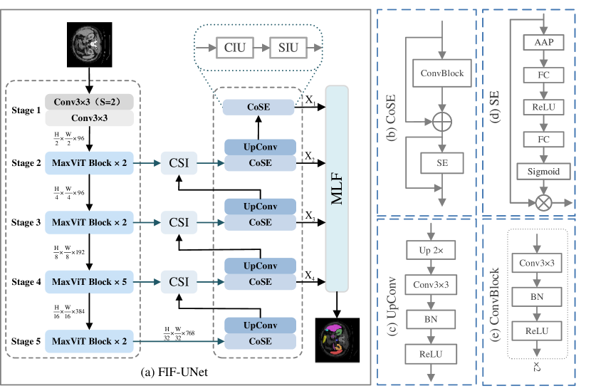
The architecture of the FIF-UNet is shown in Figure 1(a), implemented by a U-shaped architecture with symmetric encoder-decoder modules. In the encoder, MaxVit-S (Tu et al. (2022)) serves as the backbone network, which is pre-trained on the ImageNet dataset utilizing an image classification task. Compared to full self-attention, the MaxViT is implemented based on blocked local and dilated global attention to capture both the local and global features, which can be calculated by only linear complexity (, is the spatial size of an input image). The encoder network consists of 5 stages, including a stem stage and four cascaded MaxViT stages. In the stem stage, two convolution layers are with 96 channels and a kernel size of 3. The stride of the first CNN layer is set to 2 to downsample the input image resolution. The configurations of the MaxViT are with the blocks and generate the feature maps with channels, respectively.
In the skip connections, a CSI module is proposed to dynamically recalibrate the feature maps by the designed CIU and SIU, with the objective of obtaining the informative target features. In the decoder network, each decoder stage is constructed based on the CoSE module and UpConv module. The proposed CoSE module aims to enhance the representation of critical features by incorporating the SENet mechanism into CNNs. The UpConv module upsamples the resolution of the CoSE outputs by bilinear interpolation, followed by a convolution layer to refine the up-sampled feature maps, as in Figure 1(c). Instead of predicting the segmentation tasks based only on the last decoder stage, in this work, an MLF module is innovatively proposed to effectively fuse the outputs of the decoder stages to enhance the segmentation details by integrating intra- and inter-class features.
3.2 CSI Module
In general, the CSI module is based on the sequential channel interaction unit (CIU) and spatial interaction unit (SIU), as in Figure 1(a). The CSI module is performed on the skip connections of the UNet architecture, by taking feature maps of encoder stage () and decoder stage as inputs. The purpose of the design is listed below:
-
•
The CIU focuses on achieving semantic alignment between the encoder and decoder features along the channel dimension by interactively adjusting the channel weights.
-
•
The SIU is designed to capture spatial correlations among pixels, which is expected to enable semantic complementation to support feature learning.
Finally, the output of the CSI module is fed into the decoder stage . It is believed that the CSI can be integrated into any UNet architecture, enabling skip connections to effectively fuse different levels of semantics to obtain informative feature maps.
3.2.1 CIU
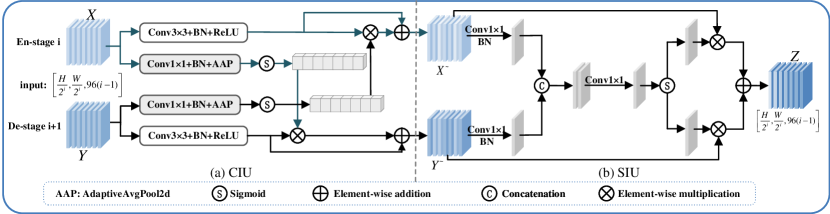
As mentioned before, the inputs of the CIU are the feature maps of encoder stage () and decoder stage (). The core idea of the CIU is to adjust the importance of input features ( or ) based on another counterpart feature map ( or ), implemented by parallel paths for the inputs separately (Figure 2(a)).
To be specific, for each path, the 1x1 convolution and batch normalization operations are first performed to integrate the global information along the channel dimension, followed by the adaptive average pooling (AAP) operation to generate feature weights. The correlation weights and are obtained by sigmoid activation functions, which indicate the importance of different feature channels. The above process can be denoted by:
| (1) |
| (2) |
where AAP denotes the adaptive average pooling operation, and denotes the sigmoid activation.
Similarly, for each path, the input feature maps are further recalibrated by a block of (CNN, BN, and ReLU), which is subsequently fused by the learned correlation weights to reweight the importance of each channel to obtain the interacted feature maps.
| (3) |
| (4) |
| (5) |
| (6) |
Finally, the residual mechanism is utilized to fuse the interacted feature maps while retaining the original feature inputs. The outputs of the CIU module are the feature maps and :
| (7) |
| (8) |
3.2.2 SIU
In general, the outputs of the CIU are fed into the SIU module to generate a fused feature map as the output of the CSI module. The SIU focuses on reweighting the importance of spatial pixels by an X-shaped path (as in Figure 2(b)), where all feature channels share a single weight matrix.
To be specific, for the left part of the X-shaped path, the 1×1 convolution and BN operations are utilized to squeeze the channels of feature maps ( and ) to 1, aiming to generate global contexts. The concatenation operation is applied to generate an initial weight matrix by fusing both the encoder and decoder features along the channel dimension, as shown below:
| (9) |
where Cat denotes the concatenation operation along the channel dimension.
In succession, the initial weight matrix is further recalibrated by the 1×1 convolution operation to generate the sample-dependent weights, as in:
| (10) |
In the right part of the X-shaped path, the sigmoid activation function is performed to project the weight elements to , which is further performed on the SIU inputs to reweight their pixel-wise importance. Finally, the output feature map is formulated by addition operations, as in:
| (11) |
In summary, the interaction and fusion operations consider the correlation among pixels in input feature maps and reassign their importance weights to achieve semantic alignment and complementation. By cascading the CIU and SIU modules, the CSI module is expected to enhance the skip connections of the UNet architecture to deliver rich features to the decoder.
3.3 CoSE Module
In the original UNet, each decoder stage consists of only two groups of (CNN, batch normalization (BN), and rectified linear unit (ReLU) activation), as in Figure 1(e). The inference rules are illustrated below:
| (12) |
| (13) |
where denotes a convolution operation with a kernel size of K. BN and ReLU denote the batch normalization and rectified linear unit activation function, respectively.
For medical images, the target regions are usually intersected with complicated background organs, presenting similar textures and shapes. It is required to improve the representation of critical features and suppress the impact of irrelevant background features. To this end, in this work, the channel attention mechanism (SENet) is introduced to the original decoder stages to capture the interdependency among channels by integrating the information of channel context. The SENet can adaptively adjust the importance of each feature channel through learnable attention weights, thus highlighting the important regions and suppressing the background regions.
As shown in Figure 1(b), the CoSE module is constructed by a convolution block and a SENet, in which corresponding residual connections are added before the convolution block and the SENet, respectively. The residual connection directly applies the addition operation to transmit the learned features into deeper layers, which can effectively solve the gradient vanishing and explosion by providing additional paths to enhance the information propagation. The mentioned process can be expressed as the equations:
| (14) |
| (15) |
where SENet is implemented by referring to (Hu et al. (2018)).
3.4 MLF Module
In the proposed model, the pre-trained encoder is applied to extract the features from input images by reducing the feature resolution with higher channels, while the decoder generates the pixel-wise segmentation masks by recovering the resolution to the raw image. In the original UNet, the interpolation algorithm is applied to implement the up-sampling operation, limiting the feature interactions among different scales and losing key details in the segmentation results.
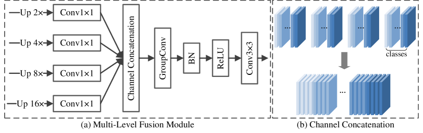
To address this issue, in this work, an MLF module is proposed to fuse the learned features in decoder stages with different scales, as in Figure 3. The primary purpose is to enhance feature details by interaction and fusion operations, which helps the model to better identify different regions with similar features. The output feature maps from decoder stages 1-4 serve as the MLF inputs, which are up-sampled by 2, 4, 8, and 16 to generate image size-liked masks. Then, the channel number of the up-sampled feature maps is mapped to the class number relevant to the task by a point-wise convolution. The inference rules are listed as:
| (16) |
where denotes the output of the decoder stage , and denotes up-sampling by a factor of . , where is the batch size, is the class number, and are the height and width of the original image, respectively. In this process, each feature channel is regarded as the feature representation for the corresponding class, which guides the model to capture discriminative features for certain classes of the segmentation targets.
To further optimize the feature representations, the corresponding channels of , , and are concatenated to combine the features of each class, as in Figure 3(b).
| (17) |
where ChannelCat denotes the channel concatenation operation.
Finally, a two-step convolution block is employed to integrate intra- and inter-class features to generate the segmentation mask.
-
•
Intra-class: the group convolution performs convolution operations on feature channels for each class separately by setting the group number to the class number, which generates the feature maps with channel 1 for each class.
-
•
Inter-class: the standard convolution is then used to integrate the learned intra-class features to obtain the final segmentation mask by fusing the intra-class features and inter-class features.
| (18) |
| (19) |
where , , denotes the group convolution with a kernel size of 3.
3.5 Loss Function
Based on the segmentation mask, the loss function of the FIF-UNet is obtained by a weighted Dice loss and the cross-entropy (CE) loss, which are designed for segmentation and classification tasks, as in:
| (20) |
where and are the weights for the Dice loss () and CE loss (), respectively.
To enhance the model convergence, the multi-stage feature mixing loss aggregation (MUTATION) method proposed by MERIT (Rahman and Marculescu (2024)) is introduced in this work. To be specific, for the feature maps (, , , ) from the MLF module, a total of 15 () nonempty subsets are first obtained, based on which 15 predicted masks are generated by element-wise addition on the feature maps in each set. Consequently, for each mask in the 15 predictions, the in Equation 20 is calculated over the ground truth to measure multi-stage prediction errors. In addition, the loss between the MLF output and the ground truth is also considered to formulate the final loss, as in:
| (21) |
| (22) |
| (23) |
where denotes a function that takes nonempty subsets of a list. denotes the element number in each subset.
4 Experiments and results
4.1 Datasets and evaluation metrics
Synapse multi-organ segmentation dataset: 30 abdominal CT scans are included in the Synapse (Landman et al. (2015)), with a total of 3779 axial contrast-enhanced abdominal CT images, which is provided by the MICCAI 2015 Multi-Atlas Abdomen Labeling Challenge. Each CT scan consists of 85-198 slices of 512 × 512 pixels, and the voxel spatial resolution is ([0:54-0:54] × [0:98-0:98] × [2:5-5:0]). Following TransUNet (Chen et al. (2021)), the dataset is randomly divided into 18 scans (2212 axial slices) for training, and 12 for validation. A total of 8 anatomical structures are segmented, including the aorta, gallbladder, left kidney, right kidney, liver, pancreas, spleen, and stomach. DICE scores and 95% Hausdorff Distance (95HD) are used as the evaluation metrics in the experiments on this dataset.
| Methods | Average | Aorta | GB | KL | KR | Liver | PC | SP | SM | ||
| DICE↑ | HD95↓ | ||||||||||
| CNN | UNet | 70.11 | 44.69 | 84.00 | 56.70 | 72.41 | 62.64 | 86.98 | 48.73 | 81.48 | 67.96 |
| AttnUNet | 71.70 | 34.47 | 82.61 | 61.94 | 76.07 | 70.42 | 87.54 | 46.70 | 80.67 | 67.66 | |
| ViT | SwinUNet | 79.13 | 21.55 | 85.47 | 66.53 | 83.28 | 79.61 | 94.29 | 56.58 | 90.66 | 76.60 |
| TransDeepLab | 80.16 | 21.25 | 86.04 | 69.16 | 84.08 | 79.88 | 93.53 | 61.19 | 89.00 | 78.40 | |
| MISSFormer | 81.96 | 18.20 | 86.99 | 68.65 | 85.21 | 82.00 | 94.41 | 65.67 | 91.92 | 80.81 | |
| Hybrid | TransUNet | 77.48 | 31.69 | 87.23 | 63.13 | 81.87 | 77.02 | 94.08 | 55.86 | 85.08 | 75.62 |
| SSFormerPVT | 78.01 | 25.68 | 82.78 | 63.74 | 80.72 | 78.11 | 93.53 | 61.53 | 87.07 | 76.61 | |
| PolypPVT | 78.08 | 25.61 | 82.34 | 66.14 | 81.21 | 73.78 | 94.37 | 59.34 | 88.05 | 79.40 | |
| MT-UNet | 78.59 | 26.59 | 87.92 | 64.99 | 81.47 | 77.29 | 93.06 | 59.46 | 87.75 | 76.81 | |
| HiFormer | 80.29 | 18.85 | 85.63 | 73.29 | 82.39 | 64.84 | 94.22 | 60.84 | 91.03 | 78.07 | |
| PVT-CASCADE | 81.06 | 20.23 | 83.01 | 70.59 | 82.23 | 80.37 | 94.08 | 64.43 | 90.10 | 83.69 | |
| CASTformer | 82.55 | 22.73 | 89.05 | 67.48 | 86.05 | 82.17 | 95.61 | 67.49 | 91.00 | 81.55 | |
| TransCASCADE | 82.68 | 17.34 | 86.63 | 68.48 | 87.66 | 84.56 | 94.43 | 65.33 | 90.79 | 83.52 | |
| Cascaded MERIT | 84.90 | 13.22 | 87.71 | 74.40 | 87.79 | 84.85 | 95.26 | 71.81 | 92.01 | 85.38 | |
| Ours | Tiny FIF-UNet | 84.40 | 19.89 | 89.19 | 74.56 | 85.22 | 81.39 | 95.19 | 70.69 | 92.11 | 86.42 |
| Small FIF-UNet | 86.05 | 15.82 | 89.49 | 76.15 | 88.23 | 86.26 | 95.87 | 74.14 | 91.31 | 86.97 | |
Automated cardiac diagnosis challenge: The ACDC dataset (Bernard et al. (2018)) consists of 100 cardiac MRI scans collected from different patients, provided by the MICCAI ACDC challenge 2017. Each scan contains three organs: the right ventricle, left ventricle, and myocardium. Following TransUNet (Chen et al. (2021)), 70 cases (1930 axial slices) are used for training, 10 for validation, and 20 for testing. DICE score serves as the evaluation metric in the experiments on this dataset.
4.2 Implementation details
The pre-trained MaxViT from (Tu et al. (2022)) serves as the encoder of the proposed model, with the input resolution of 256×256 and attention window size of 7×7. To consider the final performance, both the small and tiny MaxViT architectures are applied to conduct the experiments, i.e., Small FIF-UNet and Tiny FIF-UNet. To enhance the diversity of the training samples, random rotation, and flipping strategies are performed to augment the raw data (Chen et al. (2021)). The model is trained using AdamW (Loshchilov and Hutter (2017)) optimizer with the learning rate of 1e-4 for 400 epochs, applying the weight decay of 1e-4. The batch size of 16 is used for Synapse and ACDC. Following (Tu et al. (2022)), the loss weights and are set to 0.7 and 0.3, respectively. The proposed model is implemented using Pytorch 2.2.2 and all experiments are conducted on a single NVIDIA TITAN RTX GPU with 24GB of memory.
4.3 Results and Discussions
4.3.1 Experimental results on Synapse dataset
The experimental results on the Synapse multi-organ dataset are reported in Table 1, including the proposed model and other selective baselines. A total of 14 comparative baselines are selected to evaluate the model performance, as in three categories, CNN-based models (i.e., UNet (Ronneberger et al. (2015)), AttnUNet (Oktay et al. (2018))), ViT-based models (i.e., SwinUNet (Cao et al. (2022)), TransDeepLab (Azad et al. (2022a)), MISSFormer (Huang et al. (2021))) and hybrid CNN-Transformer models (i.e., TransUNet (Chen et al. (2021)), SSFormerPVT (Wang et al. (2022)), PolypPVT (Dong et al. (2021)), MT-UNet (Wang et al. (2022)), HiFormer (Heidari et al. (2023)), PVT-CASCADE (Rahman and Marculescu (2023)), CASTformer (You et al. (2022)), TransCASCADE (Rahman and Marculescu (2023)), Cascaded MERIT (Rahman and Marculescu (2024))).
As shown in Table 1, the average DICE and HD95 are reported to compare the model performance, as well as the DICE score for the certain 8 classes. In general, the Small FIF-UNet achieves the highest average DICE of 86.05% (primary metric), which significantly outperforms all the selective baselines (1.15% absolute improvement over the best baseline). Specifically, the Small FIF-UNet has the ability to harvest the best performance for 7/8 classes, confirming the performance superiority over baselines. Meanwhile, the proposed model also obtains the second-best HD95 measurement.
Compared to classical ViT-based SwinUNet and hybrid TransUNet, the Small FIF-UNet improves the performance in terms of average DICE by 6.92% and 8.57%, respectively. More importantly, the proposed FIF-UNet achieves significant performance improvements in both small and hard-to-segment organs. Specifically, the proposed model improves by 0.44% and 1.41% for small organs (KL and KR) over the best baseline, respectively, while improving by 1.75% and 2.33% for hard-to-segment organs (GB and PC).
In addition, we also consider the model complexity. Compared to the two competitive baselines TransCASCADE and Cascaded MERIT, the Small FIF-UNet can achieve the best performance with only 86.91M parameters (36.56M and 60.95M lower than baselines). It is worth noting that the proposed Tiny FIF-UNet achieves the third-best performance of 84.4% average DICE among baselines, comparable to the second-best Cascaded MERIT, with only 38.31M trainable parameters (109.55M lower).
In summary, the experimental results demonstrate that the proposed model achieves both performance and efficiency superiority over selected baselines on the Synapse dataset, which can also harvest desired enhancement on small and hard-to-segment organs.
4.3.2 Experimental results on ACDC dataset
| Methods | Avg DICE↑ | RV | Myo | LV | |
| CNN | R50 AttnUNet | 86.75 | 87.58 | 79.20 | 93.47 |
| R50 UNet | 87.55 | 87.10 | 80.63 | 94.92 | |
| ViT | SwinUNet | 90.00 | 88.55 | 85.62 | 95.83 |
| MISSFormer | 90.86 | 89.55 | 88.04 | 94.99 | |
| Hybrid | TransUNet | 89.71 | 88.86 | 84.53 | 95.73 |
| MT-UNet | 90.43 | 86.64 | 89.04 | 95.62 | |
| PVT-CASCADE | 91.46 | 88.90 | 89.97 | 95.50 | |
| TransCASCADE | 91.63 | 89.14 | 90.25 | 95.50 | |
| Cascaded MERIT | 91.85 | 90.23 | 89.53 | 95.80 | |
| Parallel MERIT | 92.32 | 90.87 | 90.00 | 96.08 | |
| Ours | Tiny FIF-UNet | 92.37 | 91.04 | 90.10 | 95.96 |
| Small FIF-UNet | 92.58 | 91.30 | 90.24 | 96.19 |
The experimental results of comparative methods on the ACDC dataset are reported in Table 2, in terms of the average DICE score. Similarly, three categories of models are selected as the comparative baselines, including CNN-based models(i.e., R50 UNet (Chen et al. (2021)), R50 AttnUNet (Chen et al. (2021))), ViT-based models (i.e., SwinUNet (Cao et al. (2022)), MISSFormer (Huang et al. (2021))) and hybrid CNN-Transformer models (i.e., TransUNet (Chen et al. (2021)), MT-UNet (Wang et al. (2022)), PVT-CASCADE (Rahman and Marculescu (2023)), TransCASCADE (Rahman and Marculescu (2023)), Parallel MERIT (Rahman and Marculescu (2024)), Cascaded MERIT (Rahman and Marculescu (2024))).
As shown in Table 2, the proposed Small FIF-UNet outperforms recent state-of-the-art (SOTA) methods with an average DICE score of 92.58%, over the best baseline Parallel MERIT (92.32%). Specifically, the Small FIF-UNet achieves the best DICE score in RV (91.30%) and LV (96.19%) segmentation and the second best DICE score in Myo (90.24%, only 0.01% inferior) segmentation. In addition, the proposed Tiny FIF-UNet achieves the second-best results (92.37%), while the number of parameters is 109.55M lower than the previous best result.
Notably, the proposed FIF-UNet employs the same pre-trained encoder with Parallel MERIT, our model has the ability to achieve higher performance for all three organs. This can be attributed that the proposed technical modules on both the decoder and skip connections enhance the model performance. In summary, the results not only confirm the effectiveness and efficiency of the proposed model but also support our motivations to improve the skip connections and decoder.
4.4 Ablation studies
| CoSE | CSI | MLF | Average | Aorta | GB | KL | KR | Liver | PC | SP | SM | |
| DICE↑ | HD95↓ | |||||||||||
| 84.57 | 18.58 | 88.61 | 75.78 | 84.87 | 82.74 | 95.31 | 72.60 | 91.81 | 84.81 | |||
| ✓ | 85.47 | 19.50 | 89.28 | 74.83 | 86.86 | 85.12 | 95.05 | 75.04 | 91.46 | 86.11 | ||
| ✓ | 85.46 | 15.37 | 90.06 | 73.51 | 88.31 | 85.36 | 95.30 | 73.45 | 91.12 | 86.55 | ||
| ✓ | 85.45 | 12.95 | 89.07 | 76.80 | 88.80 | 85.73 | 95.14 | 72.62 | 90.73 | 84.68 | ||
| ✓ | ✓ | 85.92 | 15.33 | 89.27 | 76.74 | 87.43 | 86.05 | 95.57 | 75.02 | 92.06 | 85.25 | |
| ✓ | ✓ | 85.57 | 14.08 | 88.87 | 74.47 | 88.71 | 85.36 | 95.40 | 73.57 | 93.02 | 85.13 | |
| ✓ | ✓ | 85.90 | 11.34 | 88.50 | 74.41 | 88.62 | 86.39 | 95.76 | 72.66 | 93.86 | 87.04 | |
| ✓ | ✓ | ✓ | 86.05 | 15.82 | 89.49 | 76.15 | 88.23 | 86.26 | 95.87 | 74.14 | 91.31 | 86.97 |
To confirm the effectiveness of the three proposed modules, the Synapse dataset is selected to conduct ablation experiments, in which we also consider the model performance by utilizing the pre-trained encoder. The experimental design for ablation studies concerns the employment of the proposed technical modules separately or their combinations with the baseline. The experimental results are listed in Table 3 in terms of concerned metrics. It can be found that all the proposed technical modules contribute to expected performance improvements. To be specific, by incorporating each module separately into the baseline model, the performance is improved by 0.9%, 0.89% and 0.88% in DICE score, respectively. In addition, the combinations of each two modules further enhance the segmentation performance with absolute improvements of 1.35%, 1.00% and 1.33%. Finally, the proposed FIF-UNet with all three modules harvests a 1.48% improvement over the baseline.
4.5 Generalization studies
| CoSE | CSI | MLF | Average | Aorta | GB | KL | KR | Liver | PC | SP | SM | |
| DICE↑ | HD95↓ | |||||||||||
| 75.65 | 40.32 | 85.74 | 64.75 | 80.07 | 70.04 | 91.63 | 60.92 | 86.59 | 65.47 | |||
| ✓ | 77.91 | 36.62 | 86.24 | 65.13 | 78.44 | 75.44 | 94.11 | 59.06 | 86.88 | 77.95 | ||
| ✓ | 77.64 | 42.18 | 88.59 | 64.86 | 79.70 | 75.90 | 92.00 | 60.84 | 87.93 | 71.30 | ||
| ✓ | 77.85 | 29.66 | 87.61 | 71.11 | 82.89 | 70.60 | 94.12 | 52.77 | 88.39 | 75.28 | ||
| ✓ | ✓ | 79.12 | 31.00 | 89.63 | 69.25 | 84.60 | 77.02 | 93.00 | 53.34 | 89.10 | 76.99 | |
| ✓ | ✓ | 78.35 | 23.77 | 86.64 | 68.28 | 85.35 | 78.92 | 93.83 | 54.67 | 87.95 | 71.15 | |
| ✓ | ✓ | 78.45 | 40.91 | 87.94 | 66.66 | 82.96 | 78.37 | 93.20 | 57.89 | 85.00 | 75.58 | |
| ✓ | ✓ | ✓ | 79.57 | 25.57 | 86.13 | 72.90 | 85.86 | 78.32 | 93.07 | 62.66 | 86.15 | 71.47 |
To evaluate the generalization capability of the proposed modules, the original UNet is selected as the baseline to conduct the generalization experiments. Similar to the ablation studies, the experimental design concerns the employment of the proposed technical modules separately or their combinations with the baseline to comprehensively evaluate the efficacy and applicability of the modules. As shown in Table 4, it can be seen that all the proposed technical modules provide desired performance improvements compared to the UNet baseline. Note that, instead of 70.11%, our re-implementation result of the UNet is 75.65% in Table 4 due to the changed experimental configurations. To be specific, the CoSE, CSI, and MLF modules improve the DICE score with 2.26%, 1.99% and 2.20%, respectively. The DICE score is improved by 3.47%, 2.70% and 2.80% by employing every two modules in UNet. Finally, the UNet yields a performance improvement of 3.92% by using all the proposed modules.
In summary, the above experimental results show that all the proposed modules contribute to performance improvement even with different backbones, presenting expected generalization ability with consistent performance. Most importantly, we can also observe that the proposed modules have the ability to obtain higher performance improvements for simpler model architecture (i.e., original UNet).
4.6 Visualization results
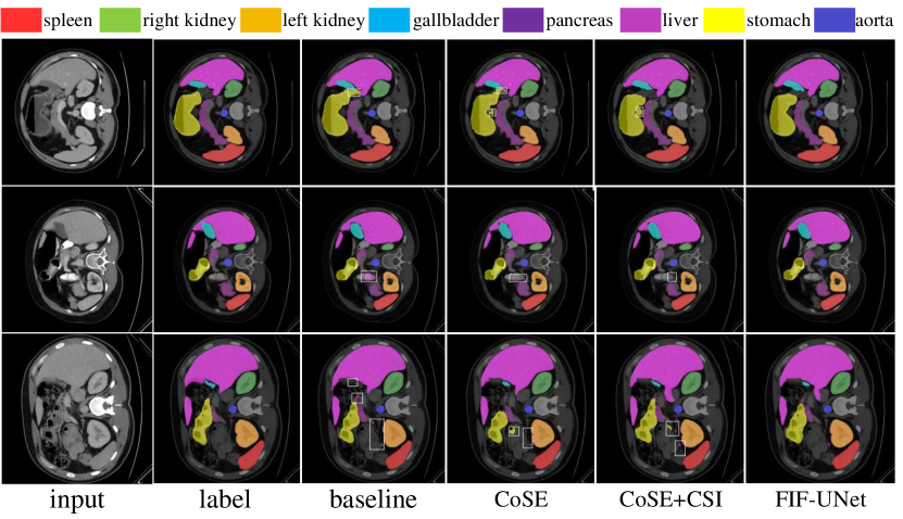
To better understand the proposed model, visualization results of ablation studies are provided in Figure 4. According to the results in Table 3, for the single module and the combination of two modules, the models with the highest DICE are selected for visualization analysis. Compared with the baseline model, the CoSE in the decoder considerably enhances the model to recognize foreground and background features, resulting in significantly reduced error regions. In particular, for the third row, the model successfully segmented the gallbladder objects, while the baseline model fails to identify them. With the combination of the CSI module, the approximate shape of each organ can be localized, suggesting that the model can accurately understand the structural characteristics of organs by obtaining informative features based on the different levels of semantic information between the encoder and the decoder. Compared with the previous model, the FIF-UNet has the ability to accurately capture the spatial distribution and morphological details of each organ by integrating semantic information of different scales, benefiting to reduce the misidentified scattering areas in the segmentation results.
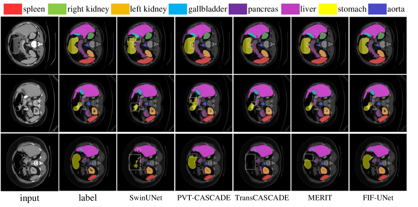
In addition, the classical SwinUNet and three recent models (PVT-CASCADE, TransCASCADE, and Cascaded MERIT) are also selected to compare the visualization results with the proposed FIF-UNet. As shown in Figure 5, compared with the selected models, the proposed FIF-UNet is capable of accurately locating all organs with precise details. For the segmentation of the stomach, the selected baselines suffer from task challenges due to its high similarity with the background, i.e., can only partially identify the organ, or even fail to detect them. Fortunately, the proposed FIF-UNet model demonstrates significant advantages in accurately segmenting the entire stomach and greatly reducing the risk of misidentifying other regions as the stomach. Furthermore, in the case of the second row, it can also be observed that compared to recent models, only the FIF-UNet can identify the hollow region in the middle of the left kidney, illustrating its advantages in capturing complex structural details. From the above analysis, except for the quantitative metrics, more qualitative results also support the performance improvements over baselines.
5 Conclusion
In this work, a novel model called FIF-UNet is proposed to achieve accurate medical image segmentation, which effectively leverages the semantic information of both the encoder and decoder stages by feature interaction and fusion. Three modules, CoSE, CSI and MLF, are designed for the FIF-UNet to enhance the skip connections and decoder. Experimental results on two public datasets demonstrate that the proposed model outperforms SOTA methods in terms of certain metrics. In addition, the proposed three modules contribute to desired performance improvements. Most importantly, they can also be adapted to other similar architectures to improve the overall performance, indicating the desired generalization ability in the segmentation tasks.
In the future, we will attempt to apply the proposed model to other modalities of medical images to construct a generalized model for medical image segmentation.
Acknowledgements
This work is supported by the National Natural Science Foundation of China under Grant U2333209, 62371323.
References
- Azad et al. (2022a) Azad, R., Aghdam, E.K., Rauland, A., Jia, Y., Avval, A.H., Bozorgpour, A., Karimijafarbigloo, S., Cohen, J.P., Adeli, E., Merhof, D., 2022a. Medical image segmentation review: The success of u-net. arXiv:2211.14830 .
- Azad et al. (2022b) Azad, R., Heidari, M., Wu, Y., Merhof, D., 2022b. Contextual attention network: Transformer meets u-net, in: International Workshop on Machine Learning in Medical Imaging, pp. 377–386.
- Bernard et al. (2018) Bernard, O., Lalande, A., Zotti, C., Cervenansky, F., Yang, X., Heng, P.A., Cetin, I., Lekadir, K., Camara, O., Ballester, M.A.G., others., 2018. Deep learning techniques for automatic mri cardiac multi-structures segmentation and diagnosis: is the problem solved? IEEE transactions on medical imaging 37, 2514–2525.
- Cao et al. (2022) Cao, H., Wang, Y., Chen, J., Jiang, D., Zhang, X., Tian, Q., Wang, M., 2022. Swin-unet: Unet-like pure transformer for medical image segmentation, in: European conference on computer vision, pp. 205–218.
- Chen et al. (2021) Chen, J., Lu, Y., Yu, Q., Luo, X., Adeli, E., Wang, Y., Lu, L., Yuille, A.L., Zhou, Y., 2021. Transunet: Transformers make strong encoders for medical image segmentation. arXiv:2102.04306 .
- Dong et al. (2021) Dong, B., Wang, W., Fan, D.P., Li, J., Fu, H., Shao, L., 2021. Polyp-pvt: Polyp segmentation with pyramid vision transformers. arXiv:2108.06932 .
- Dosovitskiy et al. (2020) Dosovitskiy, A., Beyer, L., Kolesnikov, A., Weissenborn, D., Zhai, X., Unterthiner, T., Dehghani, M., Minderer, M., Heigold, G., Gelly, S., others., 2020. An image is worth 16x16 words: Transformers for image recognition at scale. arXiv:2010.11929 .
- Hatamizadeh et al. (2022) Hatamizadeh, A., Tang, Y., Nath, V., Yang, D., Myronenko, A., Landman, B., Roth, H.R., Xu, D., 2022. Unetr: Transformers for 3d medical image segmentation, in: Proceedings of the IEEE/CVF winter conference on applications of computer vision, pp. 574–584.
- Heidari et al. (2023) Heidari, M., Kazerouni, A., Soltany, M., Azad, R., Aghdam, E.K., Cohen-Adad, J., Merhof, D., 2023. Hiformer: Hierarchical multi-scale representations using transformers for medical image segmentation, in: Proceedings of the IEEE/CVF winter conference on applications of computer vision, pp. 6202–6212.
- Hu et al. (2018) Hu, J., Shen, L., Sun, G., 2018. Squeeze-and-excitation networks, in: Proceedings of the IEEE conference on computer vision and pattern recognition, pp. 7132–7141.
- Huang et al. (2021) Huang, X., Deng, Z., Li, D., Yuan, X., 2021. Missformer: An effective medical image segmentation transformer. arXiv:2109.07162 .
- Jin et al. (2019) Jin, Q., Meng, Z., Pham, T.D., Chen, Q., Wei, L., Su, R., 2019. Dunet: A deformable network for retinal vessel segmentation. Knowledge-Based Systems 178, 149–162.
- Landman et al. (2015) Landman, B., Xu, Z., Igelsias, J., Styner, M., Langerak, T., Klein, A., 2015. Miccai multi-atlas labeling beyond the cranial vault–workshop and challenge, in: Proc. MICCAI Multi-Atlas Labeling Beyond Cranial Vault—Workshop Challenge, p. 12.
- Li et al. (2024) Li, Z., Lyu, H., Wang, J., 2024. Fusionu-net: U-net with enhanced skip connection for pathology image segmentation, in: Asian Conference on Machine Learning, pp. 694–706.
- Liu et al. (2022) Liu, Z., Mao, H., Wu, C.Y., Feichtenhofer, C., Darrell, T., Xie, S., 2022. A convnet for the 2020s, in: Proceedings of the IEEE/CVF conference on computer vision and pattern recognition, pp. 11976–11986.
- Loshchilov and Hutter (2017) Loshchilov, I., Hutter, F., 2017. Decoupled weight decay regularization. arXiv:1711.05101 .
- Oktay et al. (2018) Oktay, O., Schlemper, J., Folgoc, L.L., Lee, M., Heinrich, M., Misawa, K., Mori, K., McDonagh, S., Hammerla, N.Y., Kainz, B., others., 2018. Attention u-net: Learning where to look for the pancreas. arXiv:1804.03999 .
- Punn and Agarwal (2020) Punn, N.S., Agarwal, S., 2020. Inception u-net architecture for semantic segmentation to identify nuclei in microscopy cell images. ACM Transactions on Multimedia Computing, Communications, and Applications (TOMM) 16, 1–15.
- Rahman and Marculescu (2023) Rahman, M.M., Marculescu, R., 2023. Medical image segmentation via cascaded attention decoding, in: Proceedings of the IEEE/CVF Winter Conference on Applications of Computer Vision, pp. 6222–6231.
- Rahman and Marculescu (2024) Rahman, M.M., Marculescu, R., 2024. Multi-scale hierarchical vision transformer with cascaded attention decoding for medical image segmentation, in: Medical Imaging with Deep Learning, pp. 1526–1544.
- Ronneberger et al. (2015) Ronneberger, O., Fischer, P., Brox, T., 2015. U-net: Convolutional networks for biomedical image segmentation, in: Medical image computing and computer-assisted intervention–MICCAI 2015: 18th international conference, Munich, Germany, October 5-9, 2015, proceedings, part III 18, pp. 234–241.
- Ruan et al. (2023) Ruan, J., Xie, M., Gao, J., Liu, T., Fu, Y., 2023. Ege-unet: an efficient group enhanced unet for skin lesion segmentation, in: International conference on medical image computing and computer-assisted intervention, pp. 481–490.
- Tan and Le (2019) Tan, M., Le, Q., 2019. Efficientnet: Rethinking model scaling for convolutional neural networks, in: International conference on machine learning, pp. 6105–6114.
- Tragakis et al. (2023) Tragakis, A., Kaul, C., Murray-Smith, R., Husmeier, D., 2023. The fully convolutional transformer for medical image segmentation, in: Proceedings of the IEEE/CVF Winter Conference on Applications of Computer Vision, pp. 3660–3669.
- Tu et al. (2022) Tu, Z., Talebi, H., Zhang, H., Yang, F., Milanfar, P., Bovik, A., Li, Y., 2022. Maxvit: Multi-axis vision transformer, in: European conference on computer vision, pp. 459–479.
- Valanarasu et al. (2021) Valanarasu, J.M.J., Oza, P., Hacihaliloglu, I., Patel, V.M., 2021. Medical transformer: Gated axial-attention for medical image segmentation, in: Medical image computing and computer assisted intervention–MICCAI 2021: 24th international conference, Strasbourg, France, September 27–October 1, 2021, proceedings, part I 24, pp. 36–46.
- Vaswani et al. (2017) Vaswani, A., Shazeer, N., Parmar, N., Uszkoreit, J., Jones, L., Gomez, A.N., Kaiser, Ł., Polosukhin, I., 2017. Attention is all you need. Advances in neural information processing systems 30.
- Wang et al. (2022) Wang, H., Cao, P., Wang, J., Zaiane, O.R., 2022. Uctransnet: rethinking the skip connections in u-net from a channel-wise perspective with transformer, in: Proceedings of the AAAI conference on artificial intelligence, pp. 2441–2449.
- Wang et al. (2021) Wang, W., Chen, C., Ding, M., Yu, H., Zha, S., Li, J., 2021. Transbts: Multimodal brain tumor segmentation using transformer, in: International Conference on Medical Image Computing and Computer-Assisted Intervention, Springer, pp. 109–119.
- Wu et al. (2023) Wu, Y., Liao, K., Chen, J., Wang, J., Chen, D.Z., Gao, H., Wu, J., 2023. D-former: A u-shaped dilated transformer for 3d medical image segmentation. Neural Computing and Applications 35, 1931–1944.
- You et al. (2022) You, C., Zhao, R., Liu, F., Dong, S., Chinchali, S., Topcu, U., Staib, L., Duncan, J., 2022. Class-aware adversarial transformers for medical image segmentation. Advances in Neural Information Processing Systems 35, 29582–29596.
- Zhou et al. (2021) Zhou, D., Kang, B., Jin, X., Yang, L., Lian, X., Jiang, Z., Hou, Q., Feng, J., 2021. Deepvit: Towards deeper vision transformer. arXiv:2103.11886 .
- Zhou et al. (2019) Zhou, Z., Siddiquee, M.M.R., Tajbakhsh, N., Liang, J., 2019. Unet++: Redesigning skip connections to exploit multiscale features in image segmentation. IEEE transactions on medical imaging 39, 1856–1867.