11email: z.alhajhemidi@uni-luebeck.de
https://github.com/MDL-UzL/MICCAI24_IMMoCo.git
IM-MoCo: Self-supervised MRI Motion Correction using Motion-Guided Implicit Neural Representations
Abstract
Motion artifacts in Magnetic Resonance Imaging (MRI) arise due to relatively long acquisition times and can compromise the clinical utility of acquired images. Traditional motion correction methods often fail to address severe motion, leading to distorted and unreliable results. Deep Learning (DL) alleviated such pitfalls through generalization with the cost of vanishing structures and hallucinations, making it challenging to apply in the medical field where hallucinated structures can tremendously impact the diagnostic outcome. In this work, we present an instance-wise motion correction pipeline that leverages motion-guided Implicit Neural Representations (INRs) to mitigate the impact of motion artifacts while retaining anatomical structure. Our method is evaluated using the NYU fastMRI dataset with different degrees of simulated motion severity. For the correction alone, we can improve over state-of-the-art image reconstruction methods by SSIM, PSNR, and HaarPSI. Clinical relevance is demonstrated by a subsequent experiment, where our method improves classification outcomes by at least accuracy percentage points compared to motion-corrupted images.
Keywords:
Motion Correction Reconstruction Implicit Neural Representations Magnetic Resonance Imaging1 Introduction
Magnetic Resonance Imaging (MRI) is a non-invasive imaging technique providing detailed images of soft-tissue structures. However, common artifacts, particularly motion-related ones, degrade image quality, impacting clinical diagnosis and leading to high clinical costs [23].
Various retrospective strategies for motion correction (MoCo) have been developed [4, 24]. Retrospective correction handles motion artifacts post-acquisition, offering flexibility without requiring scanner modifications. This approach benefits from prior knowledge such as from navigators [7], and from the imaged object itself in so-called autofocus [3] or in data-consistency (DC) [9] methods. Unfortunately, these methods face challenges in solving poorly conditioned, non-convex optimization problems with high computational complexity. Image-based deep learning (DL) models, like convolutional neural networks (CNNs) [1, 14] and generative adversarial networks [2], have been employed for direct motion-refined image estimation to overcome the challenges mentioned earlier. While promising, image-based DL-based methods can be unstable, struggle with pathology preservation, and introduce undesired alterations or hallucinations [4], raising concerns about their suitability for clinical applications. Specific approaches break down the problem into intermediate steps to address instability, maintain DC, and alleviate network hallucinations. For instance, CNNs are employed to identify subsets of k-space lines affected by motion, which can either be excluded [21] or down-weighted [5] in the context of a DC-based reconstruction. However, these methods do not explicitly model motion (transformations) during reconstruction, potentially compromising quality with many motion-corrupted lines. Efforts to improve efficiency integrate image-based MoCo CNNs into DC-based [8] or autofocus [15] iterative algorithms. Alternatively, they replace the optimization-based motion estimation with a DL network and incorporate this information into the reconstruction process [10]. However, they rely on supervised training and require large paired datasets, which are difficult to obtain in the medical domain. Recently, Implicit neural representations (INRs) were used to model images as a function, spurring significant interest in instance-optimization-based reconstruction [19]. INRs have proven successful in self-supervised MRI reconstruction, addressing issues present in prior methods [6, 11]. Despite this, the potential of INRs for MoCo remains untapped.
In this work, we introduce a novel MoCo pipeline utilizing INRs to address motion artifacts effectively while preserving anatomical structures in MRI scans. Our contributions are 1) Developing a motion-guided INR-based method called IM-MoCo, combining a k-space line motion detection network (lD-Net), an Image INR, and a motion-INR. 2) Demonstrating its efficacy on the NYU fastMRI dataset [13, 25] through significant reductions in simulated motion-induced distortions compared to competing methods. 3) Highlighting its potential by improving pathology classification of fastMRI+ annotations [26]. These findings position IM-MoCo as a promising advancement for enhancing MRI quality for scans with light and heavy motion corruption.
2 Materials & Methods
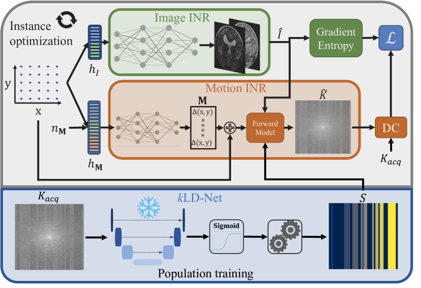
An overview of the proposed IM-MoCo pipeline is depicted in Fig. 1. Our pipeline comprises three steps: 1) Motion Detection with LD-Net, 2) Generation of motion-free intensity priors with an Image INR, and 3) Forward application of predicted motion from a Motion INR.
2.1 Physical Model & Motion Simulation
The physical forward model, , of a 2D rigid motion-induced MRI acquisition with and being the number of points in the frequency and phase encoding directions, respectively, and being the number of coils, can be described as follows:
| (1) |
where is the desired motion free image, is the motion transform at time point , are the coil sensitivity maps, is the Fourier transform, a sampling operator, and is the sampling mask at time point . The motion transform combines rotation and planar translations in and directions. The sampling mask is a binary mask indicating the -space lines acquired at time . Equation (1) describes motion in the image domain. However, the forward model can also be formulated in -space as it is mathematically equivalent [18]. Irrespective of the space in which motion is described, for the simulation of motion, it is crucial to synchronize the sampling of motion with the MR -space filling scheme to simulate realistic artifacts accurately. This work follows previously developed methods [16] and relies on the simulation of rigid-body motion in 2D single-coil cartesian sampled -spaces filled sequentially left-to-right.
2.2 IM-MoCo
2.2.1 Motion Detection.
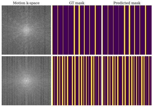
In the first step of our pipeline, we present a -space Line Detection Network [5, 21] (LD-Net) to detect motion-corrupted lines in raw -space data following a left-to-right sequential cartesian sampling scheme. The LD-Net, built on a U-Net [22], takes a complex-valued motion-corrupted -space as input, where the real and imaginary parts are concatenated in the channel dimension, and outputs a binary mask predicting the motion-corrupted lines denoted as in Eq. (1). The four layers of the U-Net consist of convolutions with a kernel size of , batch normalization, ReLU activation, and an average pooling layer with kernel size . The number of channels (starting from ) is doubled after each down-sampling operation and halved after each up-sampling operation. A final convolution outputs the predicted mask . The network is trained with the binary cross entropy loss with logits between the predicted and ground truth masks. During inference, the raw prediction of the network is activated with and thresholded () to yield a binary prediction mask. We consider a line corrupted if of its frequencies are classified as so, and we generate an index list, e.g., , where indicates a motion-corrupted line and a motion-free one. We then post-process it to yield a list of movement groups, where each group is a list of line indices for one movement, e.g., for the previously mentioned example. The following steps use the number of movements and corresponding lines to guide the motion correction process.
Motion Correction. We use from the LD-Net (step 1) in a MoCo model, which is a combination of an Image INR (step 2) that acts as an implicit image prior and a Motion INR (step 3) to search for motion that satisfies the forward model (see Sec. 2.1). We can rewrite the forward model from Eq. (1), for single-coil scans, as as follows:
| (2) |
where is the image INR with optimizable parameters , is an encoding of the 2D coordinate , is the motion INR with optimizable parameters , is an encoding of the 2D coordinate and , which is a sequence of linearly spaced numbers from to representing the number of movement groups. is the predicted motion mask at time . Each INR is built from MPLs taking encodings as input. To encode our data, we use hash grid encoding [20] for and . This encoding introduces learnable feature grids that enable a speed-up by replacing large MLPs with a look-up table of feature vectors and a much smaller MLP. The Image INR consists of three layers with channels and ReLU activations, while the Motion INR has three layers with channels and tanh activations. The predicts a 2-channel complex-valued intensity image, while predicts transformation grids for movements.
In each iteration of the pipeline, we optimize the and in an end-to-end fashion by feeding the encoding and the mask through the forward model which in term predicts a motion-free intensity image , applies the motion to the intensity image resulting in a motion corrupted image that is transferred to the -space domain by the Fourier transform yielding . Using the DC-loss in k-space, we measure the discrepancy to the acquired -space . We use the gradient entropy on for regularization in image-space. The motion correction loss is then defined as follows:
| (3) |
where is the number of data points, is the gradient operator, and is the regularization weight. The regularization is used to introduce image priors to the Image INR as a denoiser, forcing the INR to learn an artifact-free image. To ensure better motion estimates, high-frequencies are suppressed at the beginning of the optimization by setting a large weight and halving it every step after iterations [17]. The model is optimized until convergence, and the final motion-corrected image is the output of .
3 Experimental Results
3.1 Datasets & Motion Simulation
To evaluate our method, we use the MRI -weighted brain -space data from the open-source NYU fastMRI database [13, 25] in our first experiment. We employed 2D slices from different patients and split them into // for training/validation/testing. We cropped the images to and combined the coils. The second dataset contains -weighted and FLAIR brain fastMRI scans, which are part of the fastMRI+ [26] annotations and were used for the downstream classification task. We classify two pathologies, Nonspecific White Matter Lesion and Craniotomy, resulting in slices of subjects with a total of annotations. We split the slices into ( annotations) for training, ( annotations) for validation, and () for testing. For all experiments, two motion scenarios were simulated [16] with random rotations and translations between and in and , respectively. In the Light Motion scenario between and movements were introduced during simulation, whereas the Heavy Motion scenario contained between and movements.
3.2 Experimental Setup
Experiment I: Motion Correction in Simulated Data.
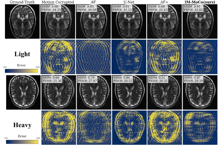
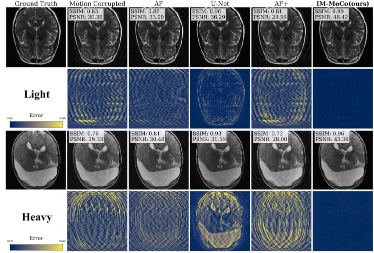
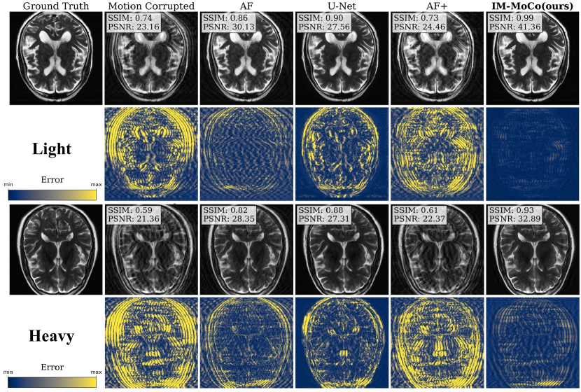
Here the image correction quality is assessed. We implemented the following methods: AF [3], U-Net [1], AF+ [15], IM-MoCo (ours). AF is an instance-wise autofocusing approach and optimizes motion parameters using Adam for iterations. The U-Net is a population-trained model, mapping motion-corrupted images to motion-free ones, for epochs using Adam with learning rate and an L1-SSIM loss. AF+ combines AF and the U-Net for deep image priors, trained with default settings using the available codebase [15]. IM-MoCo is optimized for iterations using Adam with learning rates of for the INRs and an initial , halved every step after iterations. All methods were evaluated on the -weighted corrupted test scans for the two motion scenarios. Results were evaluated using the structural similarity index (SSIM), peak-signal-to-noise-ratio (PSNR) and the Haar Perceptual Similarity Index (HaarPSI) [12].
Experiment II: Downstream Classification Task.
In this experiment, we demonstrate clinical relevance
with a classification of pathologies in motion and motion-corrected images. We extract patches of size around bounding boxes of the fastMRI+ dataset and normalized between . We used a pre-trained ResNet18 (PyTorch Hub222https://pytorch.org/hub/pytorch˙vision˙resnet/) backbone for feature extraction and added two trainable linear layers as the classification head. Using the SGD optimizer, we trained the model for epochs with a batch size of and a learning rate of . The trained model is then applied to the test set. We report the SSIM, PSNR, and HaarPSI for extracted patches around the bounding boxes and accuracy for the classification.
Implementation Details. The LD-Net was population-trained with Adam (learning rate of ) for epochs and a batch size of . We used PyTorch for implementations and an NVIDIA GeForce RTX 2080 TI with 12 GB of VRAM. The code is publicly available.
3.3 Results
Experiment I: Motion Correction in Simulated Data. Table 1 shows quantitative results from the motion correction experiment. For light and heavy motion, corrupted images yield SSIM values of and , PSNR values of and , and HaarPSI values of and , respectively. AF and U-Net improve SSIM by and , PSNR by and , and HaarPSI by and , respectively. However, AF+ worsens results by ca. across all metrics. Compared to the best baseline, AF, our method improves SSIM/HaarPSI by for SSIM, for HaarPSI, and for PSNR. A qualitative comparison is shown in Fig. 2.
Experiment II: Downstream Classification Task.
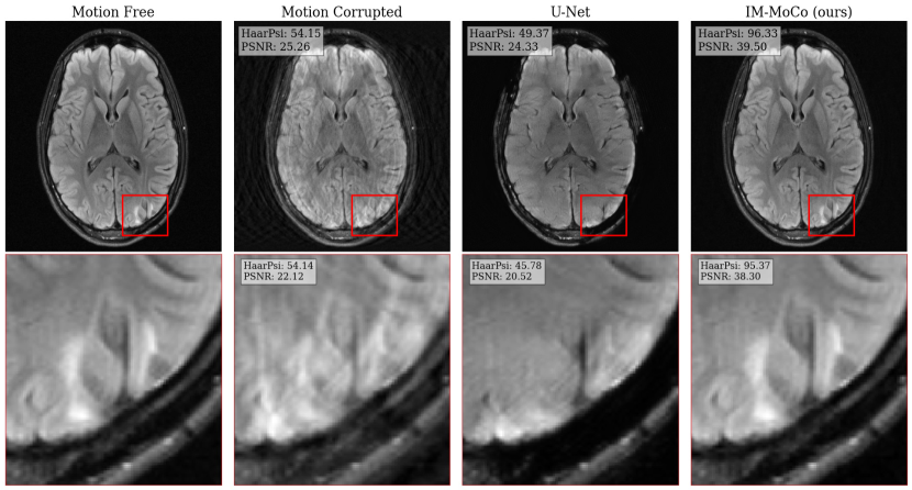
The results are summarized in Table 2. Motion-free images achieve an accuracy of . For corrupted images, the accuracy is for light and for heavy motion, which is due to image quality loss as reflected in SSIMs of and , respectively. The U-Net’s accuracy is for light and for heavy while its SSIMs are and . IM-MoCo achieves an accuracy of for light and for heavy motion, with the highest SSIMs of and , respectively.
4 Discussion & Outlook
Experiment I demonstrated that our method effectively enhanced the quality of motion-corrupted images, surpassing comparison methods, which is mainly contributed by the implicit image priors and DC with the acquired -space in the forward motion model. Notably, our approach exhibited robustness in addressing heavier motion scenarios as evidenced in Fig. 2 and Table 1. However, the efficacy of our method hinges on the accurate detection of the lD-Net, which is contingent upon the acquisition sequence and motion simulation pattern. In Experiment II, we showcased the performance enhancement of a classification task compared to both corrupted and U-Net-corrected images as indicated in Table 2. This improvement in classification accuracy can be attributed to the prevention of overfitting on "healthy" features in instance-optimization methods in contrast to population training ones like the U-Net. Although our work primarily revolved around 2D rigid motion, we are confident in the method’s potential for extension to more complex motion and multi-coil data. This can be achieved by incorporating coil sensitivity map estimation following the third step of the pipeline. Future investigations will explore the utilization of different sampling patterns, such as radial sampling, and the adaptation of the motion detection network to these patterns or pattern-independent methods. While real motion cases were not tested in this study, we are optimistic regarding the adaptability of our method through adjustments to the motion detection network.
5 Conclusion
We have introduced IM-MoCo, a pipeline that utilizes motion-guided INRs to mitigate the impact of motion artifacts in MRI scans. We tested our method on the fastMRI dataset and observed an improvement in image quality and pathology classification performance. Our results suggest that IM-MoCo is a promising solution for enhancing the quality of MRI scans in the presence of challenging motion artifacts. In the future, we plan to investigate the applicability of our method to real motion to validate its potential for clinical applications.
References
- [1] Al-Masni, M.A., Lee, S., Yi, J., Kim, S., Gho, S.M., Choi, Y.H., Kim, D.H.: Stacked U-Nets with self-assisted priors towards robust correction of rigid motion artifact in brain MRI. NeuroImage 259, 119411 (2022)
- [2] Armanious, K., Jiang, C., Fischer, M., Küstner, T., Hepp, T., Nikolaou, K., Gatidis, S., Yang, B.: MedGAN: Medical image translation using GANs. Computerized medical imaging and graphics 79, 101684 (2020)
- [3] Atkinson, D., Hill, D.L., Stoyle, P.N., Summers, P.E., Clare, S., Bowtell, R., Keevil, S.F.: Automatic compensation of motion artifacts in MRI. Magnetic Resonance in Medicine: An Official Journal of the International Society for Magnetic Resonance in Medicine 41(1), 163–170 (1999)
- [4] Chen, Z., Pawar, K., Ekanayake, M., Pain, C., Zhong, S., Egan, G.F.: Deep learning for image enhancement and correction in magnetic resonance imaging—state-of-the-art and challenges. Journal of Digital Imaging 36(1), 204–230 (2023)
- [5] Eichhorn, H., Hammernik, K., Spieker, V., Epp, S.M., Rueckert, D., Preibisch, C., Schnabel, J.A.: Physics-Aware Motion Simulation For T2*-Weighted Brain MRI. In: International Workshop on Simulation and Synthesis in Medical Imaging. pp. 42–52. Springer (2023)
- [6] Feng, R., Wu, Q., Feng, J., She, H., Liu, C., Zhang, Y., Wei, H.: IMJENSE: Scan-specific Implicit Representation for Joint Coil Sensitivity and Image Estimation in Parallel MRI. IEEE Transactions on Medical Imaging (2023). https://doi.org/10.1109/TMI.2023.3342156
- [7] Gallichan, D., Marques, J.P., Gruetter, R.: Retrospective correction of involuntary microscopic head movement using highly accelerated fat image navigators (3D FatNavs) at 7T. Magnetic resonance in medicine 75(3), 1030–1039 (2016)
- [8] Haskell, M.W., Cauley, S.F., Bilgic, B., Hossbach, J., Splitthoff, D.N., Pfeuffer, J., Setsompop, K., Wald, L.L.: Network accelerated motion estimation and reduction (NAMER): convolutional neural network guided retrospective motion correction using a separable motion model. Magnetic resonance in medicine 82(4), 1452–1461 (2019)
- [9] Haskell, M.W., Cauley, S.F., Wald, L.L.: TArgeted Motion Estimation and Reduction (TAMER): data consistency based motion mitigation for MRI using a reduced model joint optimization. IEEE transactions on medical imaging 37(5), 1253–1265 (2018)
- [10] Hossbach, J., Splitthoff, D.N., Cauley, S., Clifford, B., Polak, D., Lo, W.C., Meyer, H., Maier, A.: Deep learning-based motion quantification from k-space for fast model-based magnetic resonance imaging motion correction. Medical Physics 50(4), 2148–2161 (2023). https://doi.org/10.1002/mp.16119
- [11] Huang, W., Li, H.B., Pan, J., Cruz, G., Rueckert, D., Hammernik, K.: Neural Implicit k-Space for Binning-Free Non-Cartesian Cardiac MR Imaging. In: International Conference on Information Processing in Medical Imaging. pp. 548–560. Springer (2023)
- [12] Kastryulin, S., Zakirov, J., Pezzotti, N., Dylov, D.V.: Image Quality Assessment for Magnetic Resonance Imaging. IEEE Access 11, 14154–14168 (2023). https://doi.org/10.1109/access.2023.3243466
- [13] Knoll, F., Zbontar, J., Sriram, A., Muckley, M.J., Bruno, M., Defazio, A., Parente, M., Geras, K.J., Katsnelson, J., Chandarana, H., et al.: fastmri: A publicly available raw k-space and dicom dataset of knee images for accelerated mr image reconstruction using machine learning. Radiology: Artificial Intelligence 2(1), e190007 (2020)
- [14] Küstner, T., Armanious, K., Yang, J., Yang, B., Schick, F., Gatidis, S.: Retrospective correction of motion-affected MR images using deep learning frameworks. Magnetic resonance in medicine 82(4), 1527–1540 (2019)
- [15] Kuzmina, E., Razumov, A., Rogov, O.Y., Adalsteinsson, E., White, J., Dylov, D.V.: Autofocusing+: Noise-resilient motion correction in magnetic resonance imaging. In: International Conference on Medical Image Computing and Computer-Assisted Intervention. pp. 365–375. Springer (2022)
- [16] Lee, S.B., Jung, S., Jung, K.J., Kim, D.H.: Deep Learning in MR Motion Correction: a Brief Review and a New Motion Simulation Tool (view2Dmotion). Investigative Magnetic Resonance Imaging 24, 196–206 (2020)
- [17] Lin, C.H., Ma, W.C., Torralba, A., Lucey, S.: Barf: Bundle-adjusting neural radiance fields. In: Proceedings of the IEEE/CVF International Conference on Computer Vision. pp. 5741–5751 (2021)
- [18] Loktyushin, A., Nickisch, H., Pohmann, R., Schölkopf, B.: Blind retrospective motion correction of MR images. Magnetic resonance in medicine 70(6), 1608–1618 (2013)
- [19] Molaei, A., Aminimehr, A., Tavakoli, A., Kazerouni, A., Azad, B., Azad, R., Merhof, D.: Implicit Neural Representation in Medical Imaging: A Comparative Survey. In: 2023 IEEE/CVF International Conference on Computer Vision Workshops (ICCVW). pp. 2373–2383 (2023). https://doi.org/10.1109/ICCVW60793.2023.00252
- [20] Müller, T., Evans, A., Schied, C., Keller, A.: Instant neural graphics primitives with a multiresolution hash encoding. ACM Transactions on Graphics (ToG) 41(4), 1–15 (2022)
- [21] Oksuz, I., Clough, J., Ruijsink, B., Puyol-Antón, E., Bustin, A., Cruz, G., Prieto, C., Rueckert, D., King, A.P., Schnabel, J.A.: Detection and correction of cardiac MRI motion artifacts during reconstruction from k-space. In: International conference on medical image computing and computer-assisted intervention. pp. 695–703. Springer (2019)
- [22] Ronneberger, O., Fischer, P., Brox, T.: U-net: Convolutional networks for biomedical image segmentation. In: Medical Image Computing and Computer-Assisted Intervention–MICCAI 2015: 18th International Conference, Munich, Germany, October 5-9, 2015, Proceedings, Part III 18. pp. 234–241. Springer (2015)
- [23] Slipsager, J.M., Glimberg, S.L., Søgaard, J., Paulsen, R.R., Johannesen, H.H., Martens, P.C., Seth, A., Marner, L., Henriksen, O.M., Olesen, O.V., et al.: Quantifying the financial savings of motion correction in brain mri: a model-based estimate of the costs arising from patient head motion and potential savings from implementation of motion correction. Journal of Magnetic Resonance Imaging 52(3), 731–738 (2020)
- [24] Zaitsev, M., Maclaren, J., Herbst, M.: Motion artifacts in MRI: A complex problem with many partial solutions. Journal of Magnetic Resonance Imaging 42(4), 887–901 (2015). https://doi.org/10.1002/jmri.24850
- [25] Zbontar, J., Knoll, F., Sriram, A., Murrell, T., Huang, Z., Muckley, M.J., Defazio, A., Stern, R., Johnson, P., Bruno, M., et al.: fastMRI: An open dataset and benchmarks for accelerated MRI. arXiv preprint arXiv:1811.08839 (2018)
- [26] Zhao, R., Yaman, B., Zhang, Y., Stewart, R., Dixon, A., Knoll, F., Huang, Z., Lui, Y.W., Hansen, M.S., Lungren, M.P.: fastMRI+, Clinical pathology annotations for knee and brain fully sampled magnetic resonance imaging data. Scientific Data 9(1), 152 (2022)
Appendix 0.A Supplementary Material