Mechanisms of Exciton Formation and Long-Lived States in Polymeric Carbon Nitrides
Abstract
Polymeric carbon nitrides (PCNs) exhibit intriguing optical properties and exceptional performance in (photo)catalysis, optoelectronics, and energy storage. Nevertheless, the intricate phenomena involving light absorption extending to the visible range, long-lived excitons, photo-charging, and photochemical processes observed in PCN derivatives remain poorly understood. Our theoretical investigation elucidates the role of distinct dark and bright exciton species, exciton lifetimes, and exciton stability, correlating these findings with the microstructural attributes of PCNs. Furthermore, we elucidate the origin of trapped excitons within PCNs and deliberate on the role of trapped excitons in catalytic reactivity.
Introduction.—Polymeric carbon nitrides (PCNs), commonly referred to as graphitic carbon nitrides or melon, along with their (semi)crystalline derivatives such as poly(heptazine imide) (PHI) have emerged as compelling materials for photocatalysis, optoelectronics, and energy storage applications.[1, 2, 3, 4, 5] This can be attributed to their straightforward synthesis processes, robust chemical stability, and optical properties that span from the UV to visible light range.[5] Research on PCNs has demonstrated their efficacy in photocatalytic applications such as hydrogen production under near-visible light, water splitting, \ceCO2 reduction, and \ceH2O2 production.[6] Highly crystalline parts of carbon nitrides are typically obtained using molten salt methods, resulting in highly ordered 2D structures.[7] Particularly, these structure derivatives, containing cations, are of significant interest due to enhanced photocatalytic activity and the photo-charging behavior that can be utilized in time-delayed photocatalysis.[8, 9] Despite the plethora of useful and intriguing properties, the knowledge of the relationship between structure and optical properties in PCN structures is still largely underdeveloped.[5] Merschjann et al. reported that charge transport in PCN structures occurs between interplanar layers.[10] Godin et al. discussed the charge trapping phenomena in PCNs, and Yang et al. reported that the observed electron accumulation could lead to poor quantum yield attributed to the accelerated recombination.[11, 12] Our previous studies reported strong exciton binding energy[13] and have examined the effects of PCN microstructures, such as corrugation, degree of condensation, and stacking, focusing on their thermodynamic stability, electronic structure, and optical properties.[14] We particularly noted that the interlayer interactions in PCNs play a significant role in both thermodynamic and electronic properties.[15] However, the studies of exciton dynamics in PCNs using time-resolved spectroscopy remain limited due to the lack of detailed descriptions of the structure-induced excited state properties.[11, 12] Particularly, the frequently observed trapped excitons in PCNs are generally known to be attributed to structural defects, but little else is identified. Therefore, utilizing many-body perturbation theory based on Green’s function formalism can provide accurate descriptions of excited state properties such as accurate bandgap, exciton energy, and absorption spectra.
In this Letter, we elucidate fundamental insights into how the microstructure of PCNs influences exciton formation and stability, radiative lifetime, and how these stable excitons affect photocatalytic activity utilizing the first-principles calculations based on GW and the Bethe–Salpeter equation (GW-BSE).[16] This includes considering the actual electron–hole interactions, showing how these interactions vary with the degree of poly-condensation reaction and the presence or absence of stacking in the PCN structures. The optical characteristics of dark and bright excitons and strong interlayer interactions are identified. BSE calculations are employed to investigate the lifetime and the stability of excitons within different PCN structures. Our findings elucidate, from a structural perspective, how strongly localized electrons in PCN structures form stable excitons with long lifetimes. Additionally, these findings demonstrate how these long-lived excitons can exhibit photo-charging effects and induce surface reactions in photocatalytic processes.
Method.—All ab initio calculations were performed using VASP (6.4.1), which uses the projector augmented wave potentials (PAWs).[17] We considered PCN structures optimized by the local density approximation (LDA) and generalized gradient approximation (GGA) functionals, respectively, to compare the description of the highly localized electrons within the heptazine units.[15] The Perdew–Zunger parametrization of Ceperley–Alder Monte-Carlo correlation (CA) functional[18, 19] with the many-body dispersion energy method (MBD@rsSCS)[20, 21] is used within LDA, while PBE-D3 is adopted for the GGA calculations.[22, 23] To describe the single-particle excitations of the PCN structure, we conduct a single-shot GW calculation to avoid the bandgap overestimation.[24, 25] In brief, the GW approximation neglects the vertex functions in Hedin’s formalisms, and the self-energy () is given as the product of the Green function () with the screened self interactions (). Then, the quasi-particle energy is given by:
| (1) |
where k and are the wave vector and the band number, is the renormalization function obtained from the self-energy with DFT eigenvalues.[26] In the calculations, we include 256 bands for the 2D PCN-structure and 512 bands for 3D PCN-structure, respectively. The -centered and k-space meshes are used for the 2D and 3D PCN structures, respectively. The quasi-particle energy is updated by using BSE to include e–h interactions within the Tamm–Dancoff approximation (TDA)[27], given by:
| (2) |
where and are indices of conduction and valance bands, respectively, are – coupling coefficients, are the BSE eigenvalues, and is the – interaction kernel.[28, 29] For the BES calculations we considered 32 eigenvalues. Then, the independent quasi-particle (IP) energy considering e–e interactions is obtained as GW@IP, and the BSE is used to include e–h interactions, finally denoted as GW@BSE. The real and the imaginary parts of the macroscopic dielectric functions are computed including local field effect, respectively.[30] From the calculated oscillator strengths, the bright excitons are selected having the 5 of the highest intensity, and the rest are classified as the dark states. We modified the VASP source code to obtain the transitional dipole momentum kernel () to calculate the radiative decay rate of the exciton state () at wavevector using the equation, given by:[31]
| (3) |
where is the exciton energy at Q, is the area of the unit cells, is the speed of light, and is the exciton lifetime. Furthermore, we calculate the effective radiative lifetime at temperature () assuming a neglectable exciton momentum in PCN structures at high symmetry points.[32] Therefore, the effective radiative lifetime using the Boltzmann average of is given by:
| (4) |
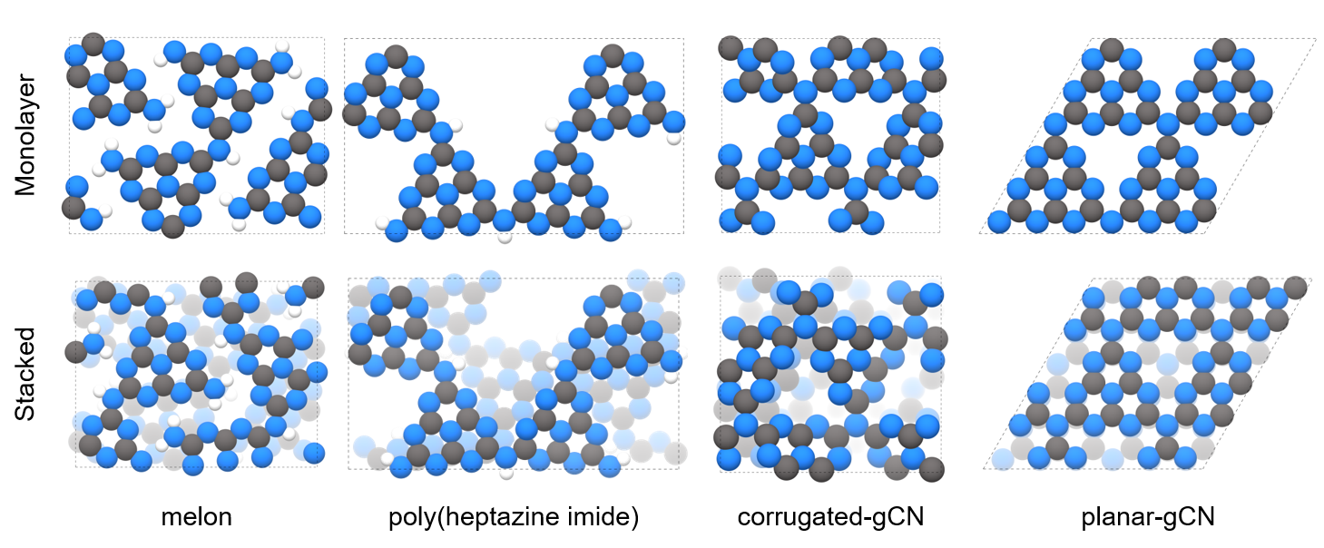
Models.—Figure 1 illustrates the different PCN structures considered in this work. These models encompass variations in the degree of poly-condensation of heptazine units, corrugations, and stacking configurations.[14] Increased poly-condensation leads to higher carbon and nitrogen contents, but a reduced hydrogen ratio. The planar-gCN (p-gCN) is known to be energetically less stable compared to corrugated-gCN (c-gCN), which represents a local minimum in the energy landscape.[33] PCN structures, composed of these heptazine units, typically exhibit highly-localized electrons. Thus, we primarily present results for optimized geometries using CA (LDA) functionals. These functionals provide an efficient description of structures with highly-localized electrons, considered as key properties. To address the limitations of the MBD@rsSCS dispersion correction, which depends on the exchange–correlation energy from the LDA functionals, we also included results obtained with the PBE-D3 xc-functional. This approach offers a better description of interlayer properties. Results of the optimized geometry with PBE-D3 are available in the supplementary information (SI) for comparison (Figs. S9–S20). The lattice parameters of the optimized geometries obtained with both functionals are summarized in Table S1.
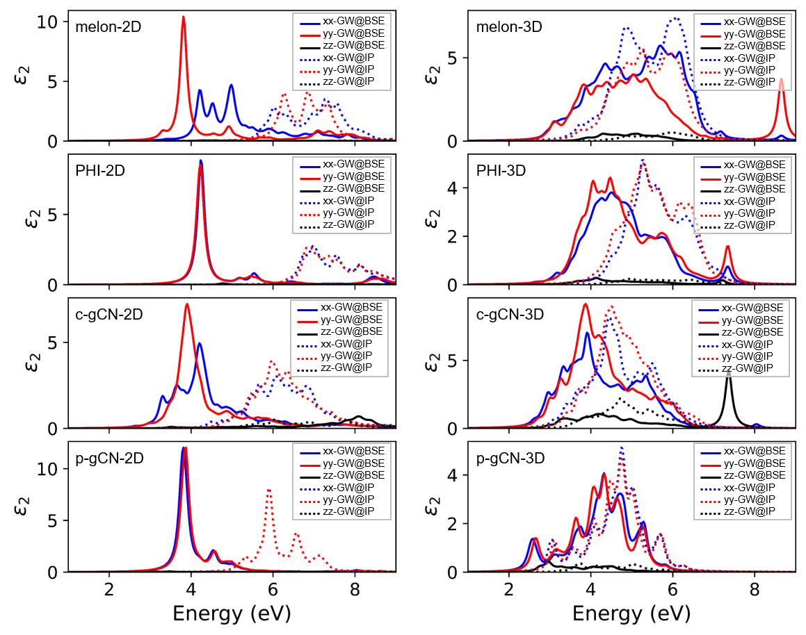
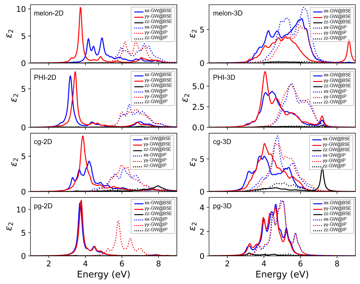
Optical spectra.—The sole use of density functional theory (DFT) to accurately describe optical properties has numerous limitations.[24, 30] To address these challenges, we use the Bethe–Salpeter Equations (BSE) to address the energy originating from electron–hole (e–h) interactions on top of the quasi-particle energy derived from calculations. This -based quasi-particle energy incorporates precisely screened electron–electron (e–e) interactions, thereby enhancing the accuracy of single excitation property predictions. Notably, the quasi-particle energy adjusted by the BSE method typically exhibits a red shift compared to those predicted by the IP approximation, which omits e–h interactions. By analyzing the imaginary part of the macroscopic dielectric functions from both GW@BSE and GW@IP calculations, we can estimate the exciton binding energy in 2D and 3D PCN structures, as illustrated in Figure 2. Spatially-projected absorption coefficients reveal distinct patterns: 2D PCN structures show strong exciton binding energy (> 2 eV).[13] However, this significant exciton binding energy diminishes to less than 1 eV in 3D stacked structures. Moreover, the transition from a 2D monolayer to a stacked 3D configuration alters the absorption edges and reduces the optical anisotropy observed in PHI-2D and c-gCN-2D structures. Since the optical anisotropy is attributed to the in-plane joint patterns of the heptazine units formed during poly-condensation, the reduced optical anisotropy implies that the fractions of the 2D PCN micro-structure can indeed interact with each other throughout the 3D assembly. In brief, this means that diverse forms of PCN fragments within the PCN structure interact electronically and optically through their stacking configurations, regardless of size or shape. Our findings provide clear evidence of strong interlayer interactions, resulting in dramatically decreased exciton binding energy and changes in absorption patterns including altered optical anisotropy and absorption edges. Consequently, these interlayer interactions strongly influence the optical properties of the different PCN configurations.
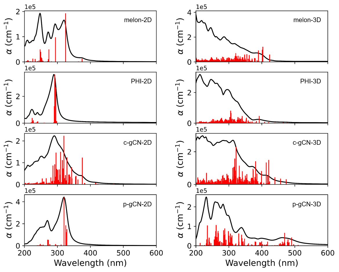
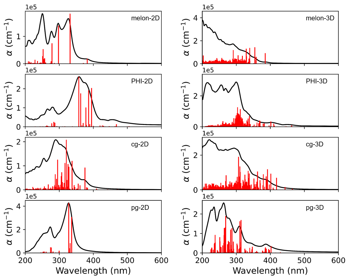
To better elucidate the origins of the absorption features near the absorption edge, in Figure 3 we depict the oscillator strengths and averaged absorption spectra by spatial directions for PCN 2D and 3D structures obtained from GW@BSE calculations. This presents the trends and changes in absorption peaks of PCNs in relation to their microstructure and stacking formation, including exciton binding energy. As mentioned above, PCN structures show distinctly different patterns in the absorption spectra of 2D monolayers compared to stacked 3D structures. The absorption peaks near 200 nm in PCN 3D structures highlight the limitations of a computational model that assumes an infinite size of 2D structures, since using actual finite-sized ribbon structures would result in a decrease in the intensity of absorption peaks corresponding to 2D structures.[15] Notably, while PCN 2D structures have major absorption peaks below 400 nm, PCN 3D structures show various absorption patterns at and beyond 400 nm. These absorption peaks, in contrast to those calculated using GGA and hybrid functionals,[34] closely align with the range reported in experimental absorption spectra of PCN.[35, 36] The peaks appearing beyond 400 nm originate from the interactions between layers, which will be discussed below. As heptazine units undergo more poly-condensation reactions, the energy levels produced by interlayer interactions lead to a red shift in absorption peaks. The absorption edges of the different PCN structures are obtained as 2.93 eV for melon-3D, 2.60 eV for PHI-3D, 2.62,eV for c-gCN-3D, and 1.69 eV for p-gCN-3D. These PCN structures and band gaps are completely in line with the experimentally observed band gaps of PCNs, which range from 2.6 to 2.8 eV.[5] Especially, the frequently observed long absorption tails can be attributed not only to thermal vibrational motion but also to a distinct structural origin, primarily from the graphitic (i.e. fully condensed) domains.[37]
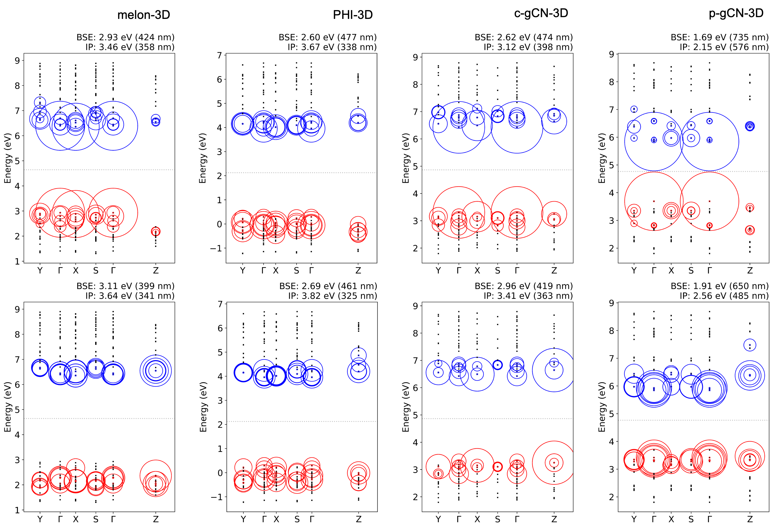
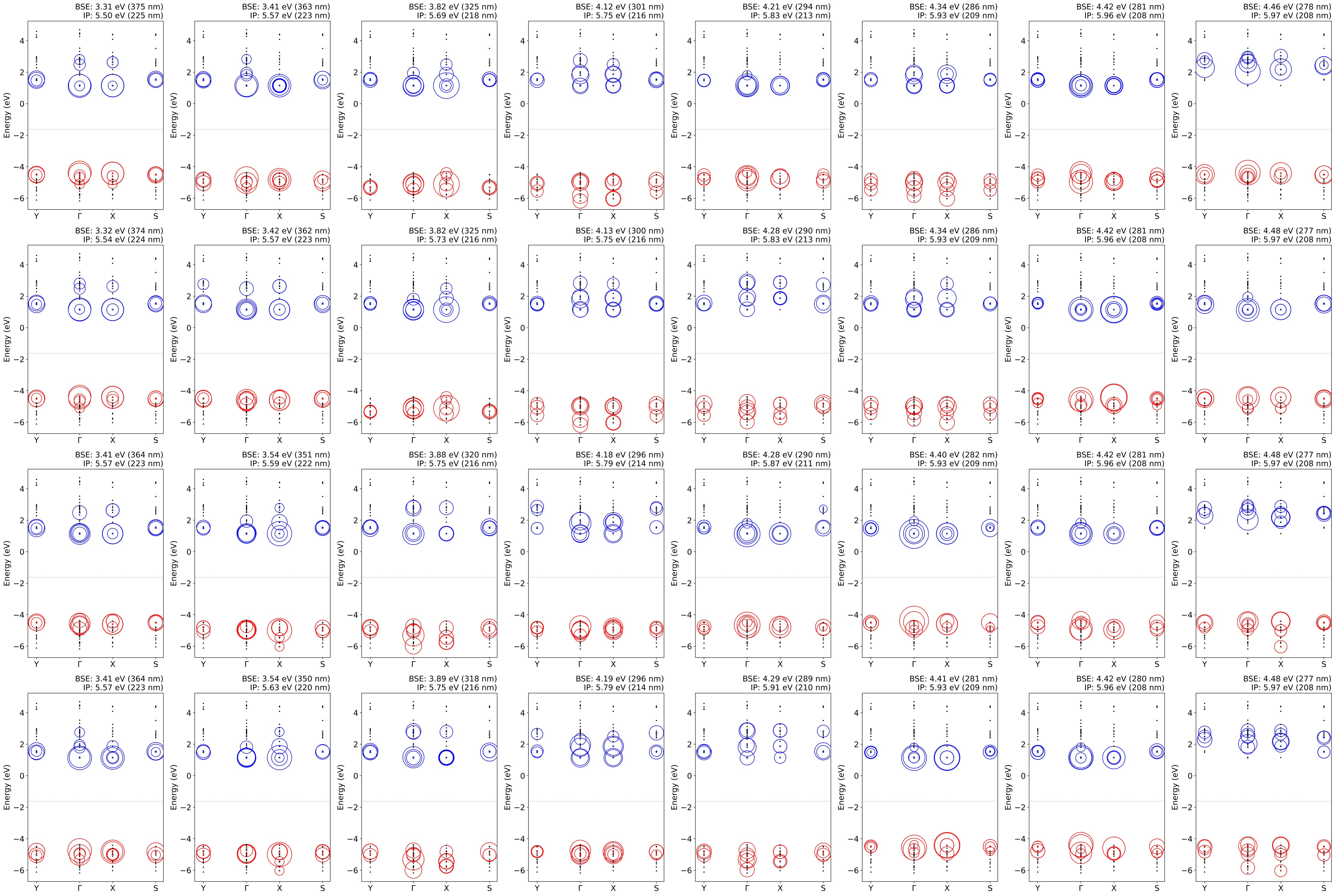
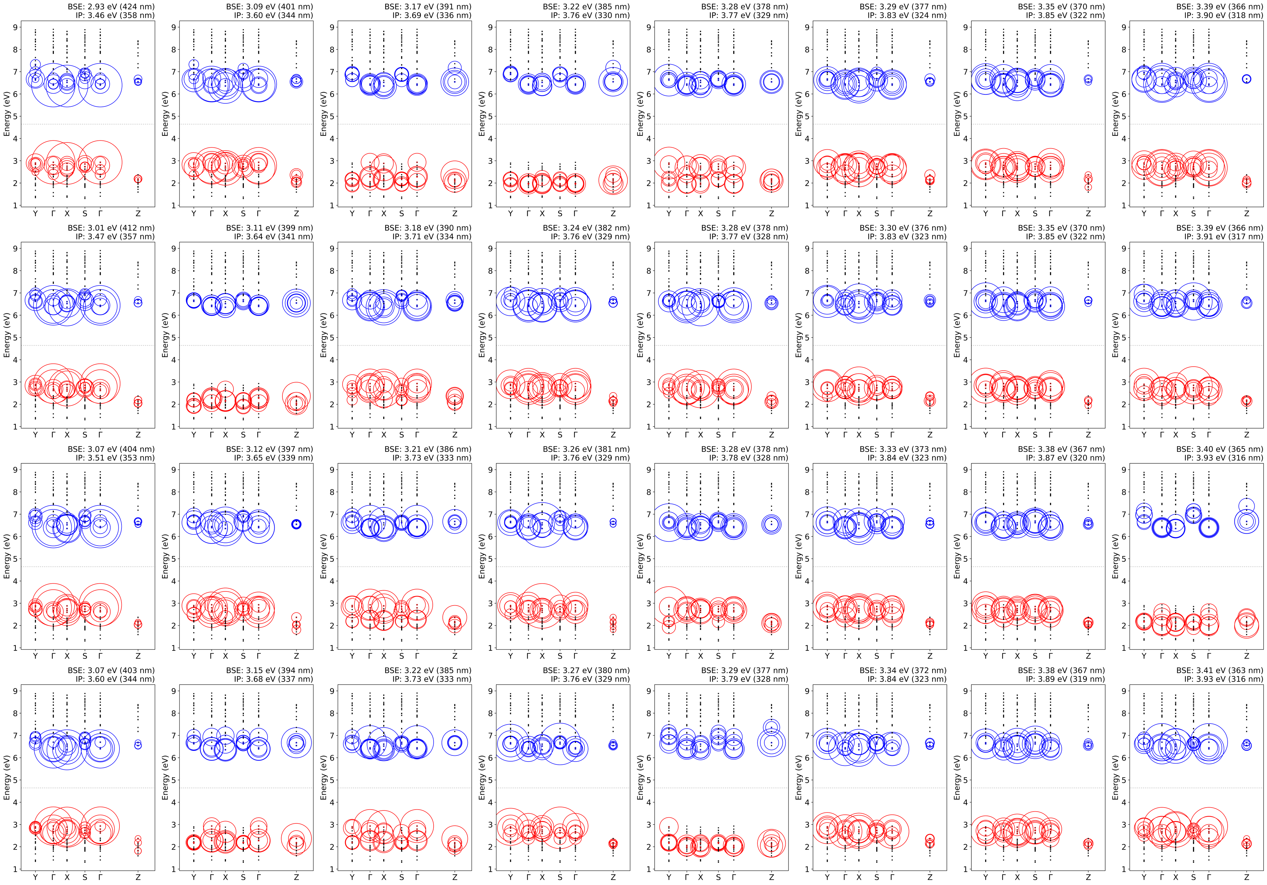
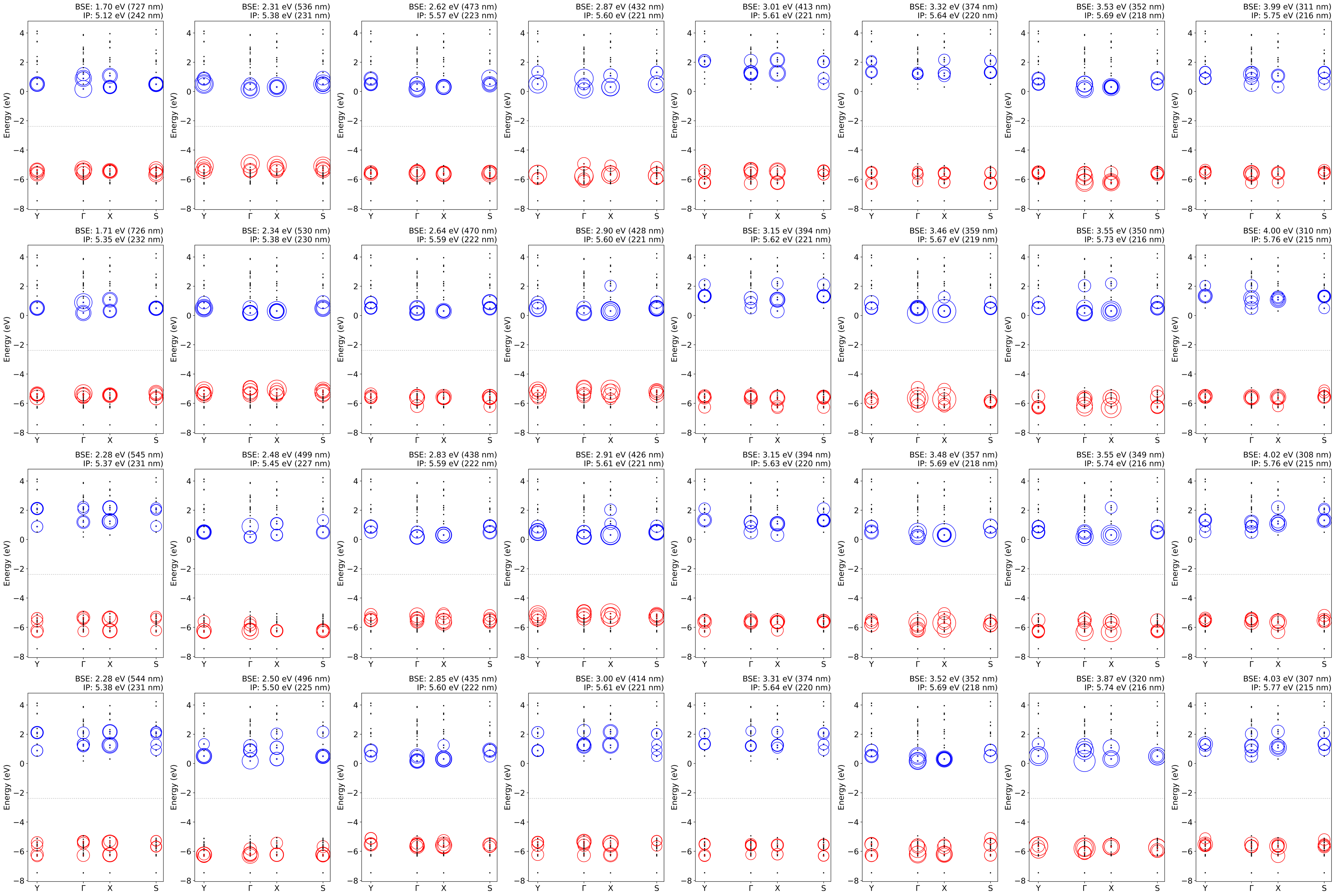
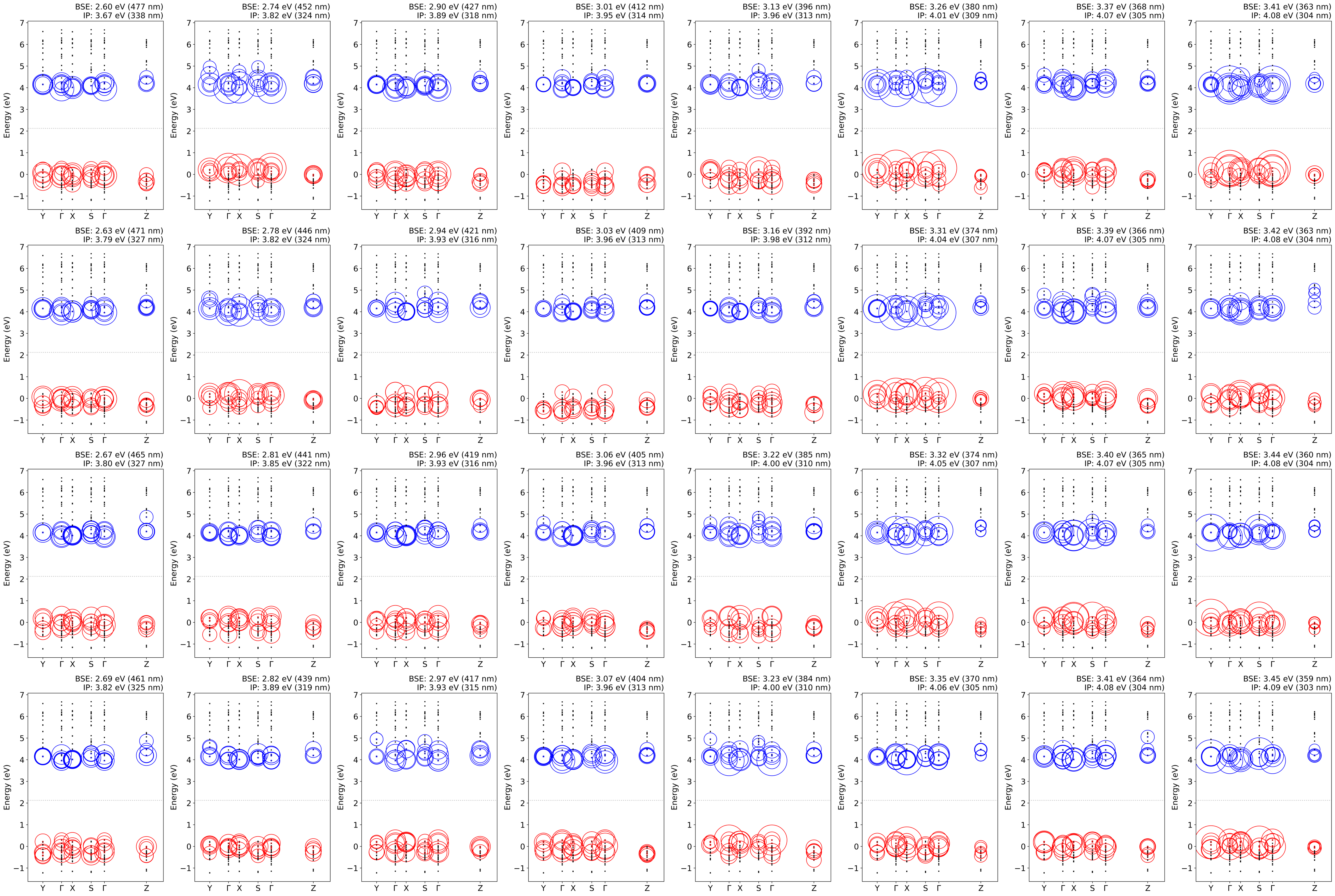
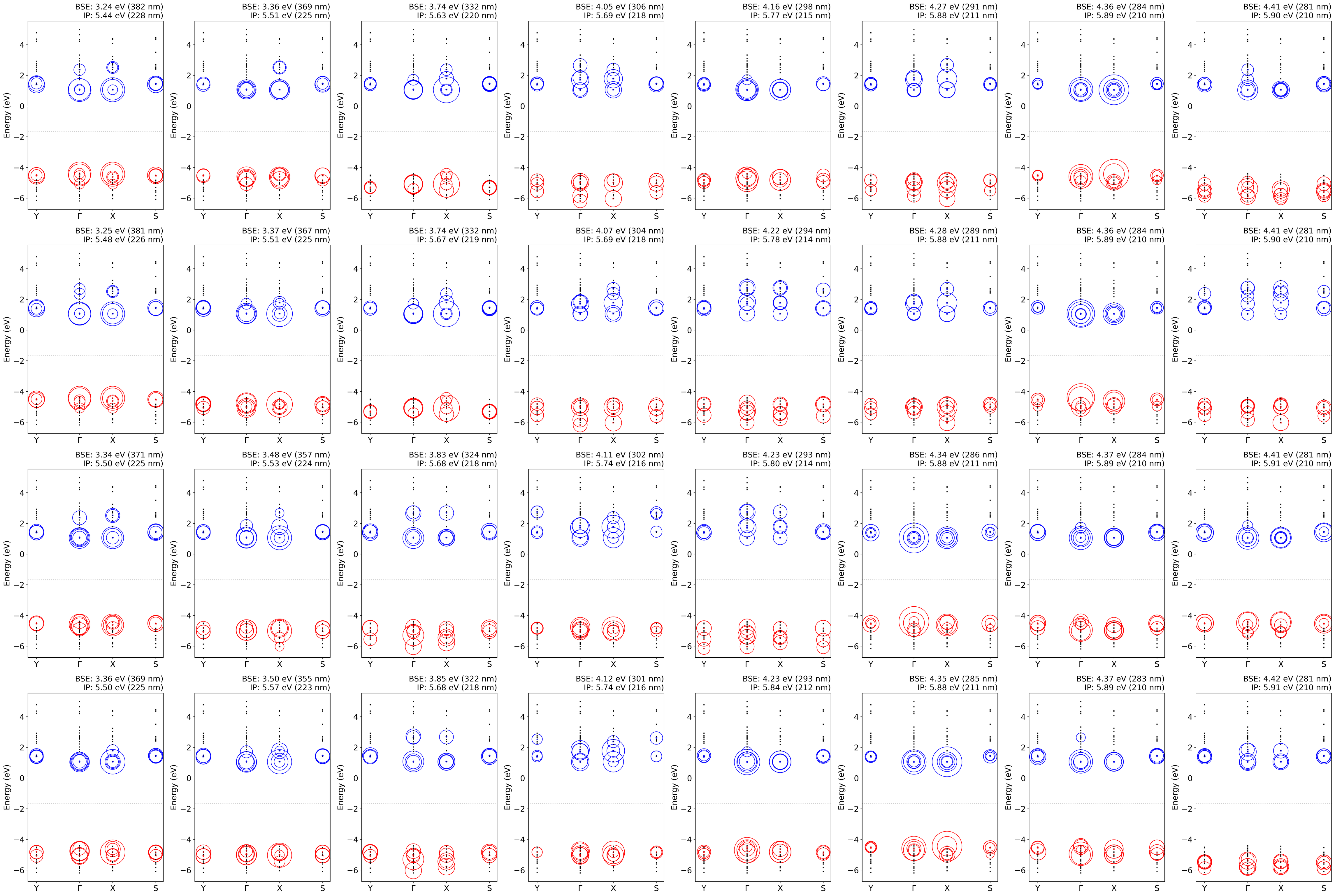
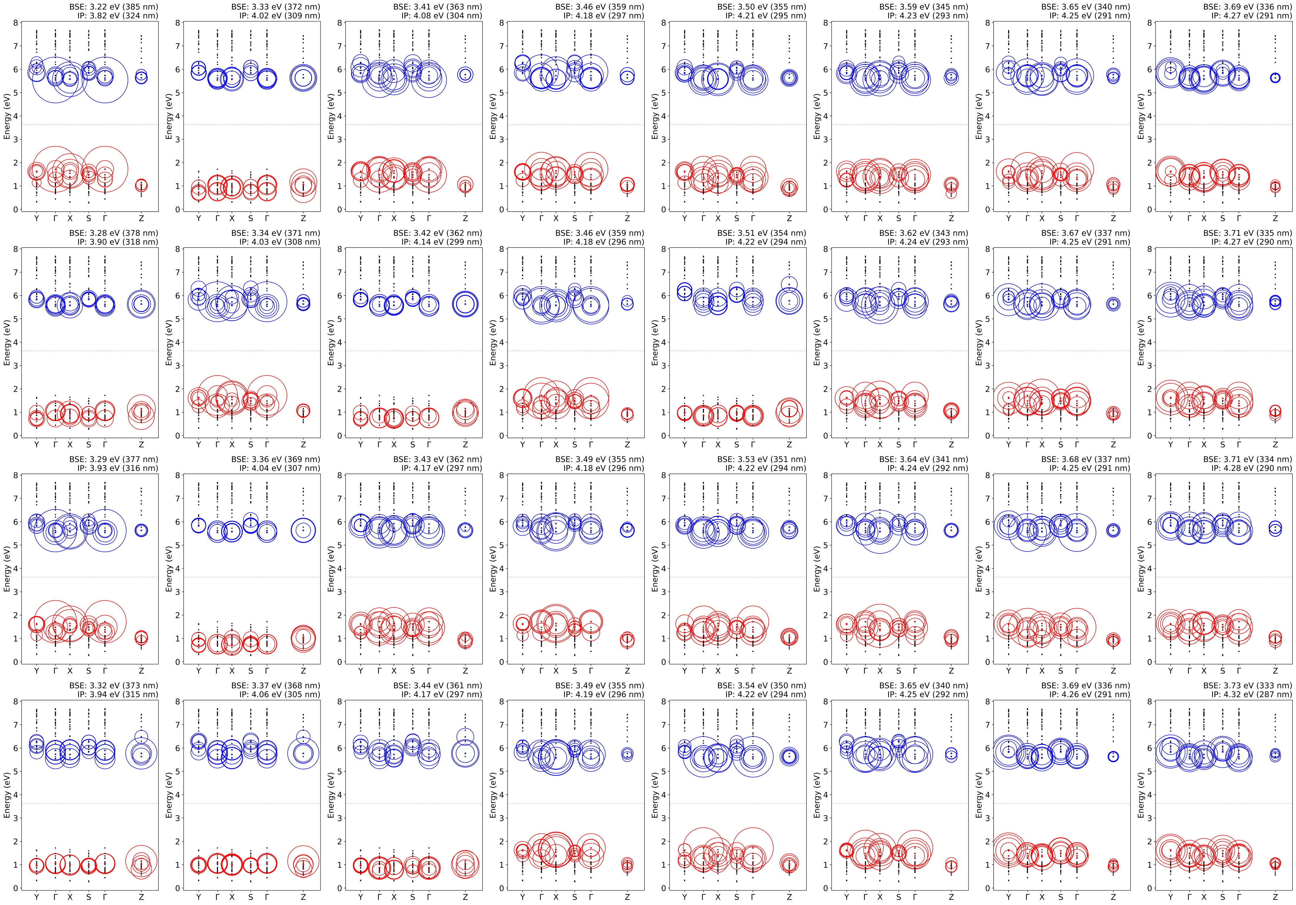
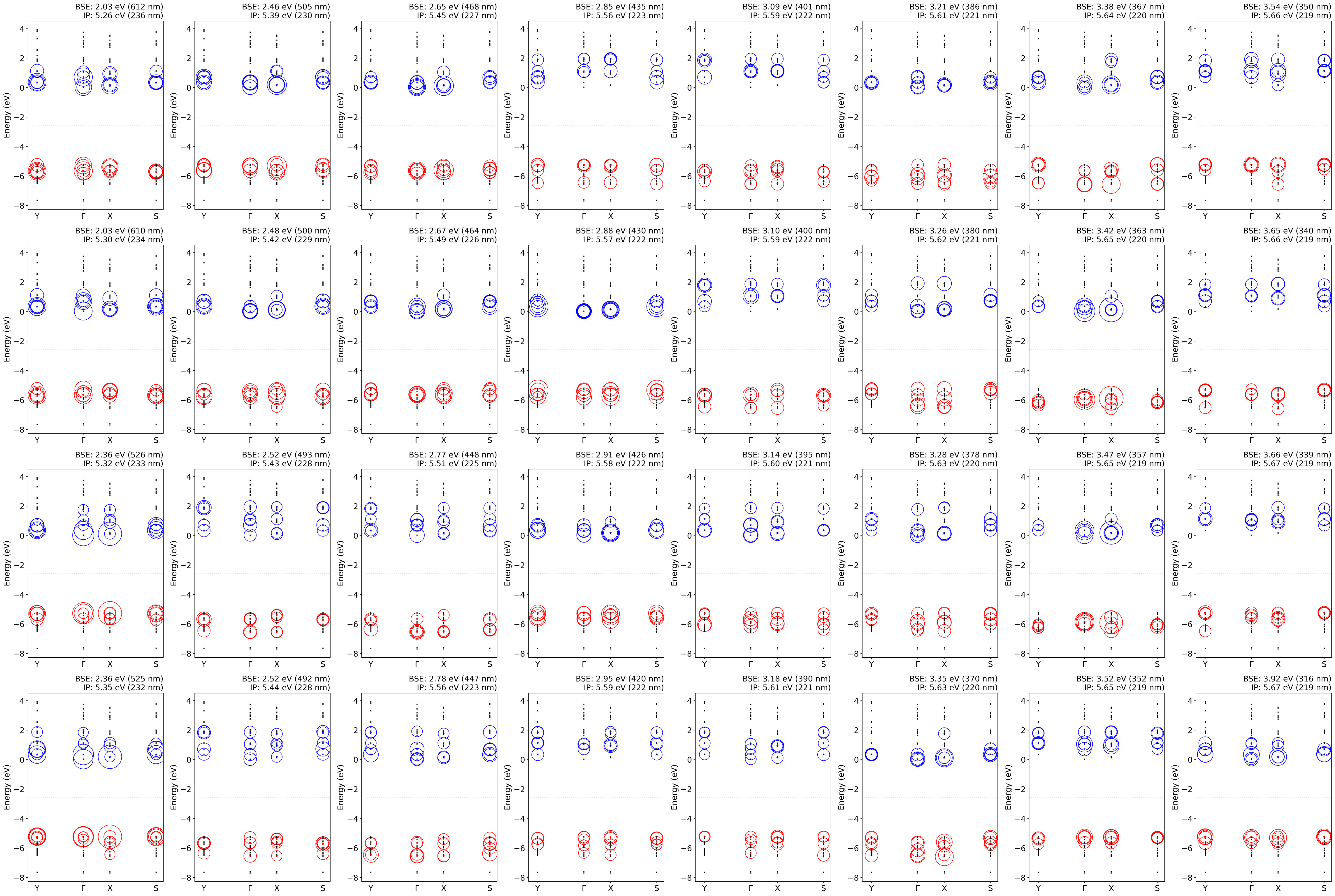
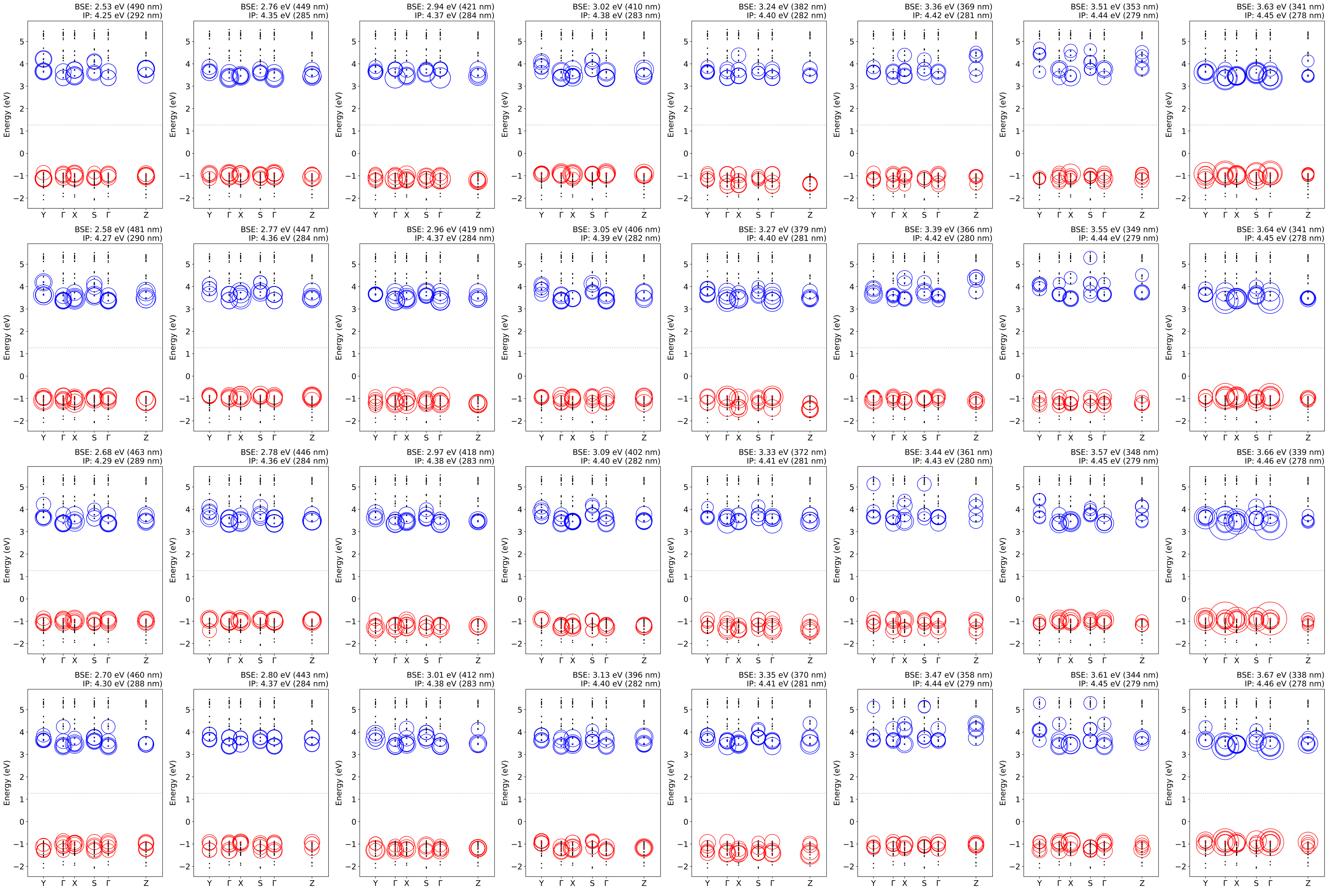
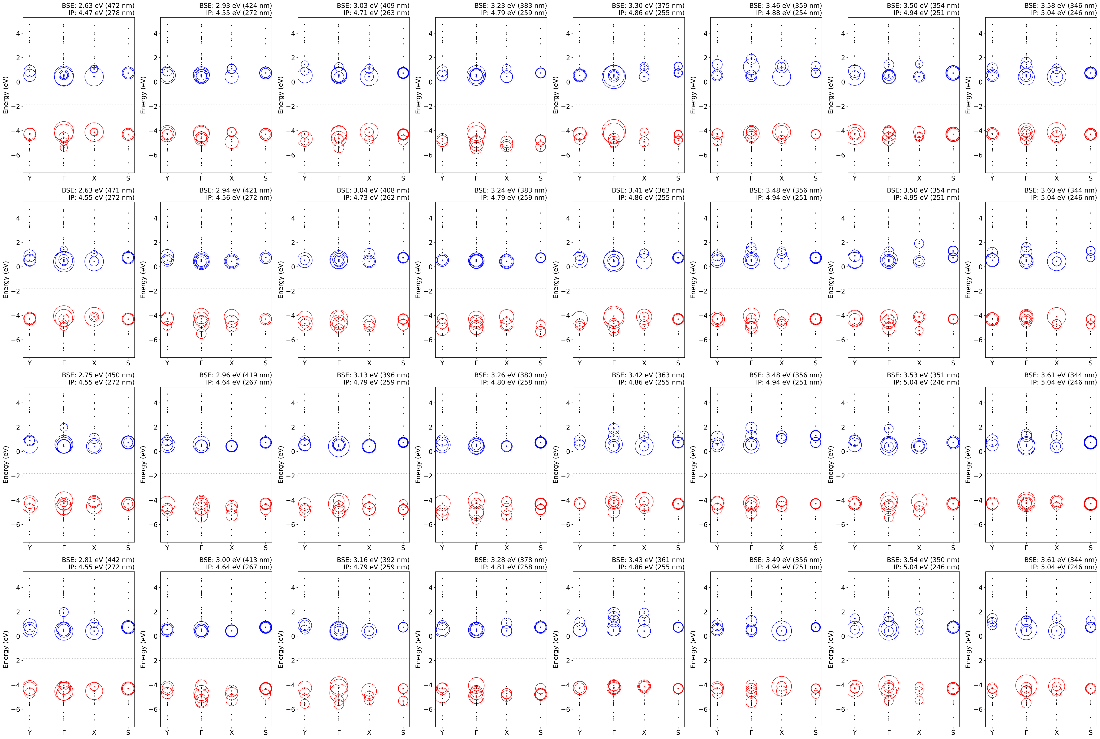
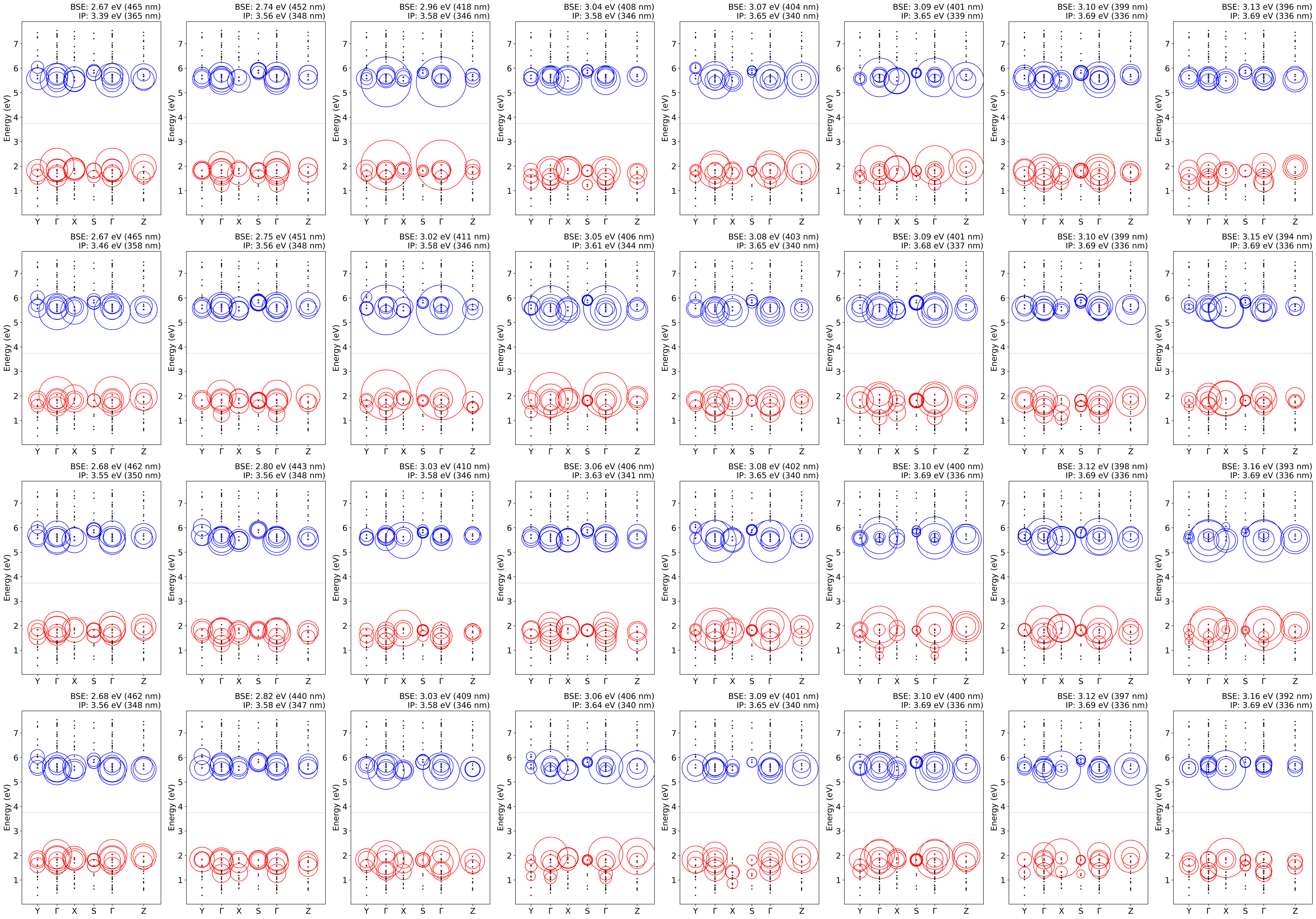
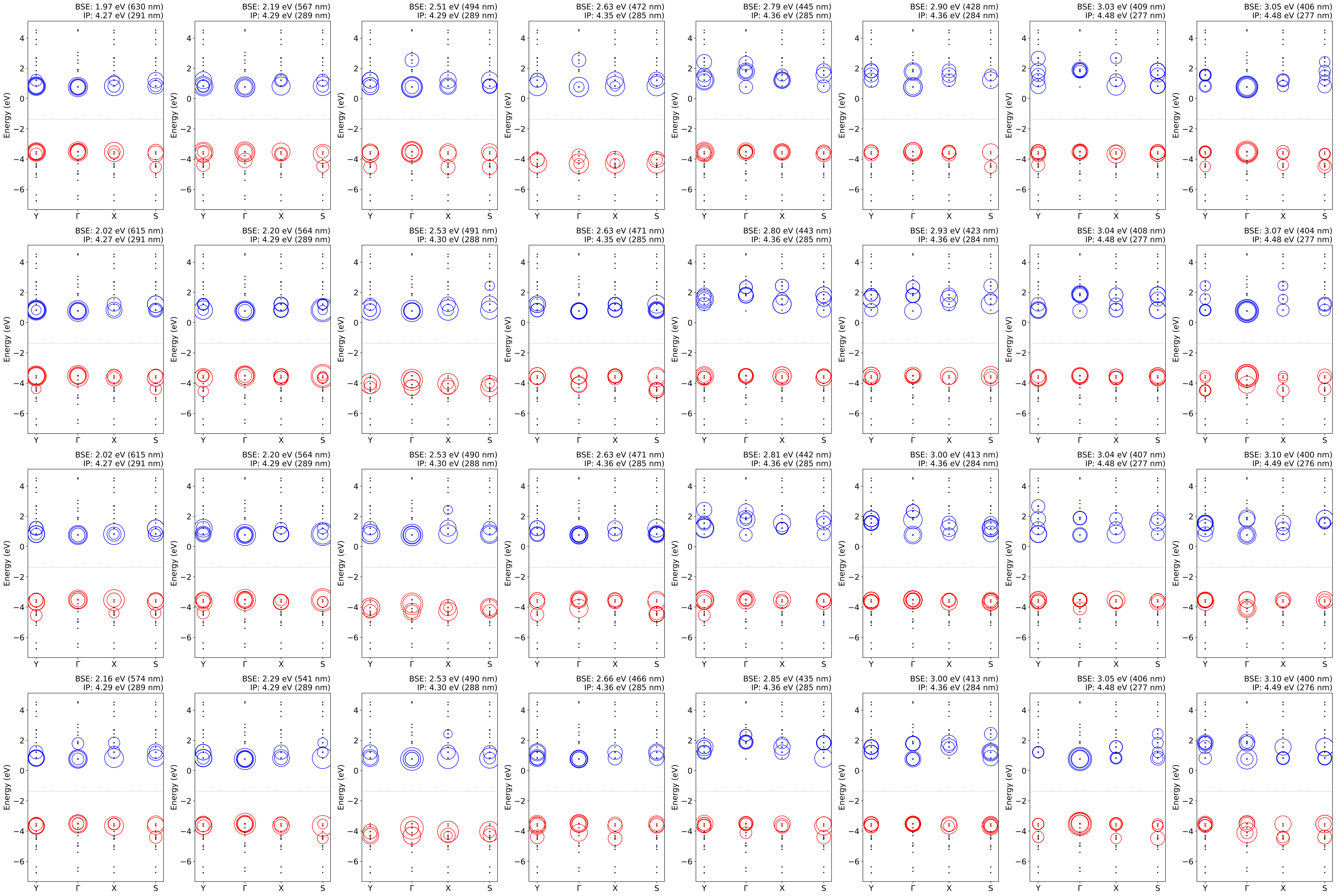
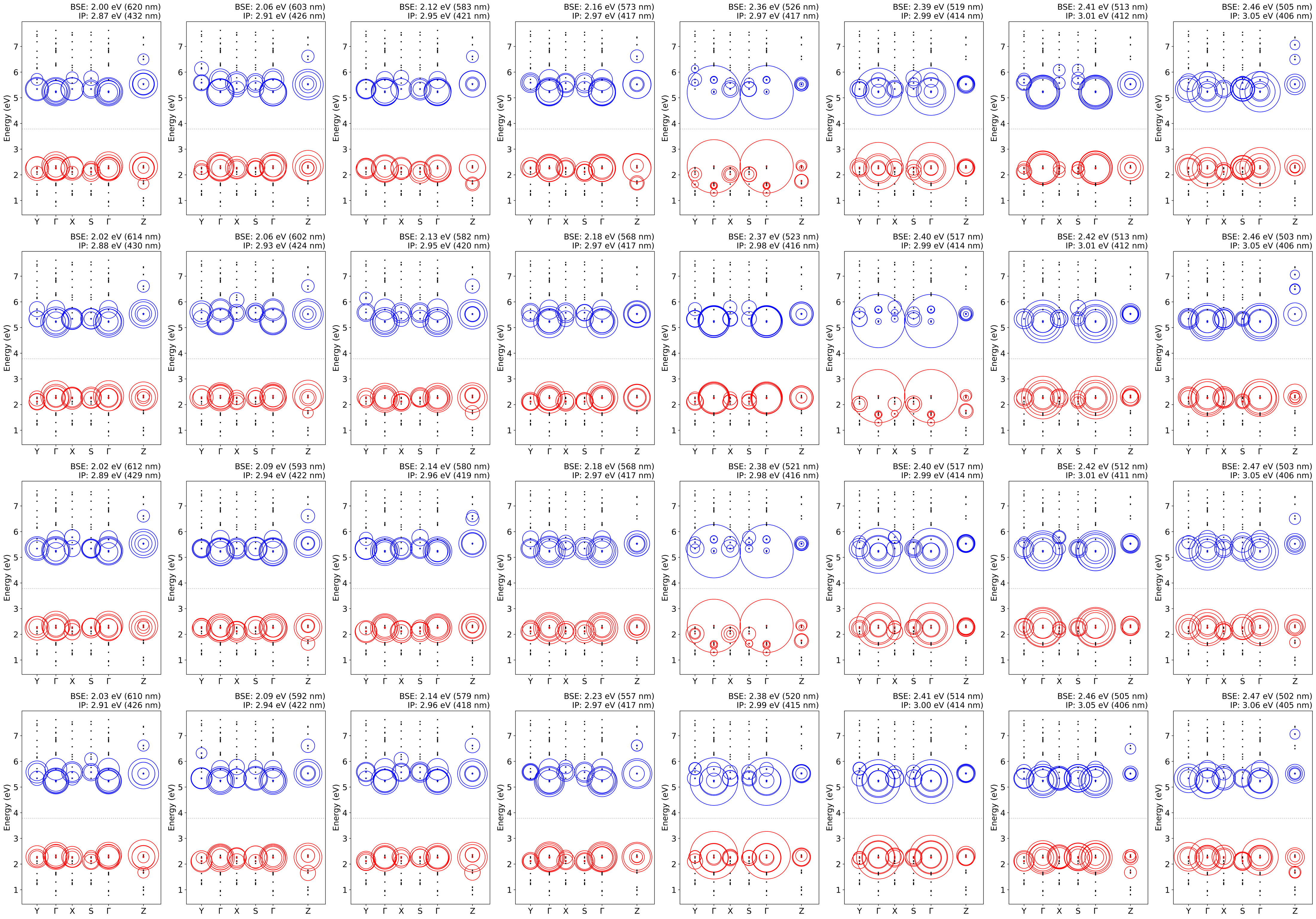
Exciton formation.—Figure 3 demonstrates the oscillator strengths that, according to the Franck–Condon principle, represent the probability of electronic transitions at each energy level. Cases of high oscillator strengths with relatively low absorption peaks occur in the absorption spectrum due to consideration of the dipole momentum and selection rules. Therefore, by comparing absorption spectra with oscillator strengths, we can predict the types of electronic transitions. Notably, most electronic transitions near the absorption edge in PCN 3D structures are forbidden transitions. Our results from the BSE calculations provide the coupling coefficients corresponding to transitions between energy levels at each high symmetry point, as shown in Figure 4. The coupling constants of the other lowest 32 excited states are presented in the Supplemental Material. The coupling coefficients (radius of circles) at the band edges for each PCN 3D structure are displayed at the top line of the figure, and the coupling coefficients that are most pronounced for interlayer interactions are displayed at the bottom line. At the absorption edges, stable excitons are related to transitions between the band edges in melon, c-gCN, and p-gCN structures while relatively more degenerate excitons are formed in the PHI structure. The coupling constants at the bottom line of Figure 4 represent the exciton formation at the -point in each structure, indicating a direction toward the other layer from the -point. These interlayer transitions are identified as the electron transitions from the sub-levels of the valence band to near the conduction band edge, indicating that the edge absorption peaks are composed of different types of electron excitations. Our previous study found that absorption peaks of PCN structure can be sorted as in-plane – transitions and – interlayer transitions.[15] When comparing the relative stability of excitons formed in the PCN structures at the interlayer, the c-gCN structure exhibits the most stable exciton with an energy gap of 2.96 eV. Melon and PHI also show stabilization of interlayer excitons at 3.11 eV and 2.97 eV, respectively, showing similar stability for other simultaneously formed excitons. The differences in exciton stability at the interlayer are determined by the degree of overlap between orbitals of different layers, specifically being regulated by the magnitude of the transition momentum off-diagonal components. These off-diagonal components turn out to be the degree of interaction of the nitrogen electrons, which impact the electrons at the interlayers. This explains how the interlayer distances are related to the configurations of PCN structures. This is more clearly understood when comparing the coupling constant plots of LDA-optimized geometries with different interlayer distances (see Table S1) to those of GGA-optimized geometry results (Figs. S11,S13–S20). Furthermore, in our previous study we found that the thermodynamic contributions from vibrational motion due to the flexible layered structure of PCNs are non-negligible.[14] Thus, we expect that enhanced interlayer interactions may be evident due to interlayer vibration modes.
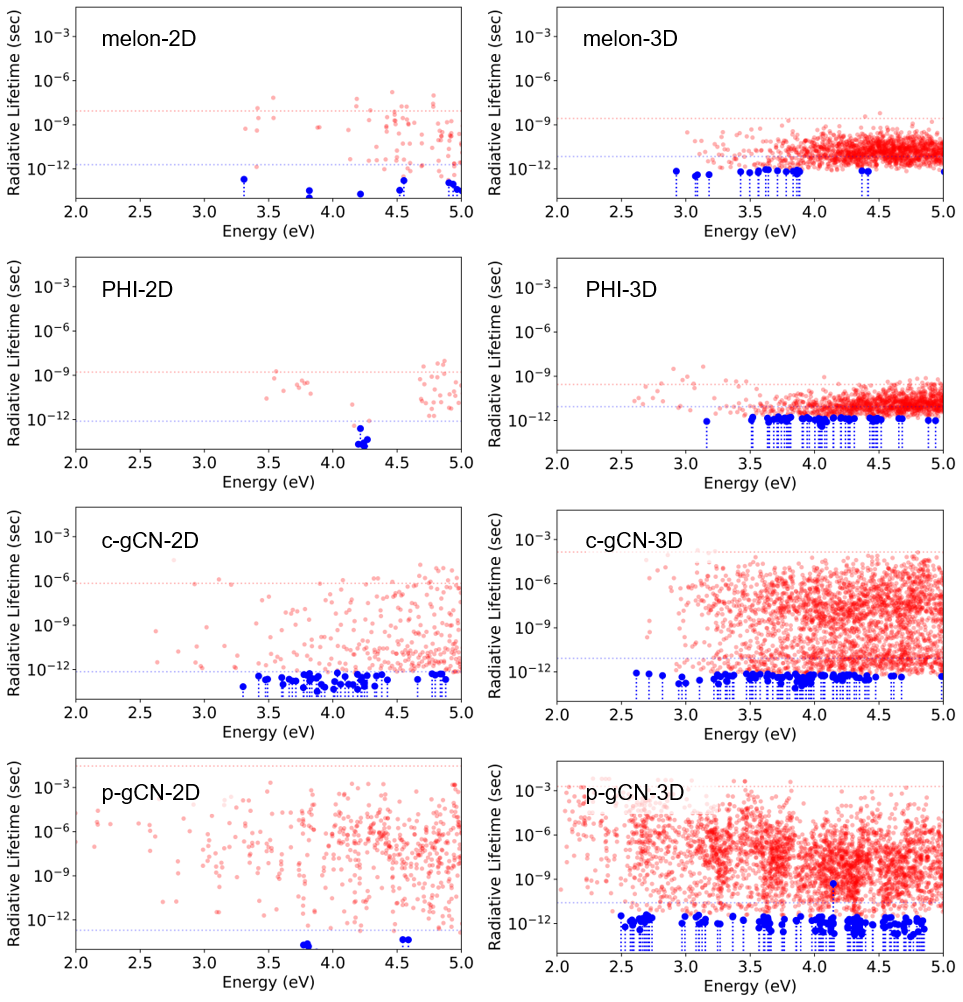
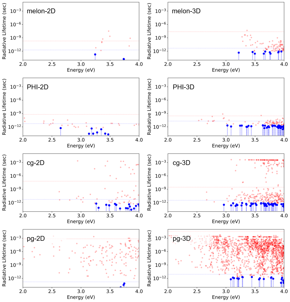
Prolonged exciton lifetime and photocatalytic activity.—We have calculated the exciton lifetime and effective lifetime at 300 K based on the energy required for each excited state, transition probability, and transition dipole momentum. The obtained behavior is shown in Figure 5 and summarized in Table 1. Here, we define the energy states with the top 5 oscillator strengths as bright states (blue) and the rest as dark states (red) (see Method section). Firstly, the effective lifetimes of bright excitons in 3D PCN structures are slightly increased compared to 2D structures. This suggests that most bright excitons in PCN structures are formed by the direct in-plane transitions of strongly localized electrons and the interlayer transition. Therefore, the stacking configuration helps to stabilize these bright excitons. In contrast, the dark excitons formed in 3D structures are destabilized by interlayer interactions. Most dark excitons are related to the in-plane – transitions and interlayer interaction cannot sufficiently stabilize these states due to the lack of interaction. Notably, the effective lifetime of dark excitons increases dramatically in graphitic structures (c-gCN and p-gCN). This indicates the presence of a new form of excitons completely different from those in melon and PHI structures. We identify these long-lived dark excitons as trapped excitons known from the literature[11, 38] and suggest that this stabilized exciton in (semi)crystalline PCN structures, such as PHI, is closely related to the photo-charging phenomenon.[11, 9, 39, 8] It has also been reported that the phenomenon persists in the H-PHI structure, which is a proton-exchanged cation-PHI structure, suggesting that the phenomenon is more attributed to the high symmetry crystalline regions rather than the influence of existing cations. However, the principle and the nature of charge accumulation in PHIs have not been elucidated so far. Our results suggest that the extended lifetime of excitons is observed in both 3D stacked and 2D graphitic structures (c-gCN and p-gCN), indicating that these long lifetimes are primarily due to structural features rather than interlayer interactions.
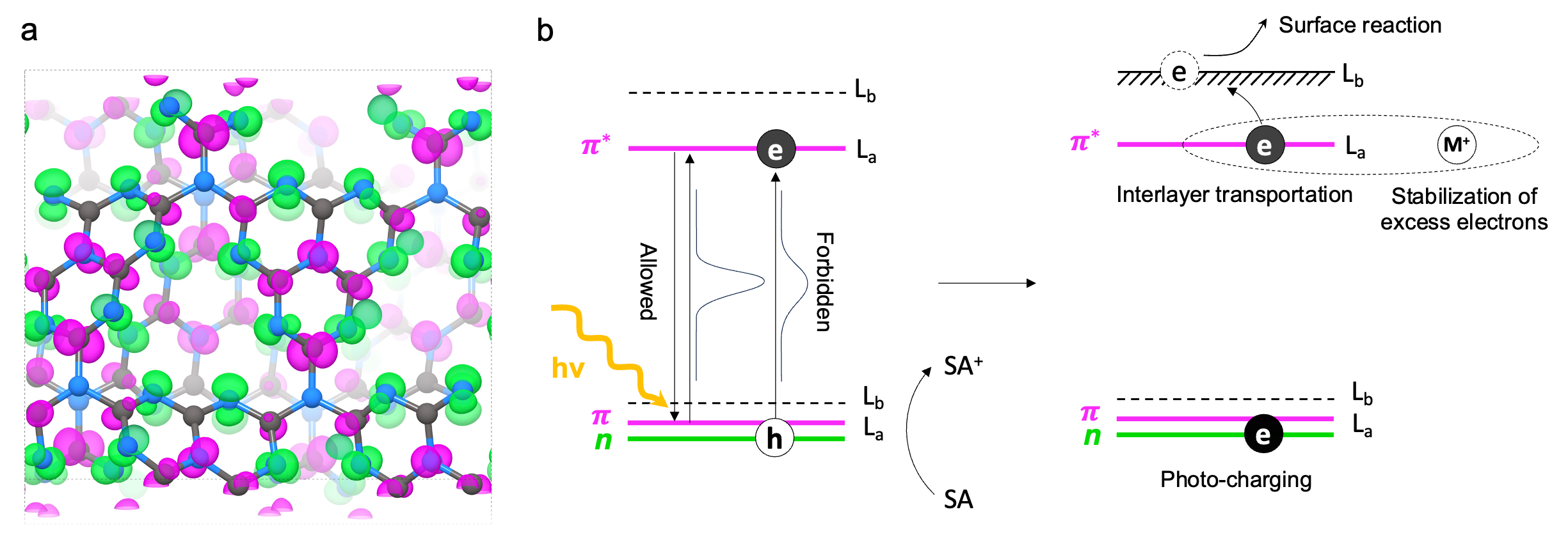
In order to further identify the long-lived excitonic states, Figure 6a shows the electronic density corresponding to the electron and the electron hole for the exciton of the longest lifetime in the c-gCN-3D structure. It clearly shows that this long-living exciton is indeed composed of the non-bonding states () and the states of the carbons, indicating the – forbidden transition. We explain that the repulsive force between heptazine units causes a distorted orientation of the electrons, enabling overlap with the electrons and thus facilitating this forbidden transition. Unlike c-gCN, in p-gCN, the high symmetry structure leads to the strongest repulsion among non-bonding electrons without structural distortion, resulting in minimal overlap with the electrons. Consequently, p-gCN forms long-lived dark excitons at lower energy levels, while most bright excitons are formed primarily through interlayer interactions.(See Figure 5) This indicates that – transition attributed to the strong repulsive force between non-bonding nitrogen electrons is the origin for long-lived excitons.
| Structure | G0W0@IP (eV) | G0W0@BSE (eV) |
|
||||
| bright | dark | ||||||
| 2D | melon | 5.50 (D) | 3.31 | 1.96 ps | 8.70 ns | ||
| PHI | 5.83 (I) | 3.48 | 0.78 ps | 1.65 ns | |||
| c-gCN | 4.48 (D) | 2.62 | 8.36 ps | 141.00 s | |||
| p-gCN | 4.26 (I) | 1.95 | 0.20 ps | 28.73 ms | |||
| 3D | melon | 3.46 (I) | 2.93 | 6.96 ps | 2.72 ns | ||
| PHI | 3.67 (D) | 2.60 | 8.77 ps | 277.34 ps | |||
| c-gCN | 3.12 (D) | 2.62 | 8.36 ps | 141.00 s | |||
| p-gCN | 2.15 (I) | 1.69 | 26.04 ps | 1.92 ms | |||
Figure 6b schematically summarizes the anticipated impact of such long-lived excitons on photochemical processes and photocatalytic reactions. The results from our recent studies clearly demonstrate that photoexcitations in PCN structures predominantly involve bright in-plane – transitions as well as interlayer – transitions.[15] Furthermore, for a PCN structure to enable typically forbidden transitions, the non-bonding electron of nitrogen have to undergo sufficient distortion to facilitate the – transition. In this case, the excitons can more readily accept an electron from the electron donor due to the stabilized and prolonged lifetime formed by the – transitions. These phenomena manifest as photo-induced electron charging observed in carbon nitrides, and this electron accumulation is more stably observed when carbon nitrides include cations. [9, 39, 8] This photo-charging effect will boost the electron transport to the surface through the highly-ordered stacking configuration by PCN corrugation.
As a result, photo-charging in the crystalline regions of carbon nitrides can enhance photocatalytic performances compared to the conventional PCNs.[8] We predict, however, that these long-lived excitons, also known as deep trapped excitons, could pose an obstacle to quantum efficiency since they do not directly participate in reactions but only assist in surface charge transport.[11, 12] Therefore, innovative approaches in the structural design of 2D carbon nitrides should be further investigated to achieve better quantum yields with enhanced photocatalytic performance.
Conclusion.—Our study elucidates the principles underlying exciton formation, transition,
and lifetime within PCN structures. These results provide valuable insights into the optical properties of 2D nitrogen-carbon compounds and the potential
to tune their electronic structures for optimal photocatalytic performance.
Utilizing GW@BSE calculations, we report that the strong exciton binding energy of PCNs, originating from the highly localized nature of electrons, is mitigated by – interlayer interactions, emphasizing robust optical interactions between layers. Additionally, the corrugation of heptazine units caused by the interaction of non-bonding nitrogen electrons reveals how forbidden (–) transitions become possible, resulting in the formation of stable and long-lived excitons. This mechanism of stable exciton formation successfully explains the charge accumulation mechanism and its effect on the subsequent surface reaction in carbon nitrides. Furthermore,
our findings aid in exploring new structures of organic carbon nitrides that could enhance the
interplay between electronic and optical properties, potentially paving the way for advanced applications
in photonic and electronic devices.
This work was funded by the Deutsche Forschungsgemeinschaft (DFG – German Research Foundation) through TRR-234 CataLight (project no. 364549901) as well as JA 1072/27-1 and BE 5102/5-1 (project no. 428764269). R.B. acknowledges funding from the European Union’s Horizon Europe programme for research and innovation under grant agreement No. 101122061 (SUNGATE). The authors acknowledge support by the state of Baden-Württemberg through bwHPC and the German Research Foundation (DFG) through grant no INST 40/575-1 FUGG (JUSTUS 2 cluster). C.I. acknowledges the German Academic Exchange Service (DAAD, Ref. No. 91676720).
References
- Kessler et al. [2017] F. K. Kessler, Y. Zheng, D. Schwarz, C. Merschjann, W. Schnick, X. Wang, and M. J. Bojdys, Functional carbon nitride materials—design strategies for electrochemical devices, Nature Reviews Materials 2, 1 (2017).
- Podjaski and Lotsch [2021] F. Podjaski and B. V. Lotsch, Optoelectronics meets optoionics: light storing carbon nitrides and beyond, Advanced Energy Materials 11, 2003049 (2021).
- Gouder et al. [2023] A. Gouder, F. Podjaski, A. Jiménez-Solano, J. Kröger, Y. Wang, and B. V. Lotsch, An integrated solar battery based on a charge storing 2d carbon nitride, Energy & Environmental Science 16, 1520 (2023).
- Liu et al. [2015] J. Liu, Y. Liu, N. Liu, Y. Han, X. Zhang, H. Huang, Y. Lifshitz, S.-T. Lee, J. Zhong, and Z. Kang, Metal-free efficient photocatalyst for stable visible water splitting via a two-electron pathway, Science 347, 970 (2015), https://www.science.org/doi/pdf/10.1126/science.aaa3145 .
- Cao et al. [2015] S. Cao, J. Low, J. Yu, and M. Jaroniec, Polymeric photocatalysts based on graphitic carbon nitride, Advanced Materials 27, 2150 (2015).
- Cheng et al. [2021] L. Cheng, H. Zhang, X. Li, J. Fan, and Q. Xiang, Carbon–graphitic carbon nitride hybrids for heterogeneous photocatalysis, Small 17, 2005231 (2021).
- Chen et al. [2017] Z. Chen, A. Savateev, S. Pronkin, V. Papaefthimiou, C. Wolff, M. G. Willinger, E. Willinger, D. Neher, M. Antonietti, and D. Dontsova, “the easier the better” preparation of efficient photocatalysts—metastable poly (heptazine imide) salts, Advanced Materials 29, 1700555 (2017).
- Schlomberg et al. [2019] H. Schlomberg, J. Kröger, G. Savasci, M. W. Terban, S. Bette, I. Moudrakovski, V. Duppel, F. Podjaski, R. Siegel, J. Senker, et al., Structural insights into poly (heptazine imides) a light-storing carbon nitride material for dark photocatalysis, Chemistry of Materials 31, 7478 (2019).
- Adler et al. [2021] C. Adler, S. Selim, I. Krivtsov, C. Li, D. Mitoraj, B. Dietzek, J. R. Durrant, and R. Beranek, Photodoping and fast charge extraction in ionic carbon nitride photoanodes, Advanced Functional Materials 31, 2105369 (2021).
- Merschjann et al. [2015] C. Merschjann, S. Tschierlei, T. Tyborski, K. Kailasam, S. Orthmann, D. Hollmann, T. Schedel-Niedrig, A. Thomas, and S. Lochbrunner, Complementing graphenes: 1d interplanar charge transport in polymeric graphitic carbon nitrides., Advanced Materials (Deerfield Beach, Fla.) 27, 7993 (2015).
- Godin et al. [2017] R. Godin, Y. Wang, M. A. Zwijnenburg, J. Tang, and J. R. Durrant, Time-resolved spectroscopic investigation of charge trapping in carbon nitrides photocatalysts for hydrogen generation, Journal of the American Chemical Society 139, 5216 (2017).
- Yang et al. [2019] W. Yang, R. Godin, H. Kasap, B. Moss, Y. Dong, S. A. Hillman, L. Steier, E. Reisner, and J. R. Durrant, Electron accumulation induces efficiency bottleneck for hydrogen production in carbon nitride photocatalysts, Journal of the American Chemical Society 141, 11219 (2019).
- Wei and Jacob [2013] W. Wei and T. Jacob, Strong excitonic effects in the optical properties of graphitic carbon nitride g-C3N4 from first principles, Phys. Rev. B 87, 1 (2013).
- Im et al. [2023] C. Im, B. Kirchhoff, I. Krivtsov, D. Mitoraj, R. Beranek, and T. Jacob, Structure and optical properties of polymeric carbon nitrides from atomistic simulations, Chem. Mater. 35, 1547 (2023).
- Im et al. [2024] C. Im, B. Kirchhoff, D. Mitoraj, I. Krivtsov, A. Farkas, R. Beránek, and T. Jacob, Unraveling the optical signatures of polymeric carbon nitrides: Insights into stacking-induced excitonic transitions, arXiv preprint arXiv:2403.13685 (2024).
- Rohlfing and Louie [1998] M. Rohlfing and S. G. Louie, Electron-hole excitations in semiconductors and insulators, Physical review letters 81, 2312 (1998).
- Hafner [2008] J. Hafner, Ab-initio simulations of materials using vasp: Density-functional theory and beyond, Journal of computational chemistry 29, 2044 (2008).
- Ceperley and Alder [1980] D. M. Ceperley and B. J. Alder, Ground state of the electron gas by a stochastic method, Phys. Rev. Lett. 45, 566 (1980).
- Perdew and Zunger [1981] J. P. Perdew and A. Zunger, Self-interaction correction to density-functional approximations for many-electron systems, Phys. Rev. B 23, 5048 (1981).
- Tkatchenko et al. [2012] A. Tkatchenko, R. A. DiStasio, R. Car, and M. Scheffler, Accurate and efficient method for many-body van der waals interactions, Phys. Rev. Lett. 108, 236402 (2012).
- Ambrosetti et al. [2014] A. Ambrosetti, A. M. Reilly, J. DiStasio, Robert A., and A. Tkatchenko, Long-range correlation energy calculated from coupled atomic response functions, The Journal of Chemical Physics 140, 18A508 (2014), https://pubs.aip.org/aip/jcp/article-pdf/doi/10.1063/1.4865104/15480786/18a508_1_online.pdf .
- Perdew et al. [1996] J. P. Perdew, K. Burke, and M. Ernzerhof, Generalized gradient approximation made simple, Physical review letters 77, 3865 (1996).
- Grimme et al. [2010] S. Grimme, J. Antony, S. Ehrlich, and H. Krieg, A consistent and accurate ab initio parametrization of density functional dispersion correction (dft-d) for the 94 elements h-pu, The Journal of chemical physics 132 (2010).
- Hedin [1965] L. Hedin, New method for calculating the one-particle green’s function with application to the electron-gas problem, Physical Review 139, A796 (1965).
- van Schilfgaarde et al. [2006] M. van Schilfgaarde, T. Kotani, and S. Faleev, Quasiparticle self-consistent theory, Phys. Rev. Lett. 96, 226402 (2006).
- Ljungberg et al. [2015] M. P. Ljungberg, P. Koval, F. Ferrari, D. Foerster, and D. Sanchez-Portal, Cubic-scaling iterative solution of the bethe-salpeter equation for finite systems, Physical Review B 92, 075422 (2015).
- Sander et al. [2015] T. Sander, E. Maggio, and G. Kresse, Beyond the tamm-dancoff approximation for extended systems using exact diagonalization, Physical Review B 92, 045209 (2015).
- Leng et al. [2016] X. Leng, F. Jin, M. Wei, and Y. Ma, Gw method and bethe–salpeter equation for calculating electronic excitations, Wiley Interdisciplinary Reviews: Computational Molecular Science 6, 532 (2016).
- Rohlfing and Louie [2000] M. Rohlfing and S. G. Louie, Electron-hole excitations and optical spectra from first principles, Physical Review B 62, 4927 (2000).
- Albrecht et al. [1998] S. Albrecht, L. Reining, R. Del Sole, and G. Onida, Ab initio calculation of excitonic effects in the optical spectra of semiconductors, Physical review letters 80, 4510 (1998).
- Palummo et al. [2015] M. Palummo, M. Bernardi, and J. C. Grossman, Exciton radiative lifetimes in two-dimensional transition metal dichalcogenides, Nano letters 15, 2794 (2015).
- Perebeinos et al. [2005] V. Perebeinos, J. Tersoff, and P. Avouris, Radiative lifetime of excitons in carbon nanotubes, Nano letters 5, 2495 (2005).
- Gracia and Kroll [2009] J. Gracia and P. Kroll, Corrugated layered heptazine-based carbon nitride: the lowest energy modifications of c 3 n 4 ground state, Journal of Materials Chemistry 19, 3013 (2009).
- Butchosa et al. [2014] C. Butchosa, P. Guiglion, and M. A. Zwijnenburg, Carbon nitride photocatalysts for water splitting: a computational perspective, The Journal of Physical Chemistry C 118, 24833 (2014).
- Zhang et al. [2017] G. Zhang, G. Li, Z.-A. Lan, L. Lin, A. Savateev, T. Heil, S. Zafeiratos, X. Wang, and M. Antonietti, Optimizing optical absorption, exciton dissociation, and charge transfer of a polymeric carbon nitride with ultrahigh solar hydrogen production activity, Angewandte Chemie 129, 13630 (2017).
- Wang et al. [2019] Y. Wang, P. Du, H. Pan, L. Fu, Y. Zhang, J. Chen, Y. Du, N. Tang, and G. Liu, Increasing solar absorption of atomically thin 2d carbon nitride sheets for enhanced visible-light photocatalysis, Adv. Mater. 31, 1807540 (2019).
- Wang et al. [2012] X. Wang, S. Blechert, and M. Antonietti, Polymeric graphitic carbon nitride for heterogeneous photocatalysis, ACS Catalysis 2, 1596 (2012).
- Nimbalkar et al. [2022] D. B. Nimbalkar, V.-C. Nguyen, C.-Y. Shih, and H. Teng, Melem-derived poly (heptazine imide) for effective charge transport and photocatalytic reforming of cellulose into h2 and biochemicals under visible light, Applied Catalysis B: Environmental 316, 121601 (2022).
- Kasap et al. [2016] H. Kasap, C. A. Caputo, B. C. Martindale, R. Godin, V. W.-h. Lau, B. V. Lotsch, J. R. Durrant, and E. Reisner, Solar-driven reduction of aqueous protons coupled to selective alcohol oxidation with a carbon nitride–molecular ni catalyst system, Journal of the American Chemical Society 138, 9183 (2016).
I Supplementary data
|
|
|||||||||||
| a | b | c | a | b | c | |||||||
| 2D | melon | 16.45 | 12.57 | 12 | 16.42 | 12.62 | 12 | |||||
| PHI | 22.29 | 12.82 | 12 | 21.58 | 12.46 | 12 | ||||||
| c-gCN | 13.28 | 11.8 | 12 | 13.35 | 11.88 | 12 | ||||||
| p-gCN | 14.15 | 14.15 | 12 | 14.23 | 14.23 | 12 | ||||||
| 3D | melon | 16.29 | 12.45 | 5.91 | 16.42 | 12.62 | 6.53 | |||||
| PHI | 21.71 | 12.55 | 5.6 | 21.58 | 12.46 | 6.51 | ||||||
| c-gCN | 13.14 | 11.81 | 6.5 | 13.29 | 11.84 | 7.35 | ||||||
| p-gCN | 14.13 | 14.13 | 6.04 | 14.23 | 14.23 | 6.66 | ||||||
| Structure | G0W0@IP (eV) | G0W0@BSE (eV) |
|
||||
| bright | dark | ||||||
| 2D | melon | 5.44 (D) | 3.24 | 1.87 ps | 196.36 ps | ||
| PHI | 5.26 (I) | 2.03 | 5.35 ps | 633.69 ps | |||
| c-gCN | 4.47 (D) | 2.63 | 5.18 ps | 63.82 ns | |||
| p-gCN | 4.27 (D) | 1.97 | 0.16 ps | 747.08 s | |||
| 3D | melon | 3.82 (D) | 3.22 | 4.61 ps | 23.29 ns | ||
| PHI | 4.25 (I) | 2.53 | 20.34 ps | 289.93 ps | |||
| c-gCN | 3.39 (D) | 2.67 | 3.32 ps | 2.88 ns | |||
| p-gCN | 2.87 (I) | 2.00 | 8.24 ps | 31.49 ms | |||