11email: {cox.j,plui1,skylastolte444,yunchaoyang,
kang.lui,kylebsee,ruogu.fang}@ufl.edu 22institutetext: NVIDIA, Santa Clara, CA 95051, USA
22email: hju@nvidia.com
BrainFounder: Towards Brain Foundation Models for Neuroimage Analysis
Abstract
The burgeoning field of brain health research increasingly leverages artificial intelligence (AI) to interpret and analyze neurological data. This study introduces a novel approach towards the creation of medical foundation models by integrating a large-scale multi-modal magnetic resonance imaging (MRI) dataset derived from 41,400 participants in its own. Our method involves a novel two-stage pretraining approach using vision transformers. The first stage is dedicated to encoding anatomical structures in generally healthy brains, identifying key features such as shapes and sizes of different brain regions. The second stage concentrates on spatial information, encompassing aspects like location and the relative positioning of brain structures. We rigorously evaluate our model, BrainFounder, using the Brain Tumor Segmentation (BraTS) challenge and Anatomical Tracings of Lesions After Stroke v2.0 (ATLAS v2.0) datasets. BrainFounder demonstrates a significant performance gain, surpassing the achievements of the previous winning solutions using fully supervised learning. Our findings underscore the impact of scaling up both the complexity of the model and the volume of unlabeled training data derived from generally healthy brains, which enhances the accuracy and predictive capabilities of the model in complex neuroimaging tasks with MRI. The implications of this research provide transformative insights and practical applications in healthcare and make substantial steps towards the creation of foundation models for Medical AI. Our pretrained models and training code can be found at https://github.com/lab-smile/GatorBrain.
Keywords:
Neuroimaging Foundation ModelSelf-Supervised LearningBrain Tumor SegmentationBrainFounderSec. 1 Introduction
The fusion of artificial intelligence (AI) with neuroimaging, particularly magnetic resonance imaging (MRI), is forging a pivotal role in advancing brain health ( [1], [2], [3], [4], [5], [6] [7]). The complexity of the human brain, with its elaborate anatomy and intricate functions, poses significant challenges in neuroimaging analysis ([8], [9],[10], [2], [6]). AI’s capability to interpret complex neurological data holds the promise of enhancing diagnostic precision and deepening our understanding of brain pathology. Numerous studies have aimed to develop AI models for specific brain health analyses, each contributing to the growing body of neuroimaging research.
Traditionally, neuroimaging AI models require extensive fine-tuning through supervised learning to address a specific downstream task. Modifications of the nnU-Net ([11]), DeepScan ([12]), and DeepMedic ([13]) architectures have performed well on a host of medical computer vision challenges such as the Brain Tumor Segmentation (BraTS) challenge ([14]), Medical Segmentation Decathlon (MSD) ([15]), and A tumor and liver automatic segmentation challenge (ATLAS) ([16]). Many of these advances stem from utilizing self-supervised pretraining methods on large, unlabeled datasets to transfer weights for model encoders and decoders to the smaller datasets present in the challenge ([17], [18]). Complimentary to these pretraining modifications, there has been a recent push towards developing massive medical datasets ([19], [20], [21]) to aid in the creation of these models. However, medical image analysis has yet to benefit from the recent advances in natural image analysis and language processing through models like the Segment Anything Model (SAM) ([22]) and LLaMA ([23]). However, foundation models capable of performing few- or zero-shot learning with one set of model weights have the potential to overcome this massive fine-tuning barrier.
In medical language processing, models like MI-Zero ([24]) and BioViL-T ([25]) utilize contrastive learning to make significant advancements in representational analysis and zero-shot transfer learning in medical image recognition. By leveraging different learning objectives, similar image-text pairs are pulled closer in the latent space while different pairs are pushed further apart. Though such models have pushed the boundary of histopathology research and combined text-based analysis with computer vision, these models rely on having text-based prompts accompanying their training images ([26]).
With SAM’s demonstrated success on few-shot segmentation tasks of natural images, medical models trained on purely image-based modalities have largely aimed at modifying the SAM architecture. Models like MedSAM ([27]), MedLSAM ([28]), and SAM-Med2D ([29]) focus on enhancing performance on medical segmentation tasks and bridging the gap between SAM’s generalizability on real-world images and its performance on medical tasks by adapting the SAM architecture to these medical tasks. Through a massive dataset of image-mask pairs from large medical image databases, [27] crafted MedSAM for image segmentation, a technique which was further refined by including landmark localization in MedLSAM. SAM-Med2D further improved segmentation results by increasing the dataset to multiple modalities and increasing prompt density. However, these models function in 2-dimensional space, requiring 3D modalities to be sub-sampled or solved in slices ([9]). Not only is this computationally inefficient, but also the most information dense and often most valuable information is found in 3D modalities like CT or MRI. [30] aimed to address this discrepancy by adapting the SAM models to 3D space using a visual sampler and a mask decoder to aggregate layers. Their model, dubbed 3DSAM-adapter, outperformed leading segmentation models in a variety of tasks while still utilizing an algorithm that functions in 2D space, indicating that these models would benefit from the critical anatomical and spatial information found from being fully capable of functioning in 3D space.
Despite the progress in medical imaging, accurately analyzing the vast a-mount of data generated by brain MRIs remains a formidable challenge ([9]). The intricate structure and function of the brain necessitate advancements in MRI analysis due to their critical impact on patient outcomes, especially in the early detection and treatment of brain disorders ([10]). Existing AI models in neuroimaging are hampered by their need for extensive supervised learning and their limited ability to generalize across different tasks without substantial retraining, revealing a gap for a robust, adaptable model that functions in 3D space ([9], [10]).
This study presents BrainFounder, a foundational framework intended to serve as a precursor to comprehensive neuroimage foundation models. BrainFounder is designed to pave the way towards setting new standards in the accuracy and efficiency of medical AI models. We focus our study on two key tasks - brain tumor segmentation and brain lesion segmentation. A primary obstacle in creating AI models for brain tumor and brain lesion analysis is the scarcity of brain tumors within the general populace. This scarcity significantly hampers the compilation of large diseased patient datasets, which are essential for the supervised training of AI models. In response, the development process of BrainFounder incorporates a segmented approach to feature learning, specifically engineered to mitigate the challenges posed by data scarcity.
In its initial phase, BrainFounder leverages an extensive dataset from brain scans of 41,400 participants. This foundational step enables the framework to effectively encode normal brain tissue structures, creating a detailed baseline of anatomical features from a predominantly healthy population. Subsequently, the framework’s training shifts focus towards identifying disease-specific attributes, such as geometric shapes of tumors and lesions and spatial placements within the brain. This dual-phase methodology significantly diminishes the extensive data requirements usually necessary for AI model training in tumor detection. Moreover, it naturally expands the dataset available for the AI to learn from in an efficient and straightforward manner, sidestepping the need for generating synthetic images. This approach mirrors the analytical techniques used by radiologists and has undergone thorough validation against the BraTS challenge and ATLAS 2.0 datasets, showcasing significant improvements over current models.
BrainFounder, our novel framework, represents a pivotal advance in neuroimaging analysis by laying the groundwork for a future of comprehensive foundation models in this field. BrainFounder is designed to be adaptable for a variety of neurological tasks, including brain tumor segmentation, stroke localization, brain region segmentation, and the diagnosis of Alzheimer’s disease. By utilizing a large dataset of brain imaging from a generally healthy population, BrainFounder sets the stage for transforming clinical workflows, aiming to enhance the speed and accuracy of diagnoses across a spectrum of neurological conditions. Our methodology is twofold: firstly, leverage a broad dataset of healthy brain images to create a latent-space representation of healthy brain MRI; and secondly, introduce the ability to detect anomalies by training on anomaly specific datasets. This approach overcomes the obstacle of scarce patient data in brain tumor analysis and establishes BrainFounder as a versatile, multifaceted framework in the realm of medical diagnostics. Its extensive applicability signals a move towards more integrated and flexible approaches in neurological diagnostics.
Sec. 2 Methods and Materials
2.1 Model Architecture and Pipeline
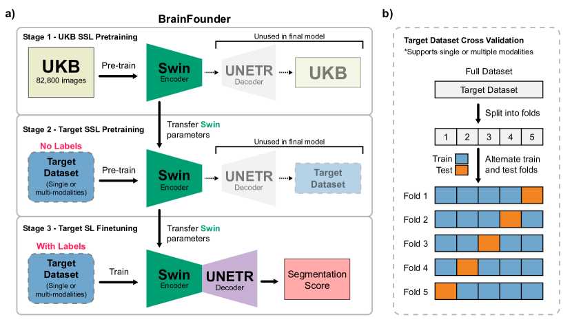
The BrainFounder framework introduces a deep learning training scheme tailored for diverse applications by showcasing a distinct approach to self-supervised pretraining followed by precise fine-tuning. This section offers a detailed examination of the framework’s architecture and its procedural pipeline and highlights the innovative phases of self-supervised pretraining, termed Stage 1 and Stage 2, before proceeding to fine-tuning for downstream tasks. The architecture and learning pathway of BrainFounder are illustrated in Figure 1. Central to BrainFounder is a vision transformer-based encoder that employs a series of self-attention mechanisms. This encoder is linked with an up-sampling decoder, tailored for segmentation tasks. This encoder based model architecture is adapted from the SwinUNETR architecture [31] with modified input channels and input hyperparemeters. BrainFounder pioneers a novel dual-phase self-supervised pretraining method, integrating self-supervised learning components within its structure. During Stage 1 pretraining, the framework is exposed to a wide-ranging dataset of brain MRIs from the UK Biobank dataset, predominantly featuring healthy individuals. This initial stage is designed to equip the model with a thorough comprehension of standard brain anatomy, utilizing self-supervised learning to enhance prediction capabilities. Stage 2 of pretraining advances the model’s proficiency by introducing it to a specialized dataset of MRIs geared towards the downstream task. This phase leverages the architecture’s refined anomaly detection skills, focusing on distinguishing deviations in brain structure.
Following the pretraining stages, BrainFounder undergoes fine-tuning on the final dataset, where the model’s encoder is enhanced through transfer learning. As depicted in Figure 1, the fine-tuning process leverages the pretrained Swin Transformer encoder from the earlier two stages. The first stage of pretraining with the UKB dataset is aimed at developing a foundational understanding of normal brain anatomy. The second stage of pretraining with unhealthy datasets builds upon this foundation by introducing pathology, thus allowing the model to learn the distinction between healthy and pathological tissues. Transfer learning is applied after each pretraining stage to retain and refine the knowledge acquired, ensuring that the model can effectively adapt to the new dataset while preserving previously learned patterns.
The culmination of this process is the integration of the U-NET decoder, which works in concert with the pretrained encoders to generate segmentation scores that delineate tumor boundaries with precision. This hybrid approach combines the strengths of the Swin Transformer and UNETR architectures, optimizing the model for the critical task of tumor segmentation and providing an authoritative score that reflects the model’s accuracy in identifying and delineating tumor regions.
In summary, the BrainFounder model’s architecture and pretraining para-digm represent a comprehensive approach to understanding and segmenting brain images, with a training pipeline that methodically builds the model’s capacity to differentiate and characterize complex patterns in MRI data. The self-supervised learning stages, coupled with the fine-tuning process, prepare BrainFounder to tackle downstream tasks with high efficiency and accuracy.
2.2 Data Acquisition and Preprocessing
Throughout our pretraining and fine-tuning, we make use of the UK Biobank (UKB), Brain Tumor Segmentation (BraTS) Challenge, and Anatomical Tracings of Lesions After Stroke v2.0 (ATLAS v2.0) datasets. The following section provides a summary of the datasets as they relate to our methodology, a summary of which can be found in Table 1.
| UK Biobank | BraTS | ATLAS | |
| Number of Subjects | 41,400 | 1,251 | 655 |
| Modalities | T1, T2-FLAIR | T1, T1-ce, T2, T2-FLAIR | T1-ce |
| Number of Images | 82,800 | 5,004 | 655 |
| Diseases | Generally Healthy | Malignant Brain Neoplasms | Stroke |
2.2.1 UK Biobank dataset
In our first stage, we utilize T1 and T2-weighted Fluid Attenuation Inversion Recovery (T2-FLAIR) from the UK Biobank (UKB) data-set ([32]). These data-points were collected starting from 2014 and preprocessed by the UKB. Utilizing a comprehensive 35-minute protocol, the UKB obtained a diverse array of brain imaging modalities, including T1-weighted and T2-FLAIR structural brain MRI images ([33]). We obtained all T1 and T2 images available between 2014 and 2022 from 44,172 participants with neuroimaging data. Raw T1 structural images were processed using a processing pipeline developed by UK Biobank researchers, generating additional images including segmentation between different types of matter and effectively reduced non-brain tissue interference. Volumetric measures of gray matter and internal structures were generated alongside the processed images, providing valuable insights into the characteristics of gray matter and internal structures. Each T1 structural image underwent further processing with FreeSurfer ([34]), followed by a quality control check for inclusion into the data made available by UK Biobank researchers. Additionally, T2-FLAIR images were defaced to uphold participant privacy and then aligned to the corresponding T1 image, resulting in two additional product images. More volumetric measures of white matter lesions were saved as an additional data measure with both T1 and T2-FLAIR data within UK Biobank. Images were reconstructed as DICOM images and converted to the NIfTI format using dcm2niix ([35]). Images were then defaced to preserve patient anonymity and broadcast to the MNI152 template space using FNIRT ([34]). Among these 44,172 participants, 43,369 participants have both T1 and T2-FLAIR images. To build 3-dimensional foundation models of neuroimages, we selected participants with both T1 and T2-FLAIR volumes more than 100 slices, resulting in 41,400 participants, and 82,800 imaging volumes. A CONSORT diagram depicting the data used in this study can be found in Figure 3, and demographic data for participants is summarized in Figure 2. For detailed information, see the Appendix.


2.2.2 BRaTS dataset
In our second stage, we perform self-supervised pretraining on MRI images from the training set of BraTS 2021 Task 1 (Tumor Segmentation) challenge. This dataset consists of 1,251 subjects each with T1, T1-contrast enhanced (T1-ce), T2, and T2-FLAIR images. We obtained all publicly available images as part of the challenge. The BraTS challenge utilizes a similar standard preprocessing pipeline as the UKB dataset. First, images are converted from DICOM images to NIfTI using dcm2niix ([35]) and tools available from the Cancer Imaging Phenomics Toolkit (CaPTk) ([36]). Images are then co-registered to the SRI24 template and resampled to a uniform resolution of 1 mm3. Finally, each modality is skull stripped; through skull stripping and conversion to NIfTI, images are both defaced, and all identifiable information is stripped from the file metadata ([14]). Imaging volumes were then segmented into three tumor classes using the STAPLE algorithm ([37]) across previous BraTS winners and refined manually. These manual annotations were further verified by multiple board-certified neuro-radiologists, resulting in quality-controlled tumor segmentation labels across all 4 modalities in 3 classes: Gd-enhancing tumor (referred to as the whole tumor: WT), edematous tissue (ED), and necrotic tumor core (TC).
2.2.3 ATLAS v2.0 Dataset
Additionally, we perform self-supervised Stage 2 pretraining and fine-tuning on MRI images from the training set of the Anatomical Tracings of Lesions After Stroke (ATLAS) v2.0 Dataset [38]. This dataset consists of 655 T1-ce MRIs aggregated from 44 research cohorts. Each MRI is from one subject, and time-points range from <24 hours to >180 days after stroke onset. The standard labeling pipeline for the ATLAS dataset consists of (1) manual quality control to exclude large motion artifacts, (2) manual lesion tracing in ITK-SNAP [39] [40], and (3) mask review by two independent raters. The data then went through a similar prepossessing pipeline to BraTS; MR images were first intensity-normalized with the MINC toolkit (https://github.com/BIC-MNI/minc-toolkit). The MINC toolkit was also used to then register the images to the MNI-152 template. Finally, FreeSurfer’s MRI deface functionality was used to deface the scans. Images were reviewed again in a final quality check at the end of the pipeline before being included in the dataset. Segmentations are evaluated on four metrics - Dice coefficient for the final segmentation (Dice), the difference between true total lesion volume and predicted total lesion volume (Volume Difference), the difference in the number of lesions between ground truth and prediction (Lesion Count), and Lesion-wise F1 Score. The Lesion-wise F1 Score is calculated by performing 3D connected-component analysis to determine true positives, false positives, and false negatives. A true positive is defined as any 3D connected-component in the ground-truth image that overlaps with at least one voxel in the prediction image. Conversely, a false positive is identified as any 3D connected-component in the prediction image that does not overlap with the ground-truth image, while a false negative is a connected-component in the ground-truth that lacks any overlapping voxels in the prediction image [38].
2.3 Stage 1: Pretraining on the UKB
The initial stage of pretraining involves the self-supervised learning of a trans-former-based neural network model using a substantial unlabeled image dataset. For this purpose, the UKB dataset ([32]) is utilized. From our 82,800 3-dimen-sional volumetric images used for pretraining, the input MRI modalities are randomly cropped into 96x96x96 sub-volumes and augmented with random inner cutout and rotation. These augmented images are then fed into the SwinUNETR encoder for processing.
Adopting the methodology from [18], the SwinUNETR encoder is pretrained through three distinct proxy tasks that serve as self-supervised fine-tuning mechanisms. The SwinUNETR architecture incorporates a Swin Transformer encoder, which is specifically designed to handle 3D input patches. This encoder operates with a patch size of 2x2x2, a feature dimension of 8, and an embedding space of 48 dimensions. It consists of four stages, with a patch merging layer introduced between stages to reduce the feature size by half.
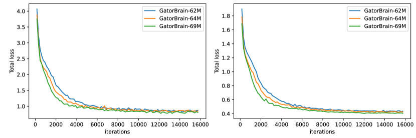
The primary objective of pretraining is to minimize the total loss function. This is achieved by training the SwinUNETR encoder using three proxy tasks: masked volume inpainting, 3D image rotation, and contrastive coding. To cater to varying complexities, three models have been developed: the foundational BrainFounder-Tiny with 62 million parameters, the intermediate BrainFounder-Small with 64 million parameters, and the advanced BrainFounder-Big, which boasts 69 million parameters. The key differentiation among these models is the variation in the number of sliding window blocks within their third stage. BrainFounder’s sliding-window encoder backbone’s parameters, number of SSL heads, and number of sliding window blocks are shown in Table 2.
For the pretraining process, a total of 64 NVIDIA DGX A100 GPUs, distributed across 8 DGX-2 nodes, are deployed at the University of Florida’s HiPerGator-AI supercomputer. Data parallelism is implemented to optimize the efficiency of model training. Both training and validation losses are monitored to track progress. The AdamW optimizer is employed with a warm-up cosine scheduler set for 500 iterations. The training employs a batch size of 2 per GPU, using 96x96x96 patches. The initial learning rate is established at 6E-6, coupled with a momentum of 0.9 and a decay of 0.1 over 15,000 iterations. These parameters are summarized in Table 3.
BrainFounder-Tiny (62M) BrainFounder-Small (64M) BrainFounder-Big (69M) # of encoder parameters 19,097,191 20,982,103 26,636,839 Encoder layer level Output size # of SSL Heads # Swin Blocks # of SSL Heads # Swin Blocks # of SSL Heads # Swin Blocks Level 1 48x(48x48x48) 3 2 3 2 3 2 Level 2 96x(24x24x24) 6 2 6 2 6 2 Level 3 192x(12x12x12) 12 2 12 6 12 18 Level 4 384x(6x6x6) 24 2 24 2 24 2 Feature size 48 48 48 Bottleneck dimension 768 768 768
2.4 Training on BraTS
2.4.1 Stage 2: Pretraining on BraTS
The pretrained models based on the UK Biobank (UKB) dataset underwent further pretraining through transfer learning on the Brain Tumor Segmentation (BraTS) dataset. 1,251 subjects were employed for a 5-fold cross-validation process. To ensure consistent performance evaluation, the data splits for these 5 folds were kept identical to those used in the baseline SwinUNETR model. During training, four of the folds were utilized for training purposes, and the remaining folds served for validation.
Given that the BraTS dataset comprises four modalities, but only two (specifically, T1 and T2) were selected for pretraining in the initial stage, the first layer of the pretrained network on UKB was modified. This modification involved expanding the number of input channels by adding two new channels, whose weights were randomly initialized using the Kaiming initialization method ([41]).
Hyperparameter settings for Stage-2 Pretraining can be found in Table 3. For pretraining on BraTS, two NVIDIA A100 GPUs, each with 32 GB of memory, were utilized. Depending on the model size, the BrainFounder models require between 48 to 72 hours for training. The batch size and learning rate were uniformly set at 2 and for all models during this pretraining phase.
2.4.2 Fine-tuning on BraTS
In the final fine-tuning stage we attach the pretrained encoder from the previous stage to a UNet decoder. This model is then finetuned directly on the BraTS dataset. We used the same hyper-parameter settings as those used in the Stage 2 pretraining phase on BraTS (in Table 3): The batch size remained at 2, mirroring the encoder-only stage, and the learning rate remained at . The number of steps for this phase was set to 50,000, with the input data having 4 channels, which indicates the typical inclusion of multi-modal MRI scans in the BraTS dataset.
2.4.3 Few-shot Learning on BraTS
To investigate our model’s performance using limited training data, we conducted a systematic comparison between BrainFounder and the baseline model, SwinUNETR, utilizing a descending percentage training approach in the context of the BraTS challenge. Using both our BrainFounder pretrained model and SwinUNETR, we finetuned on 100% of the BraTS training dataset, with subsequent incremental reductions in data availability, decreasing to a final 5% of the original dataset. This method aimed to explore the impact of training data scarcity on model performance and adaptability. Performance evaluations were carried out on the BraTS test set after each training step and evaluated with the Dice coefficient to assess segmentation accuracy.
Stage Data No. Subjects GPU Batch size Learning rate No. steps No. input channel Encoder only Pretraining UKB 43369 64 x A100 128 6E-06 2E+05 2 Encoder + Decoder Pretraining & Fine-tuning BRaTS 1251 2 x A100 2 1E-04 50000 4 Encoder + Decoder Pretraining & Fine-tuning ATLAS v2.0 655 4 x A100 4 5E-03 600 1
2.4.4 Modality Restriction and Flexibility of Modalities in Training and Inference
The proposed method is designed to be adaptable across various data modalities in downstream tasks. Specifically, we train our model using both T1 and T2 modalities, which allows it to be fine-tuned on either T1 or T2 data alone during inference without requiring any modifications to the network structure. This is achieved by simply configuring the two input channels to process the same type of data (either T1 or T2).
Furthermore, if the downstream tasks involve more modalities than those used in the initial training stage on UK Biobank data, we can accommodate this by increasing the number of input channels. The pre-trained weights are then loaded into the corresponding layers of the network.
To investigate the efficacy of this method, we performed ablation testing on BraTS by restricting the modalities available to the model. The model was given only T1 or T2 images rather than utilizing all four standard modalities (T1, T2, T1ce, FLAIR) available in BraTS. The models were then trained with our Stage 2 pretraining and finetuning pipelines with these restrictions. Hyperparameters and number of GPUs were kept the same as in our earlier pretraining steps on BraTS as summarized in Table 3.
2.5 Training on ATLAS v2.0
2.5.1 Stage 2: Pretraining on ATLAS v2.0
Similarly, the pretrained models based on the UK Biobank (UKB) underwent further self-supervised pretraining through transfer learning on the ATLAS v2.0 dataset. In this stage, a total of 665 MR images were included in the training set, intentionally avoiding cross-validation to align our methodology with that employed by the submissions in the challenge leaderboard. This approach allows for direct performance comparisons under similar training conditions, providing a robust test of our models against established benchmarks.
Since the ATLAS dataset has only one modality, T1-ce, the first layers of the Stage-1 pretrained model were modified by dropping the channel corresponding to the T2 modalitiy present in the UKB. For pretraining, four NVIDIA A100 GPUs, each with 32 GB of memory, were utilized. The stage 2 model took 35 hours to train, with a batch size and learning rate set to 4 and , respectively.
2.5.2 Fine-tuning on ATLAS v2.0
Upon completion of pretraining, the model was fine-tuned further on the ATLAS v2.0 dataset to adapt to the specific challenges of lesion detection in stroke patients. The fine-tuning employed a cosine-annealing learning rate scheduler, starting with an initial learning rate of . Batch size was set to 4, and the model was trained for a total of 600 epochs.
Training was conducted using 2 NVIDIA A100 GPUs with 32GB of RAM accessible to each GPU. We applied data augmentation techniques of random cropping, rotation, and to improve model robustness against variations in real-world data. The loss function used was Dice-loss, suited for addressing imbalance between classes. Dropout at a rate of 10% was applied to prevent overfitting.
Model performance was periodically assessed using a held-out set of 100 images from the training dataset, ensuring that the model’s improvements were generalizable and aiding with tuning the hyperparameters effectively and tracking training progress without the use of a separate validation dataset.
Sec. 3 Results
3.1 Pretraining
The pretraining of our BrainFounder models, which varied in size based on the number of parameters, took between 3 to 6 days. This process utilized a computational setup ranging from 8 to 64 NVIDIA A100 GPUs, each with 80GB capacity. Figure 4 illustrates the validation loss during the pretraining phase across different BrainFounder model sizes.
3.2 Evaluation on BraTS Challenge Dataset
3.2.1 Comparison to State-of-the-Art Methods
Table 4 summarizes our BrainFounder’s best performing model against published results from other state-of-the-art models on the BraTS challenge. In addition, we include comparison to the corresponding single-stage pretrained model that was pretrained only on the UKB. The appendix present a comparative analysis of all trained BrainFounder models (of varying sizes) against the current leading model in this field broken down by model size.
BF-S (64M) BF1-S (64M) SwinU-MRI SwinU-Res SwinU-CT nn-UNET SegResNet TransBTS Model-Zoo Fold 1 0.9032 0.8994 0.8854 0.895 0.894 0.896 0.899 0.883 0.857 Fold 2 0.9182 0.9055 0.9059 0.899 0.902 0.917 0.916 0.902 0.879 Fold 3 0.9121 0.9125 0.8981 0.894 0.898 0.910 0.909 0.889 0.820 Fold 4 0.9100 0.9133 0.8924 0.890 0.893 0.909 0.908 0.893 0.889 Fold 5 0.9139 0.9114 0.9035 0.903 0.902 0.909 0.906 0.892 0.893 Average 0.9115 0.9110 0.8971 0.896 0.898 0.908 0.907 0.891 0.868
These results show that pretraining on a large scale of healthy brain MRI data from UKB can significantly improve performance. Other observations can be made as below:
-
•
Across all folds, the BrainFounder-Small framework consistently outperformed the SwinUNETR model. This indicates that the additional training steps taken within the BrainFounder framework play a significant role in enhancing its effectiveness in brain tumor segmentation tasks.
-
•
The Small (64M) parameter version of BrainFounder achieved higher Dice coefficients on average than the 62M and 69M versions. We believe this indicates that there is an optimal range of model complexity that maximizes performance, and simply increasing the number of parameters does not necessarily lead to better results.
-
•
The one-stage Tiny model performed comparably to the two-stage BrainFounder (62M) model, which is notable implying it did not benefit considerably from the second stage pretraining on the BraTS. This might imply that the UKB dataset alone provides enough variability for effective training. Further study should be made to verify whether the benefit of pretraining on the target datasets can be found using large-scale networks.
3.2.2 Few-shot learning
Our experimental results demonstrate the performance capabilities of BrainFounder relative to the baseline model, SwinUNETR, under constrained training data conditions. As depicted in Figure 5, BrainFounder consistently matched the performance of SwinUNETR across higher levels of available training data and outperformed SwinUNETR when training data was constrained to 10% and 20%. When trained with the full 100% data, both models achieved nearly equivalent accuracy. However, as the amount of training data decreased, BrainFounder exhibited superior robustness and adaptability. Notably, at the 20% and 10% data availability levels, BrainFounder maintained higher segmentation accuracy.
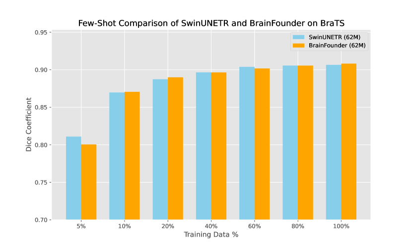
Overall, the BrainFounder (64M) model provides the best balance between complexity and performance, as evidenced by its leading average Dice coefficient. These results demonstrate the potential benefits of pretraining on the data from a large number of health subjects.
3.2.3 Modality Restriction
Models trained on T1 images alone demonstrated a significant Dice coefficient reduction of 0.168 when compared to supervised learning with all 4 modalities (mean Dice across folds of 0.8971 vs. 0.7208). However, this reduction is less than that from the same-size SwinUNETR model with the same modality restriction (mean Dice 0.678). Findings for each fold are summarized in Table 5.
BrainFounder (T1 only) SwinUNETR (T1 only) SwinUNETR (T1, T1ce, T2, T2-FLAIR) Fold 1 0.718 0.700 0.885 Fold 2 0.721 0.672 0.906 Fold 3 0.707 0.657 0.898 Fold 4 0.731 0.686 0.892 Fold 5 0.725 0.676 0.904 Average 0.721 0.678 0.897
3.3 Evaluation on ATLAS Challenge Dataset
Our model’s performance on the ATLAS v2.0 dataset was compared against the top-performing models listed on the challenge leaderboard. The results, as summarized in Table 6, demonstrate that our model achieved a Dice score of 0.712, a lesion-wise F1-score of 0.711, simple lesion count of 3.421, and volume difference of 8993.85. These scores would place our model within the top 3 models in the training set leaderboard. Worth noting again is that our training protocol did not include 5-fold cross-validation due to the lack of predetermined folds for with ATLAS can be evaluated on in the training set and the unavailability of some models from the leaderboard for independent validation. Instead, our approach focused on maximizing the comparability with the leaderboard conditions by adhering closely to their reported training setups.
Metric BrainFounder SwinUNETR CTRL(*†) HeRN(*‡) POBOTRI(*) Dice (↑) 0.712 0.703 0.663 0.718 0.663 Lesion-wise F1 Score (↑) 0.711 0.703 0.556 0.724 0.559 Simple Lesion Count (↓) 3.421 3.677 4.657 2.750 4.500 Volume Difference (↓) 8993.85 9165.18 8804.91 6162.00 9535.23
Sec. 4 Discussion
The findings from our work with BrainFounder, especially the "Small" model comprising 64 million parameters, signify a noteworthy progression in AI-driven neuroimaging technology. The framework’s novel two-stage pretraining strategy—initially utilizing a broad dataset of multi-modal neuroimages from the generally healthy population found in the UK Biobank, followed by training on pathological brain MRIs from the BraTS dataset—has demonstrated substantial efficacy. This approach enables the framework to acquire a detailed comprehension of normal brain anatomy, a vital aspect for the precise identification of anomalies, like those encountered in brain tumor segmentation tasks.
One of the key findings is the superior performance of the BrainFounder-Small (64M) model over the SwinUNETR model and other BrainFounder variants. Based on our limited explored range of parameters, our model performs best with an intermediate number of parameters. This suggests that an optimal balance of model complexity and training data is crucial. It is also indicative of the importance of large-scale datasets in training AI models for medical imaging, as even the one-stage pretraining model showed significant effectiveness. However, there is still a possibility to see higher performance using a higher number of parameters that we did not explore.
Our results from our study limiting training data indicate that BrainFounders training methods potentially offers better generalization from limited data, a crucial factor for practical applications in medical imaging where annotated data can be scarce. These findings suggest that the enhancements integrated into BrainFounder are effective in optimizing performance under varying data constraints, thereby affirming its suitability for real-world deployment in medical imaging contexts.
Further, the comparable performance of the one-stage 62M model with the two-stage approach indicates that extensive pretraining on a large and diverse dataset like UKB might be sufficient for effective model training, reducing the need for additional pretraining on targeted datasets. This insight could streamline future AI model development in medical imaging, especially in scenarios where specific pathological datasets are limited or hard to acquire.
In our modality restriction experiment, our model sees significant reduction in quality when training with multiple. This difference indicates that the multiple modalities present in BraTS contain important information not present in just T1 images about tumor segmentation. However, BrainFounder’s better performance under these difficult conditions when compared to the base SwinUNETR model validate the feasibility of our extensible approach to handling varying numbers of modalities. Our results demonstrating that the pretraining steps still have a positive effect when the information provided by the original pretraining steps requires truncation. Moreover, our results on ATLAS (discussed more in depth below) further support our method of handling multiple modalities.
The results of our study on the ATLAS dataset indicate BrainFounder’s training scheme is generalizable and effective at more than just tumor segmentation, a trait desirable for foundation models. While methods specifically adapted to optimizing results on this dataset do outperform ours, we still maintain third place in the leaderboard. Remarkably, our results were achieved without the use of ensemble learning techniques, which are commonly employed to boost performance by leveraging the strengths of multiple models. The fact that our single-model approach is competitive with ensemble models underscores the robustness and efficiency of our model in managing the intricacies of medical image analysis. We believe that the methodology used for BrainFounder can be refined and extended to move towards a Medical Foundation Model for neuroimages.
However, it’s important to note that while BrainFounder shows promise in brain tumor segmentation and brain region segmentation, its application in other neuroimaging tasks remains to be explored. One such task is brain tissue segmentation - a common task in automated analysis. Future research should investigate its adaptability to other neurological conditions, its performance in different clinical environments, and its usefulness in additional common analysis tasks.
In conclusion, BrainFounder is a significant step forward in medical AI, particularly neuroimaging. Its development underscores the potential of AI in enhancing diagnostic accuracy and efficiency, paving the way for more advanced, adaptable, and robust AI tools in healthcare.
Sec. 5 Acknowledgments
This work was partially supported by the National Science Foundation (1908299, 2318984). We greatly acknowledge the support from NVIDIA AI Technology (NVAITC) to this research project, especially the great support with GPU parallelization technology. We extend our appreciation to the contributors of the UK Biobank and BraTS datasets, whose extensive data collection efforts have been fundamental to the success of this research. Special thanks to the team at the University of Florida’s HiPerGator-AI supercomputer for providing the computational resources that were essential in training the BrainFounder models.
References
- [1] Yutong Chen et al. “AI-Based Reconstruction for Fast MRI-A Systematic Review and Meta-Analysis” In Proceedings of the IEEE 110.2, 2022, pp. 224–245 DOI: 10.1109/JPROC.2022.3141367
- [2] Alice Segato, Aldo Marzullo, Francesco Calimeri and Elena De Momi “Artificial intelligence for brain diseases: A systematic review” In APL Bioengineering 4.4, 2020, pp. 041503 DOI: 10.1063/5.0011697
- [3] Rajesh P.. Rao “Brain Co-processors: Using AI to Restore and Augment Brain Function” In Handbook of Neuroengineering Singapore: Springer Nature, 2023, pp. 1225–1260 DOI: 10.1007/978-981-16-5540-1_32
- [4] Mayowa O. Owolabi et al. “Global synergistic actions to improve brain health for human development” Number: 6 Publisher: Nature Publishing Group In Nature Reviews Neurology 19.6, 2023, pp. 371–383 DOI: 10.1038/s41582-023-00808-z
- [5] Diego Moreno-Blanco et al. “Technologies for Monitoring Lifestyle Habits Related to Brain Health: A Systematic Review” Number: 19 Publisher: Multidisciplinary Digital Publishing Institute In Sensors 19.19, 2019, pp. 4183 DOI: 10.3390/s19194183
- [6] Pranav Rajpurkar, Emma Chen, Oishi Banerjee and Eric J. Topol “AI in health and medicine” Number: 1 Publisher: Nature Publishing Group In Nature Medicine 28.1, 2022, pp. 31–38 DOI: 10.1038/s41591-021-01614-0
- [7] A.. Khachaturian et al. “Accelerating Innovations for Enhanced Brain Health. Can Artificial Intelligence Advance New Pathways for Drug Discovery for Alzheimer’s and other Neurodegenerative Disorders?” In The Journal of Prevention of Alzheimer’s Disease 10.1, 2023, pp. 1–4 DOI: 10.14283/jpad.2023.1
- [8] Michael Moor et al. “Foundation models for generalist medical artificial intelligence” Number: 7956 Publisher: Nature Publishing Group In Nature 616.7956, 2023, pp. 259–265 DOI: 10.1038/s41586-023-05881-4
- [9] Bobby Azad et al. “Foundational Models in Medical Imaging: A Comprehensive Survey and Future Vision” arXiv:2310.18689 [cs] arXiv, 2023 DOI: 10.48550/arXiv.2310.18689
- [10] Shaoting Zhang and Dimitris Metaxas “On the challenges and perspectives of foundation models for medical image analysis” In Medical Image Analysis 91, 2024, pp. 102996 DOI: 10.1016/j.media.2023.102996
- [11] Fabian Isensee et al. “nnU-Net: a self-configuring method for deep learning-based biomedical image segmentation” Number: 2 Publisher: Nature Publishing Group In Nature Methods 18.2, 2021, pp. 203–211 DOI: 10.1038/s41592-020-01008-z
- [12] Richard McKinley, Raphael Meier and Roland Wiest “Ensembles of Densely-Connected CNNs with Label-Uncertainty for Brain Tumor Segmentation” In Brainlesion: Glioma, Multiple Sclerosis, Stroke and Traumatic Brain Injuries, Lecture Notes in Computer Science Cham: Springer International Publishing, 2019, pp. 456–465 DOI: 10.1007/978-3-030-11726-9_40
- [13] Konstantinos Kamnitsas et al. “Efficient multi-scale 3D CNN with fully connected CRF for accurate brain lesion segmentation” In Medical Image Analysis 36, 2017, pp. 61–78 DOI: 10.1016/j.media.2016.10.004
- [14] Ujjwal Baid et al. “The RSNA-ASNR-MICCAI BraTS 2021 Benchmark on Brain Tumor Segmentation and Radiogenomic Classification” arXiv:2107.02314 [cs] arXiv, 2021 DOI: 10.48550/arXiv.2107.02314
- [15] Michela Antonelli et al. “The Medical Segmentation Decathlon” Number: 1 Publisher: Nature Publishing Group In Nature Communications 13.1, 2022, pp. 4128 DOI: 10.1038/s41467-022-30695-9
- [16] F. al. Quinton “A tumor and liver automatic segmentation challenge”, Zenodo, 2023 DOI: 10.5281/zenodo.7835370
- [17] Zongwei Zhou et al. “Models Genesis” In Medical Image Analysis 67, 2021, pp. 101840 DOI: 10.1016/j.media.2020.101840
- [18] Yucheng Tang et al. “Self-Supervised Pre-Training of Swin Transformers for 3D Medical Image Analysis” arXiv:2111.14791 [cs] arXiv, 2022 URL: http://arxiv.org/abs/2111.14791
- [19] Xueyan Mei et al. “RadImageNet: An Open Radiologic Deep Learning Research Dataset for Effective Transfer Learning” Publisher: Radiological Society of North America In Radiology: Artificial Intelligence 4.5, 2022, pp. e210315 DOI: 10.1148/ryai.210315
- [20] Kenneth Clark et al. “The Cancer Imaging Archive (TCIA): Maintaining and Operating a Public Information Repository” In Journal of Digital Imaging 26.6, 2013, pp. 1045–1057 DOI: 10.1007/s10278-013-9622-7
- [21] Clare Bycroft et al. “The UK Biobank resource with deep phenotyping and genomic data” Number: 7726 Publisher: Nature Publishing Group In Nature 562.7726, 2018, pp. 203–209 DOI: 10.1038/s41586-018-0579-z
- [22] Alexander Kirillov et al. “Segment Anything” arXiv:2304.02643 [cs] arXiv, 2023 DOI: 10.48550/arXiv.2304.02643
- [23] Hugo Touvron et al. “LLaMA: Open and Efficient Foundation Language Models” arXiv:2302.13971 [cs] arXiv, 2023 DOI: 10.48550/arXiv.2302.13971
- [24] Ming Y. Lu et al. “Visual Language Pretrained Multiple Instance Zero-Shot Transfer for Histopathology Images” arXiv:2306.07831 [cs] arXiv, 2023 DOI: 10.48550/arXiv.2306.07831
- [25] Shruthi Bannur et al. “Learning to Exploit Temporal Structure for Biomedical Vision-Language Processing” arXiv:2301.04558 [cs] arXiv, 2023 DOI: 10.48550/arXiv.2301.04558
- [26] Ekin Tiu et al. “Expert-level detection of pathologies from unannotated chest X-ray images via self-supervised learning” In Nature Biomedical Engineering 6.12, 2022, pp. 1399–1406 DOI: 10.1038/s41551-022-00936-9
- [27] Jun Ma et al. “Segment Anything in Medical Images” arXiv:2304.12306 [cs, eess] arXiv, 2023 DOI: 10.48550/arXiv.2304.12306
- [28] Wenhui Lei et al. “MedLSAM: Localize and Segment Anything Model for 3D CT Images” arXiv:2306.14752 [cs] arXiv, 2023 DOI: 10.48550/arXiv.2306.14752
- [29] Junlong Cheng et al. “SAM-Med2D” arXiv:2308.16184 [cs] arXiv, 2023 DOI: 10.48550/arXiv.2308.16184
- [30] S. al. Gong “3DSAM-adapter: Holistic Adaptation of SAM from 2D to 3D for Promptable Medical Image Segmentation”, arXiv, 2023 DOI: 10.48550/arXiv.2306.13465
- [31] Ali Hatamizadeh et al. “Swin UNETR: Swin Transformers for Semantic Segmentation of Brain Tumors in MRI Images” arXiv:2201.01266 [cs, eess] arXiv, 2022 DOI: 10.48550/arXiv.2201.01266
- [32] Thomas J. Littlejohns et al. “The UK Biobank imaging enhancement of 100,000 participants: rationale, data collection, management and future directions” Number: 1 Publisher: Nature Publishing Group In Nature Communications 11.1, 2020, pp. 2624 DOI: 10.1038/s41467-020-15948-9
- [33] Stephen M. Smith, Alfaro-Almagro Fidel and Miller L. Karla “UK Biobank Brain Imaging Documentation, Version 1.9” UK Biobank, 2022 URL: https://biobank.ctsu.ox.ac.uk/crystal/crystal/docs/brain_mri.pdf
- [34] Mark W. Woolrich et al. “Bayesian analysis of neuroimaging data in FSL” In NeuroImage 45.1 Suppl, 2009, pp. S173–186 DOI: 10.1016/j.neuroimage.2008.10.055
- [35] Xiangrui Li et al. “The first step for neuroimaging data analysis: DICOM to NIfTI conversion” In Journal of Neuroscience Methods 264, 2016, pp. 47–56 DOI: 10.1016/j.jneumeth.2016.03.001
- [36] Christos Davatzikos et al. “Cancer imaging phenomics toolkit: quantitative imaging analytics for precision diagnostics and predictive modeling of clinical outcome” In Journal of Medical Imaging (Bellingham, Wash.) 5.1, 2018, pp. 011018 DOI: 10.1117/1.JMI.5.1.011018
- [37] Simon K. Warfield, Kelly H. Zou and William M. Wells “Simultaneous truth and performance level estimation (STAPLE): an algorithm for the validation of image segmentation” In IEEE transactions on medical imaging 23.7, 2004, pp. 903–921 DOI: 10.1109/TMI.2004.828354
- [38] Sook-Lei Liew et al. “A large, curated, open-source stroke neuroimaging dataset to improve lesion segmentation algorithms” In Scientific Data 9.1, 2022, pp. 320 DOI: 10.1038/s41597-022-01401-7
- [39] Paul A. Yushkevich et al. “User-guided 3D active contour segmentation of anatomical structures: Significantly improved efficiency and reliability” In NeuroImage 31.3, 2006, pp. 1116–1128 DOI: 10.1016/j.neuroimage.2006.01.015
- [40] Paul A. Yushkevich and Guido Gerig “ITK-SNAP: An Intractive Medical Image Segmentation Tool to Meet the Need for Expert-Guided Segmentation of Complex Medical Images” Conference Name: IEEE Pulse In IEEE Pulse 8.4, 2017, pp. 54–57 DOI: 10.1109/MPUL.2017.2701493
- [41] Kaiming He, Xiangyu Zhang, Shaoqing Ren and Jian Sun “Delving Deep into Rectifiers: Surpassing Human-Level Performance on ImageNet Classification” arXiv:1502.01852 [cs] version: 1 arXiv, 2015 DOI: 10.48550/arXiv.1502.01852
Sec. 6 Appendix
6.1 UK Biobank Data
Table 7 presents a comprehensive summary of the participants used from the UK Biobank.
| Entire UK Biobank | 40K Cohort (%) | |
| Age at Recruitment | ||
| 45 | 51,763 (10.3%) | 4,601 (11.1%) |
| 45-49 | 51,866 (10.3%) | 5,031 (12.2%) |
| 50-54 | 74,387 (14.8%) | 7,563 (18.3%) |
| 55-59 | 86,899 (17.3%) | 8,819 (21.3%) |
| 60-64 | 118,959 (23.7%) | 9,579 (23.1%) |
| 65 | 118,435 (23.6%) | 5,796 (14.0%) |
| Unanswered | 101 (0.02%) | 11 (0.03%) |
| Gender | ||
| Male | 229,051 (45.6%) | 19,497 (47.1%) |
| Female | 273,258 (54.4%) | 21,891 (52.9%) |
| Unanswered | 101 (0.02%) | 12 (0.03%) |
| Race | ||
| White | 472521 (94.1%) | 40057 (96.8%) |
| Mixed | 2953 (0.6%) | 190 (0.5%) |
| Asian | 11447 (2.3%) | 541 (1.3%) |
| Black | 8055 (1.6%) | 268 (0.7%) |
| Other | 4555 (0.9%) | 223 (0.5%) |
| Unanswered | 2,778 (0.6%) | 121 (0.3%) |
| Data Information | ||
| # Samples | 502,309 | 41,400 |
| # Brain Tumors | 1,210 | 42 |
6.2 Fold-wise comparison of all models.
Table 8 provides fold-wise comparison of our BrainFounder models across all tested parameters.
SwinUNETR (62M) BF-T (62M) BF-S (64M) B.F (69M) One-Stage (62M) One-Stage (64M) One-Stage (69M) Fold 1 0.8854 0.9027 0.9032 0.9014 0.9019 0.8994 0.8999 Fold 2 0.9059 0.9181 0.9182 0.9164 0.9188 0.9186 0.9055 Fold 3 0.8981 0.9102 0.9121 0.9097 0.9119 0.9125 0.9002 Fold 4 0.8924 0.9103 0.9100 0.9070 0.9107 0.9133 0.9109 Fold 5 0.9035 0.9139 0.9141 0.9101 0.9132 0.9114 0.9103 Average 0.8971 0.9110 0.9115 0.9089 0.9112 0.9110 0.9054