DeNVeR: Deformable Neural Vessel Representations for Unsupervised Video Vessel Segmentation
Abstract
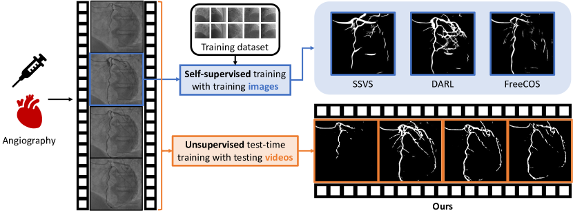
This paper presents Deformable Neural Vessel Representations (DeNVeR), an unsupervised approach for vessel segmentation in X-ray videos without annotated ground truth. DeNVeR uses optical flow and layer separation, enhancing segmentation accuracy and adaptability through test-time training. A key component of our research is the introduction of the XACV dataset, the first X-ray angiography coronary video dataset with high-quality, manually labeled segmentation ground truth. Our evaluation demonstrates that DeNVeR outperforms current state-of-the-art methods in vessel segmentation. This paper marks an advance in medical imaging, providing a robust, data-efficient tool for disease diagnosis and treatment planning and setting a new standard for future research in video vessel segmentation. See our project page for video results: kirito878.github.io/DeNVeR.
1 Introduction
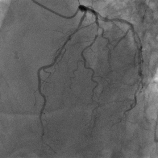
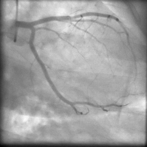
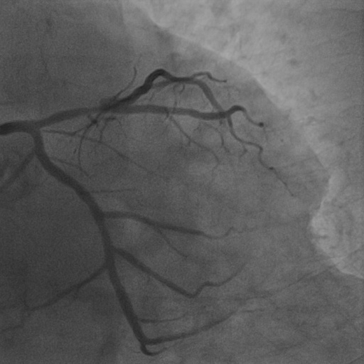
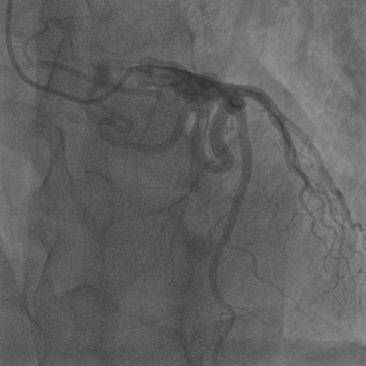
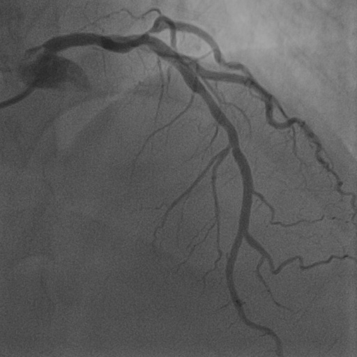
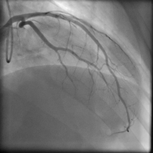
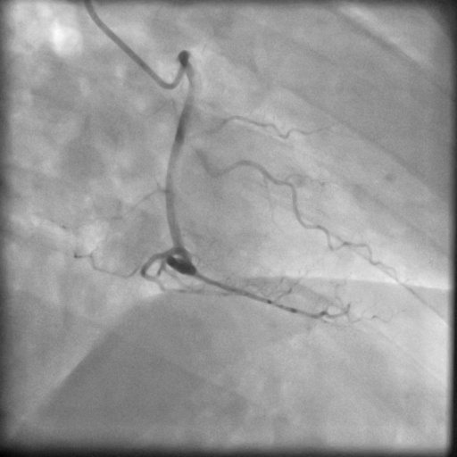
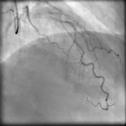
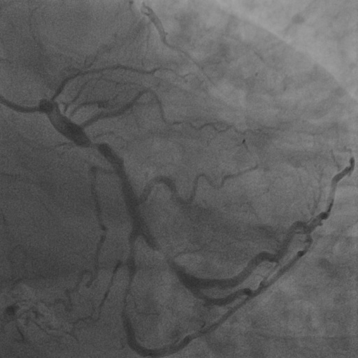
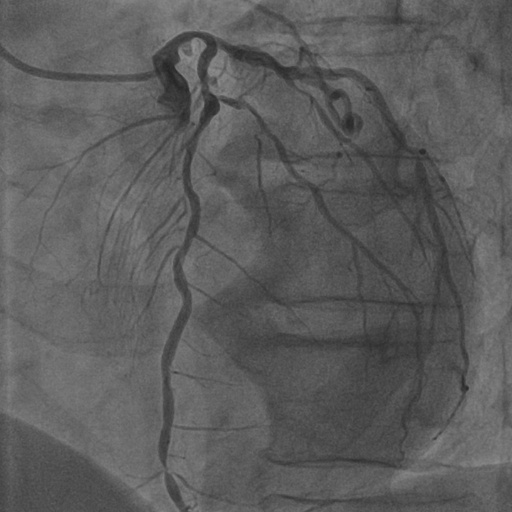
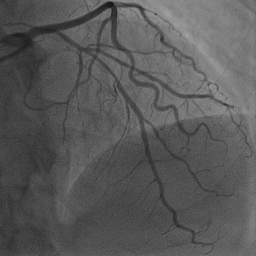
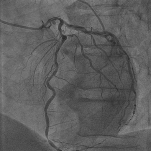
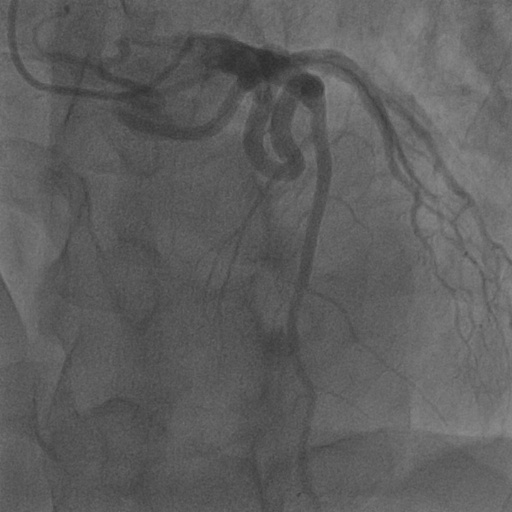
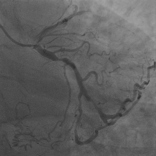
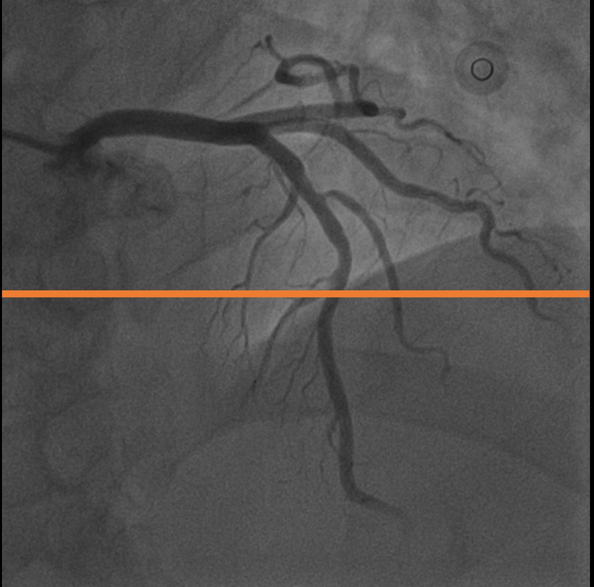
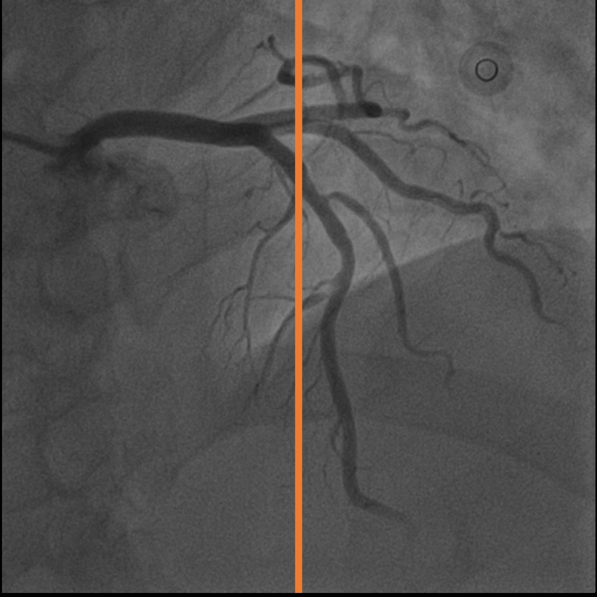
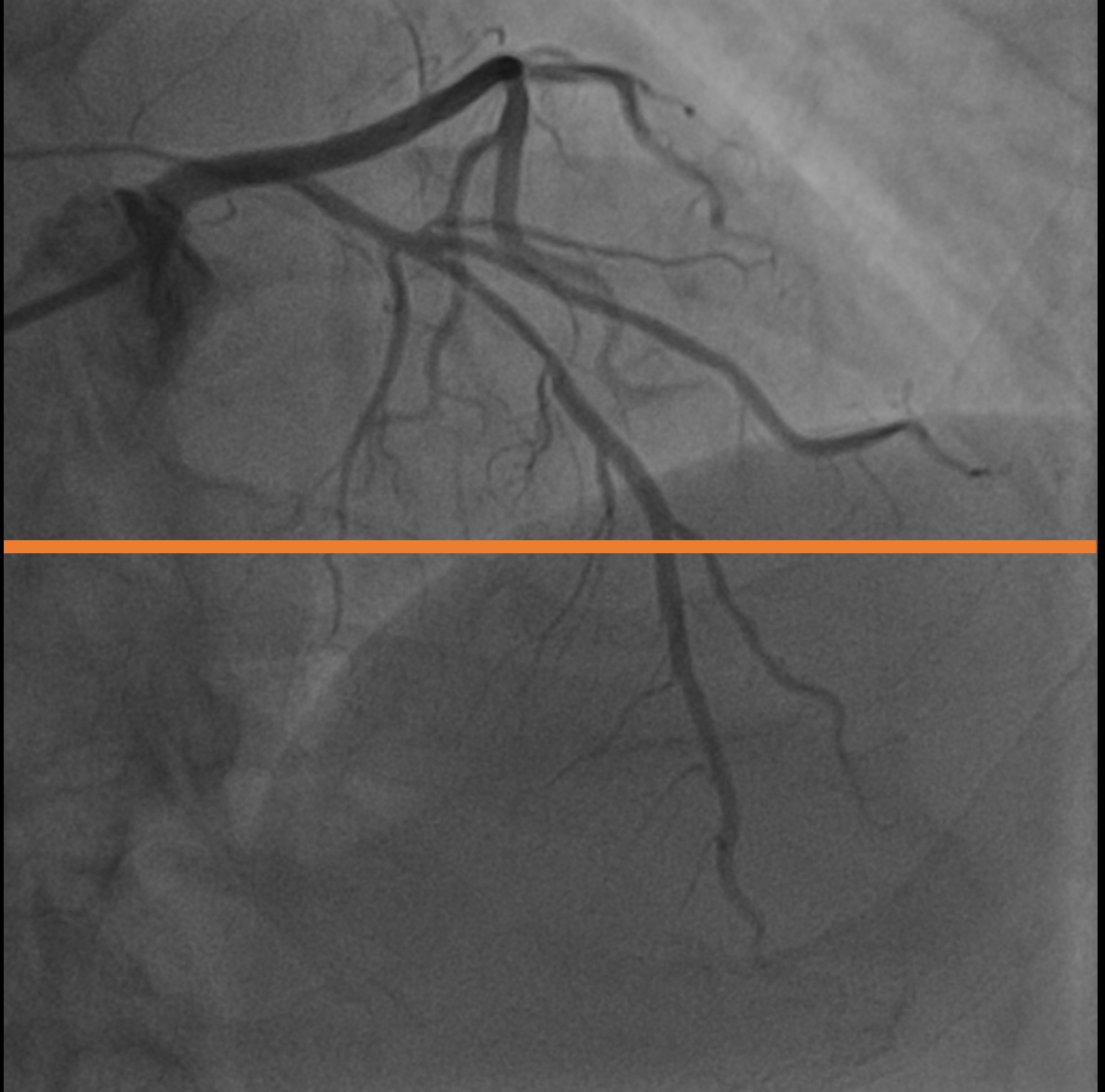
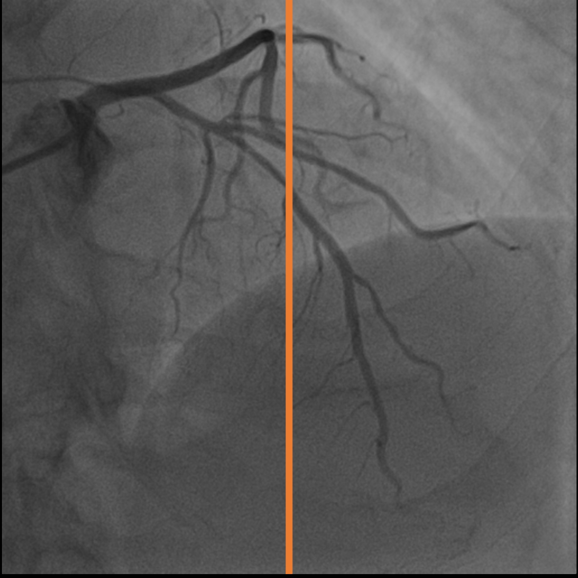
Coronary arteries (CAs) are essential for delivering oxygen-rich blood to the heart muscle [9]. To assess coronary artery circulation and diagnose disease, cardiologists use hemodynamic measures like fractional flow reserve (FFR) and instantaneous wave-free ratio to determine the severity of stenosis [15]. Since traditional pressure wire (PW)-based techniques are invasive and involve higher risks [53], cardiologists often assess stenosis severity by visually inspecting X-ray angiography (XRA) images. By injecting contrast agents into the coronary vessels and capturing the flow in the vessel structure on video, X-ray coronary angiography (XCA) is a common medical imaging method that exposes patients to less ionizing radiation and provides clear boundaries of the coronary arteries.
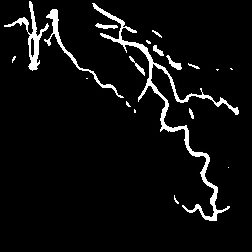
Accurate vessel segmentation remains challenging due to XCA’s inherent limitations, which can obscure the severity of stenosis [55]. These limitations include low signal-to-noise ratios, minimal radiation contrast [12], and interference from surrounding structures like catheters and bones [34]. The complexity of interpreting 2D projections of 3D vessels further complicates the task. Existing automatic angiographic vessel segmentation algorithms have several drawbacks. They often require professional user input and supervision to identify corresponding vessels or features in all input images [17]. The time-consuming annotation and knowledge-based training processes make it challenging to adopt these methods in practical settings. Models that take a single image as input discard critical information from the original XCA video and show reduced adaptability and compatibility when the imaging system changes or is unknown. In addition to the characteristics of X-ray angiography, involuntary organ motions and overlapping structures contribute to an increased ratio of ghosting artifacts [30, 29]. These supervision, generalization, and dynamics issues significantly limit the application of automatic angiographic segmentation algorithms (Figure 1).
To address these challenges, we introduce DeNVeR (Deformable Neural Vessel Representations), an unsupervised approach for segmenting cardiac vessels in X-ray videos. Inspired by Deformable Sprites and using optical flow, DeNVeR starts with traditional Hessian-based filters to establish initial vessel masks as priors. It then uses a layered separation process to decompose the foreground vessel and background layers. Acknowledging the limitations of frame-by-frame processing, we enhance foreground-background segmentation through test-time optimization. This optimization incorporates neural representations of the Eulerian motion field and introduces a novel parallel vessel motion loss, thereby improving segmentation fidelity. Our approach emphasizes dynamic adaptation to cardiac movements and vessel flow, ensuring detailed, temporally consistent, and unsupervised vessel segmentation in X-ray videos. Our experimental results show significant performance improvements in predicting vessel regions compared to state-of-the-art models. The main contributions are:
-
•
DeNVeR uses unsupervised learning on X-ray video data, leveraging the full temporal information of the videos and eliminating the need for annotated training datasets.
-
•
Using optical flow and a unique layer separation strategy, DeNVeR enhances segmentation accuracy and adjusts during test time, improving adaptability and ensuring consistent results across cardiac conditions.
-
•
We collect the first X-ray angiography coronary video dataset (XACV) with high-quality, manually labeled segmentation ground truth, serving as a new standard for training and evaluating video vessel segmentation models, making full use of video temporal information.
2 Related Work
Traditional segmentation methods.
Traditional object segmentation [21, 35] requires heuristic human design rules or filters. Several methods are proposed, one of which designs the Hessian-based filter [13] to enhance vessel filtering. Khan et al. [21] design retinal image denoising and enhancement of B-COSFIRE filters to perform segmentation. Memari et al. [35] used contrast-limited adaptive histogram equalization and designed filters to achieve the task. Another line of work proposed optimally oriented flux (OOF) [57, 26], which performs better in adjacent curvilinear object segmentation. These human strategy design methods do not need any training, which gives them the advantage of fast training. However, these methods are often confined to certain datasets and loss of generalize ability.
Supervised and self-supervised segmentation.
In the domain of the supervised segmentation method [52], Esfahani et al. [39] design a Top-Hat transformation and Convolutional Neural Networks (CNNs) for segmentation. Khowaja et al. [22] applied bidirectional histogram equalization. Another work [63, 47] utilizes image masking to reduce artifacts, which relies on paired mask datasets. The most popular vessel segmentation backbone recently is U-Net [48]. These methods [11, 52, 64] also require extensive and time-consuming human annotation, further limiting the application. Consequently, self-supervised learning methods are designed to elevate performance with large-scale unsupervised data. Some self-supervised learning research focuses on image painting [44], image colorization [25], and others [10, 40, 27, 46, 37, 62, 4, 10, 44, 14, 36, 33, 61, 3, 42, 60, 59, 2, 67]. Ma et al. [33] and Kim et al. [23] proposed vessel segmentation methods with adversarial learning.
Unsupervised segmentation methods.
Unsupervised segmentation methods fall into two categories: clustering-based and adversarial. Clustering-based approaches [18, 28, 8] like Invariant Information Clustering (IIC) by Xu et al. [18] inputs into clusters but struggle with curvilinear objects. Adversarial methods [7, 1], exemplified by Redo [1], generate object masks by guiding generators with inputs to redraw objects in new colors.
Video segmentation methods.
Our work advances video decomposition into layers, originally proposed by Wang & Adelson [58] in the 1990s, by incorporating neural network techniques [31, 32]. Unlike traditional methods [5, 20, 41, 49, 6], we operate unsupervised, learning deformable canonical layers to model vessel motion more effectively. Additionally, unlike unsupervised video segmentation methods [65, 66], we address motion segmentation by associating pixels with Eulerian motion clusters and extend our approach to unsupervised video vessel segmentation. Our method, DeNVeR, separates X-ray video into canonical foreground and background, per-frame masks, and dynamic transformations for realistic vessel motion representation optimized through specific loss functions.
3 Method
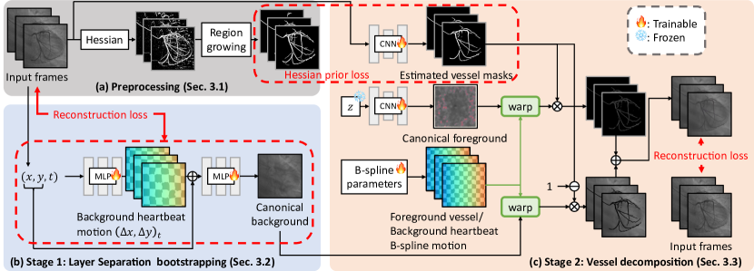
Inspired by Deformable Sprites [66], we develop a novel unsupervised algorithm for Cardiac Vessel Segmentation in X-ray video based on optical flow. Given a cardiac X-ray video, it can perform test-time optimization by computing reconstruction without any training data. Initially, we employ traditional blood vessel segmentation methods to extract vascular regions in a very coarse manner (Section 3.1). Since each input frame is processed independently, the temporal consistency of the output video is severely compromised, leading to fragmentation. To address those issues, we employ implicit neural representation to learn layer separation for segmenting blood vessels (foreground) from X-ray heart images (background) and estimate the corresponding background flow with lower degrees of freedom (Section 3.2). Subsequently, we fix the background transformation to optimize the contrast agent flow in the foreground. Here, we introduce our test-time training workflow (Section 3.3). Finally, we elaborate on the loss and regularization terms we use (Section 3.4).
3.1 Preprocessing
Segmenting vascular regions from X-ray images through unsupervised methods is a highly challenging task. To facilitate our subsequent work, we employ a Hessian-based filter [13] to generate a set of binary masks that crudely represent blood vessel regions. Specifically, our approach comprises the following two steps:
(1) Apply a Hessian-based filter to the entire sequence of images. The output pixel intensities range from 0 to 255, with higher numerical values indicating a stronger presence of tubular structural features.
(2) Based on the outputs from step (1), calculate the overall intensities of the entire image and set an appropriate threshold to convert them into binary images. The purpose of selecting the threshold is to ensure that images with higher intensities correspond to larger vascular areas. Finally, we employ a region-growing post-processing technique to eliminate noise.
Additionally, we use a pre-trained optical flow model, RAFT [54], to generate the initial optical flow between consecutive frames. We illustrate the preprocessing steps in Figure 2 (a).
3.2 Layer Separation Bootstrapping
While utilizing the Hessian-based filter allows us to quickly acquire a set of rough masks, the periodic heartbeat introduces temporal inconsistency, resulting in variations in the position of vascular regions over time. In addressing this challenge, we implement a solution by separating the input frames into foreground and background. Inspired by NIR [38], we embrace the approach of employing MLPs to learn implicit neural representations of images (Figure 2 (b)). The primary objective of each MLP is to minimize the following losses:
| (1) |
| (2) |
| (3) |
| (4) |
where and denote the original ground truth (i.e., input RGB frames) and the output of the first MLP, respectively. To ensure flow smoothness, we introduce a penalty term for the MLP computing the background, denoted as . Here, represents a Jacobian matrix comprising gradients of . Finally, since the stationary background should occupy the vast majority of the scene, we introduce an additional penalty term for which learns to represent the scene beyond the background. and are weight hyperparameters.
3.3 Test-Time Training for Vessel Decomposition
Our work introduces test-time training as a key feature of our unsupervised segmentation method, DeNVeR. This approach adapts the model directly to test data during inference without labeled training data. Using the test video’s inherent structure and patterns, the model refines its parameters, allowing it to tailor its learning to each video’s unique characteristics. This capability is particularly valuable in medical imaging, where patient variability is high and personalized diagnostics are crucial. Test-time training allows DeNVeR to adjust dynamically to new, unseen cases, significantly improving diagnostic accuracy and effectiveness.
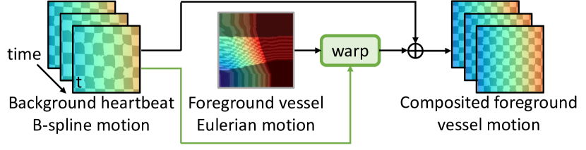
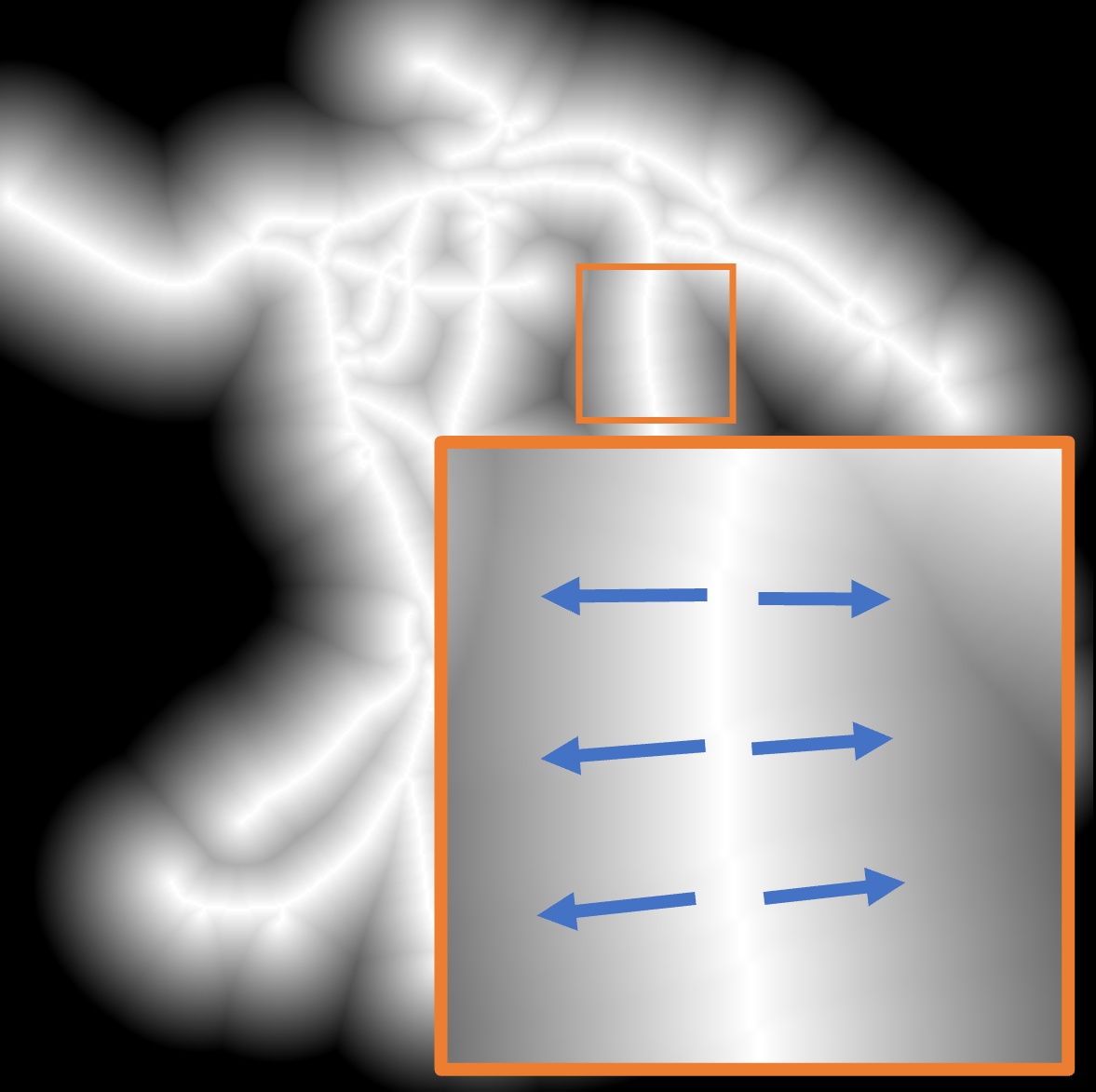
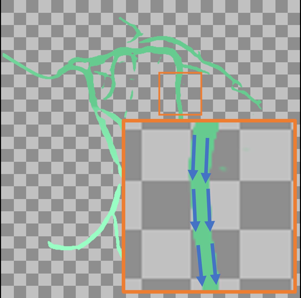
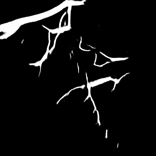
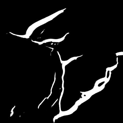
After obtaining the Hessian-based approach as prior and bootstrapped static background, we focus on utilizing a pre-trained optical flow estimator, RAFT [54], to further separate the vessel and background layer and obtain vessel segmentation masks. As shown in Figure 2 (c), for each image, we use a CNN model to predict masks for the foreground and background. Both foreground and background have their canonical images. To simplify the problem, we first compute the canonical image for the background in Stage 1 (Section 3.2) and keep it fixed. Then, in Stage 2, we optimize the canonical foreground using a CNN model with a fixed latent code [56]. Next, we use motion flow to reconstruct respective images from the canonical images. To enhance the coherence of each frame’s mask, we calculate the flow warp loss, requiring both foreground and background motion fields. Thus, we utilize the spatial and temporal B-spline to model the entire motion trajectory.
 |
 |
 |
| (a) Vessel mask | (b) Distance transform | (c) Vessel motion |
Background motion fields.
In the case of cardiac X-ray imaging, the background usually includes the heart and ribs, which don’t experience significant displacement. Therefore, we use a B-spline with lower degrees of freedom to estimate the motion flow.
Foreground motion fields.
As for the foreground, we observe that the contrast agent flows out from the catheter. Therefore, we consider the Eulerian motion field, as shown in Figure 4, to be a more reasonable specification of blood flow behavior compared to the traditional motion field.
3.4 Losses and Regularizations
Hessian prior loss.
After obtaining the initial mask from preprocess (Section 3.1), we utilize a CNN model for the initial segmentation task. In this step, we aim for the model’s predicted mask to closely resemble the mask generated by traditional algorithms. To achieve this, we employ a Hessian prior loss:
| (5) |
where represents the mask of frame t generated by the preprocessing part, denotes the background mask of frame t predicted by the mask model, and represents the foreground weight. Note that masks generated by this per-frame operation are not temporally continuous. Therefore, we will optimize continuity through subsequent methods.
Parallel loss.
Clearly, the direction of blood flow should align with the course of blood vessels (Figure 4). Hence, we design the parallel loss to achieve a parallel alignment between them. Initially, we conduct skeletonization and distance transform on the masks obtained from Section 3.1, and calculate pixel-wise cosine similarity between these transformed masks and the predicted flow:
| (6) |
| (7) |
where represents the value obtained from the distance transform at pixel coordinate , and are the image gradients from the two spatial directions, and denotes the predicted flow value at position .
Flow warp loss.
To maintain consistency in the predicted flow for both the foreground vessel and background, we introduce the flow warp loss:
| (8) |
where represents the mask for frame t, denotes RAFT optical flow computed from frame at time to , denotes the background layer or foreground layer, and is the scale of . The flow warp loss encourages the flow between nearby frames of both foreground vessel and background layers to follow the guidance from the optical flow predicted by RAFT.
Mask consistency loss.
For the predicted masks, to ensure consistency across frames, we introduce the mask consistency loss :
| (9) |
Reconstruction loss.
We use the L1 distance between the predicted image and the original image for Reconstruction loss calculation:
| (10) |
Our final loss function is described as follows:
| (11) |
4 Experiments
 |
 |
 |
 |
 |
 |
 |
 |
 |
|
 |
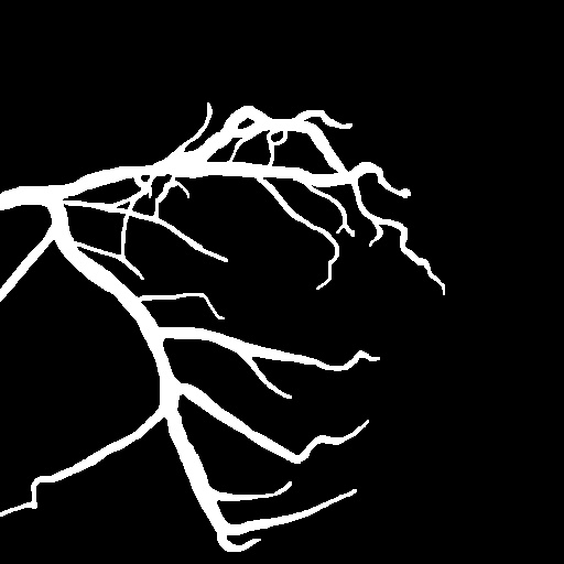 |
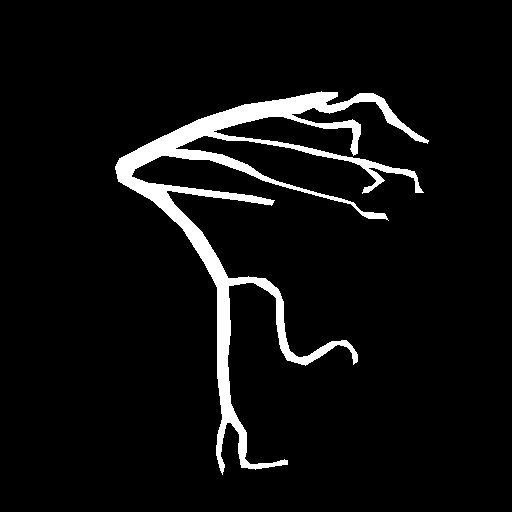
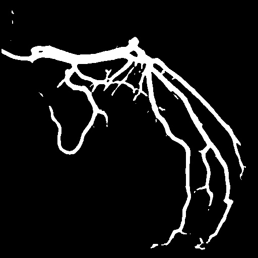
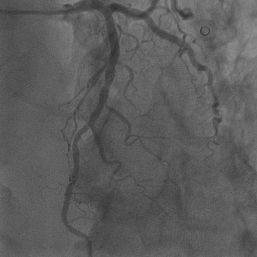
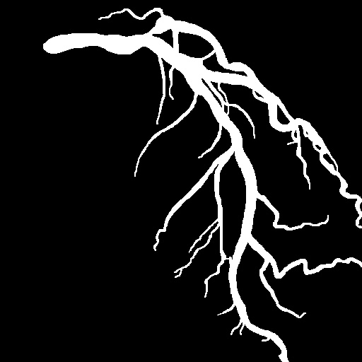
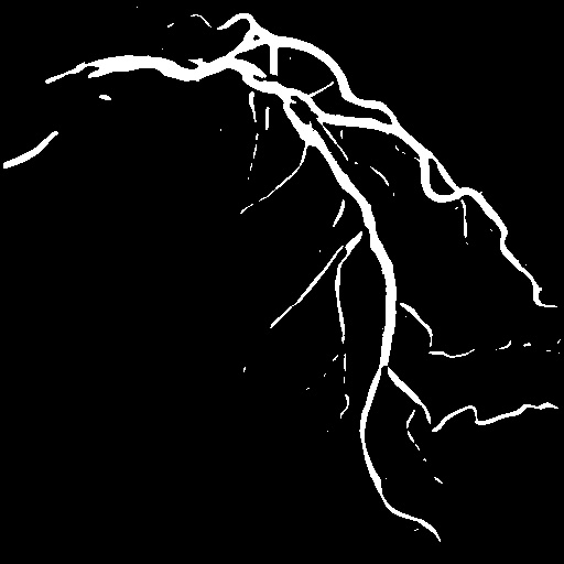
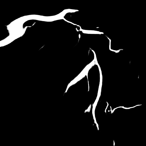
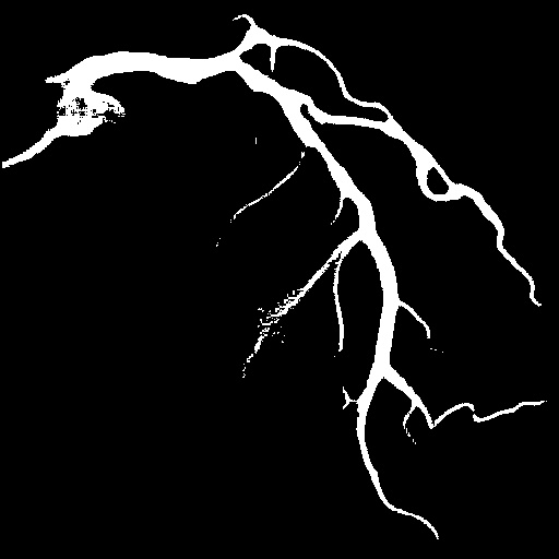
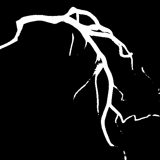
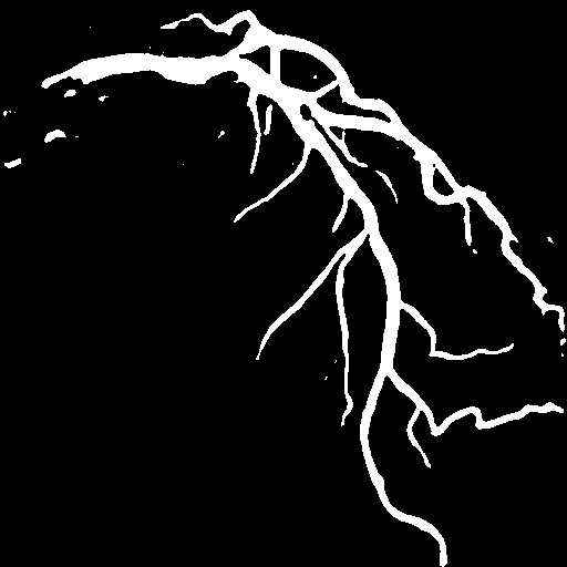
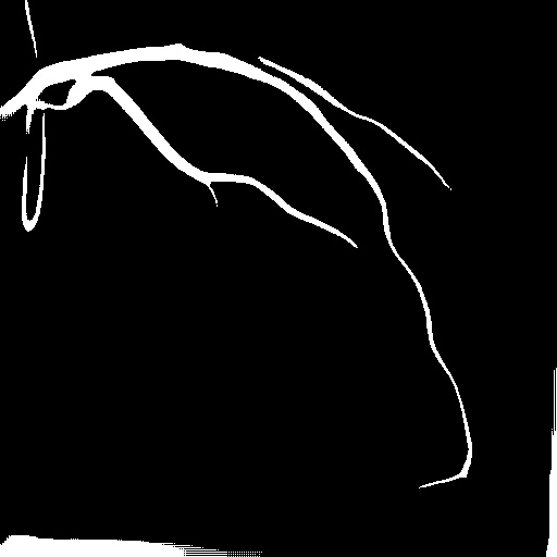
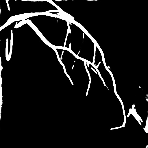
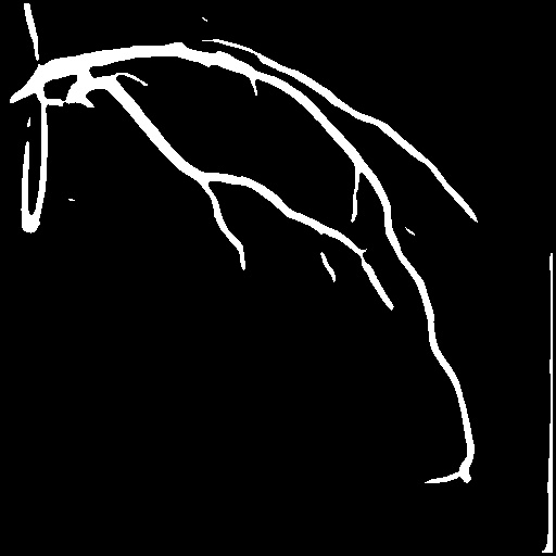
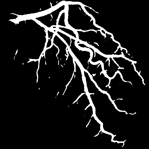
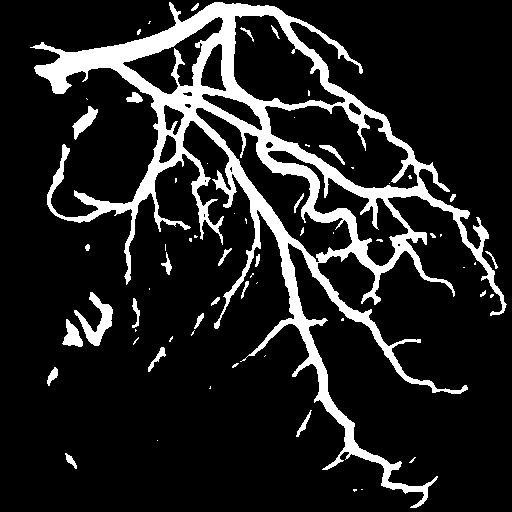
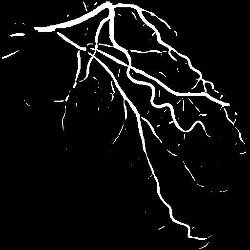
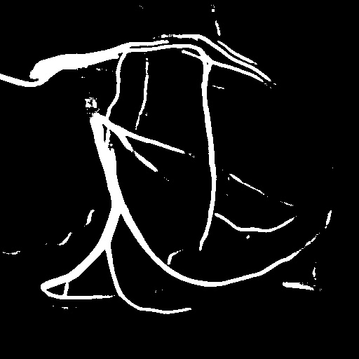
| Image | Ground truth | Video frame | Ground truth | Video frame | Ground truth |
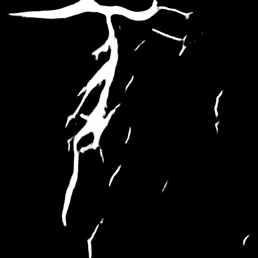
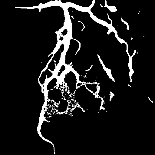
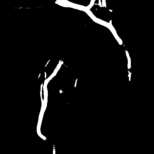
| XCAD [33] | CADICA [19] | Our XACV dataset | |||
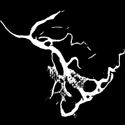
4.1 XACV Dataset
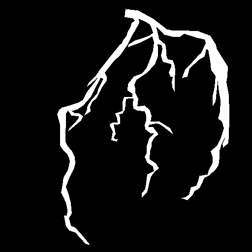
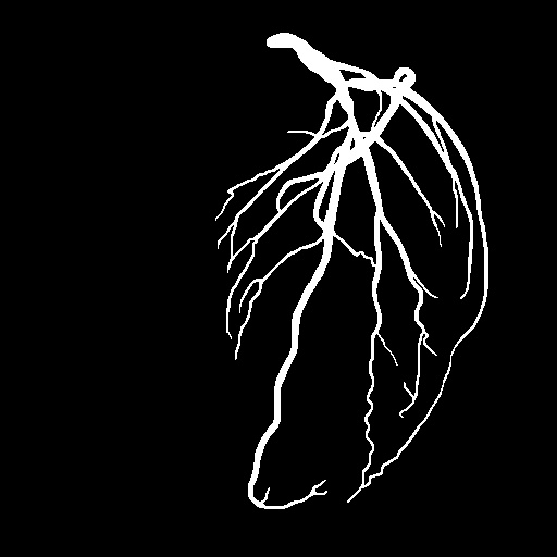
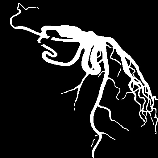
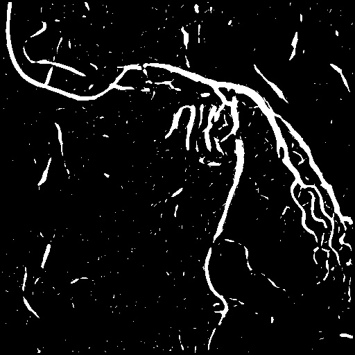
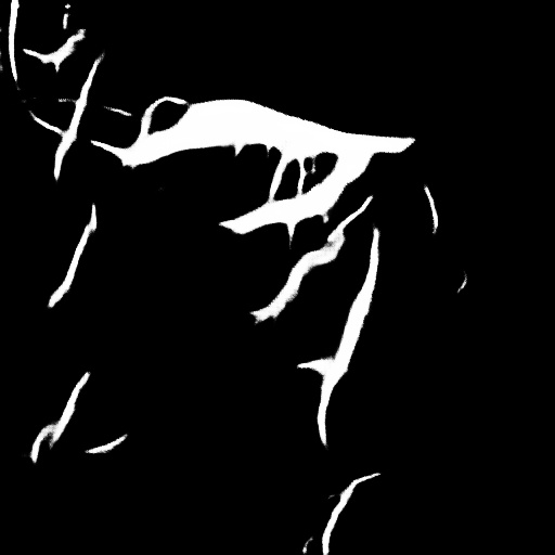
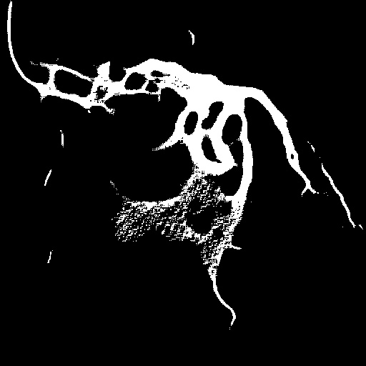
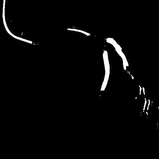
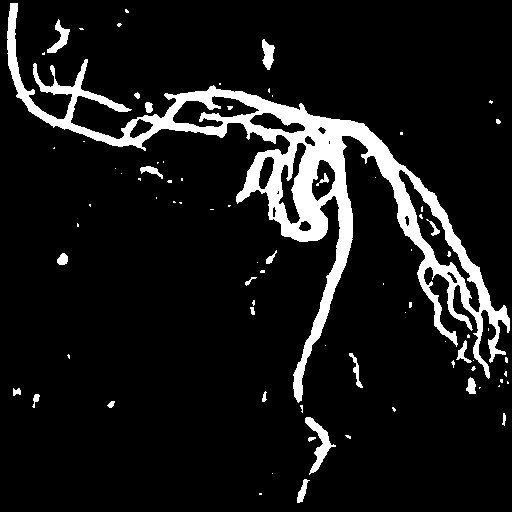
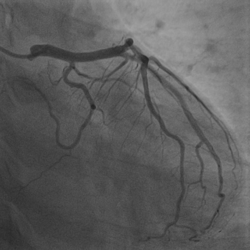
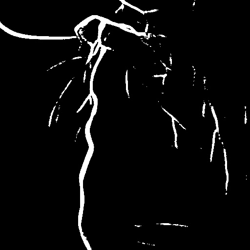
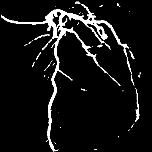
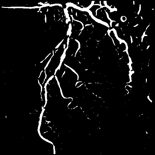
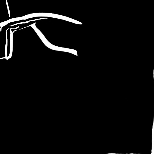
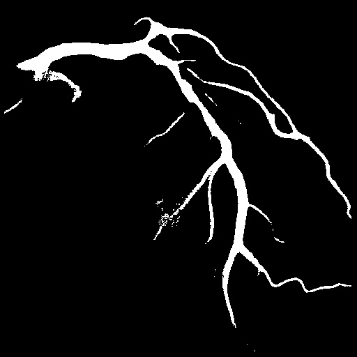
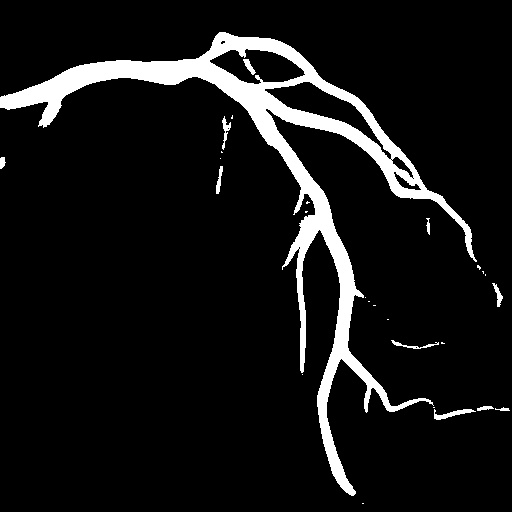
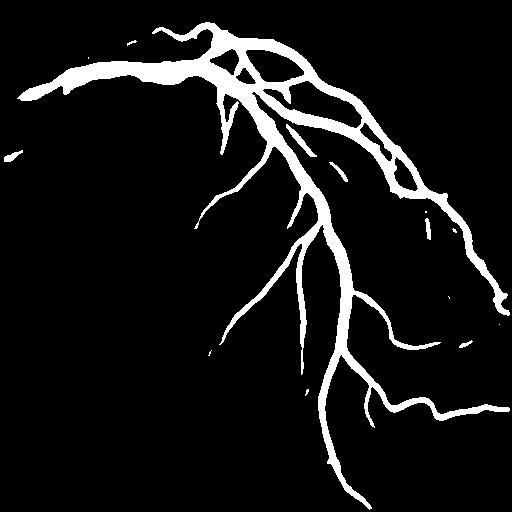
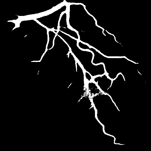
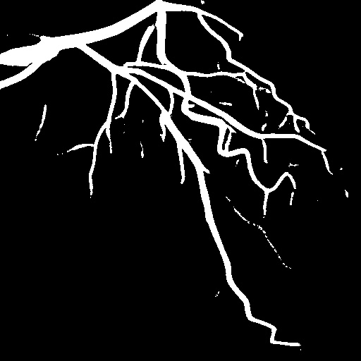
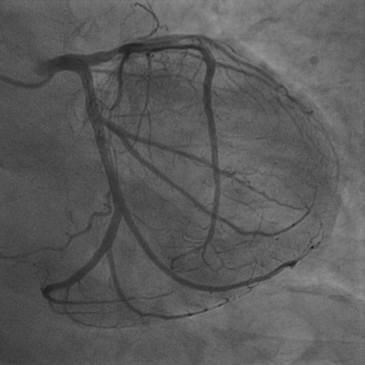
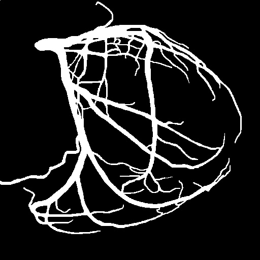
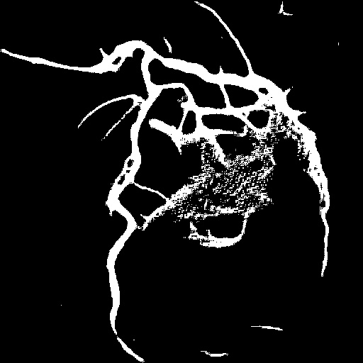
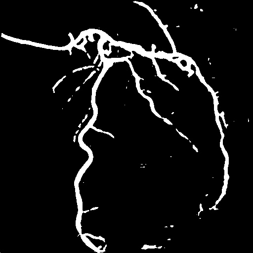
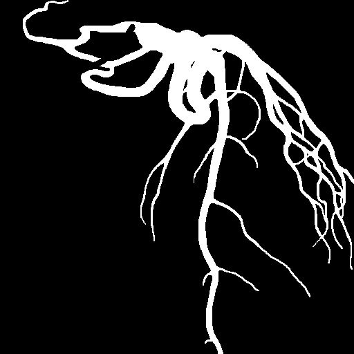
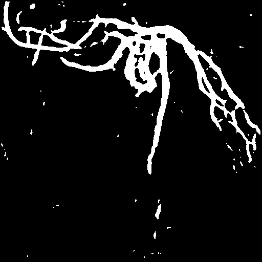
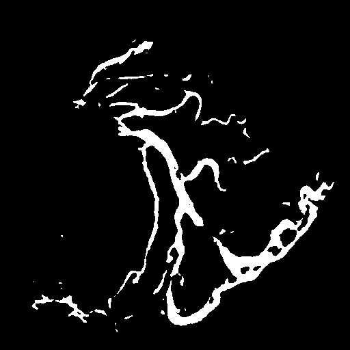
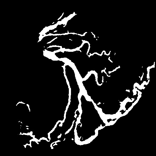
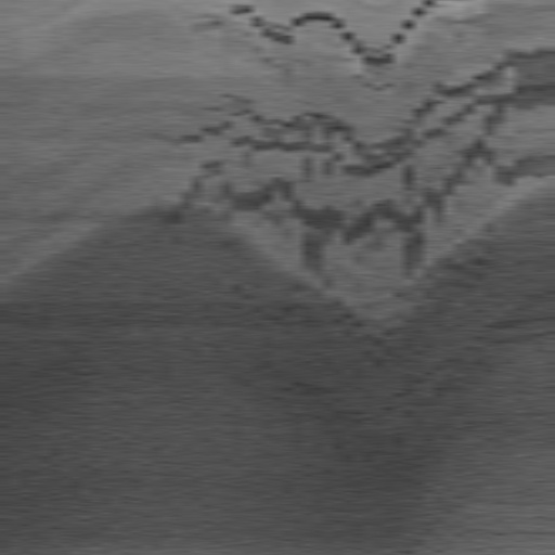
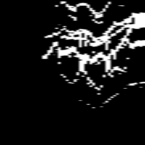
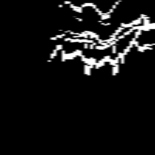
We collect 111 complete records of coronary artery X-ray videos, encompassing the injection, flow through the blood vessels around the heart, and dissipation of the contrast agent. Subsequently, we establish the XACV (X-ray Angiography Coronary Video) dataset. Each video consists of varying numbers of high-resolution coronary artery X-ray images. We invite experienced radiologists to annotate the vascular regions, focusing on one or two frames where the contrast agent is most prominent in each video. The XCAD dataset contains only a single image, and the CADICA video dataset does not provide corresponding ground truth. Therefore, in the following experiments, we conduct all the analyses on our collected XACV dataset and the corresponding GT for each sequence. In Figure 5, we show that compared to other publicly available datasets, XCAD [33] and CADICA [19], our dataset exhibits finer annotations in the vascular regions, providing an advantage for future related tasks. The development and use of our dataset have been approved by our institution’s IRB.
4.2 Baseline Methods and Evaluation Metrics
We compare the performance of video vessel segmentation on the XACV dataset between DeNVeR and other state-of-the-art methods, including the self-supervised methods SSVS [33], DARL [23] , FreeCOS [50], and the traditional method [13]. Since the mentioned self-supervised and traditional methods are image-based, we only use the very image with annotated vascular regions as input for them. To make a fair comparison for heuristic thresholds required by those methods, we will iterate through all possible values to enable them to achieve optimal performance. Finally, we adopt the widely-used metrics, including the Jaccard Index, Dice Coefficient, accuracy, sensitivity, specificity, following [33, 23, 50], as well as the advanced metrics, normalized surface distance (NSD) [45], and clDice [51] to evaluate the results.
4.3 Implementation Details
In this paper, we implement the entire deep learning architecture using PyTorch [43] and train it with Adam optimizer [24] on a single NVIDIA GeForce RTX 4090 GPU. The entire testing process, including model training and inference, takes approximately 20 minutes and utilizes 18GB of RAM. In the preprocessing stage, we compute the optical flow using RAFT [54].
Masks obtained from preprocessing are typically discontinuous and noisy. Therefore, we utilize deep learning methods for training. To simplify the task of vessel segmentation, we divide it into two stages. In stage 1, we use MLPs to acquire the background canonical image, with and . In stage 2, we employ U-Net [48] to predict masks, B-spline models, and foreground canonical images. Initially, we use a warm start U-Net [48] network with to generate a coarse mask, with weight set to 0.5. Then, we gradually incorporate (), (), (), and () to optimize DeNVeR.
 |
 |
 |
 |
 |
 |
 |
 |
 |
 |
 |
 |
 |
 |
 |
 |
 |
 |
 |
 |
 |
 |
 |
 |
 |
 |
 |
 |
 |
 |
 |
 |
 |
 |
 |
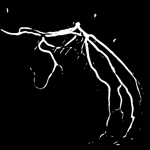
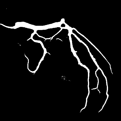
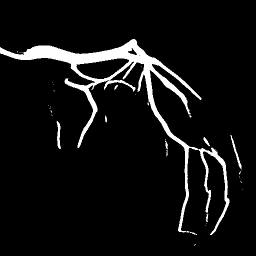
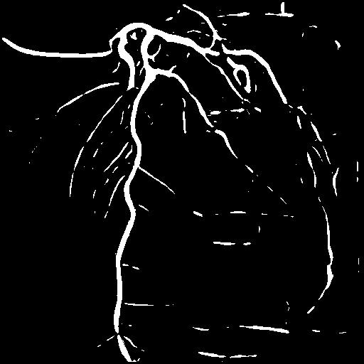
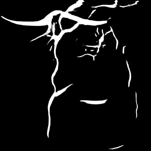
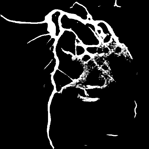
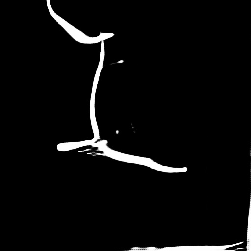
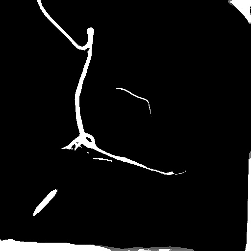
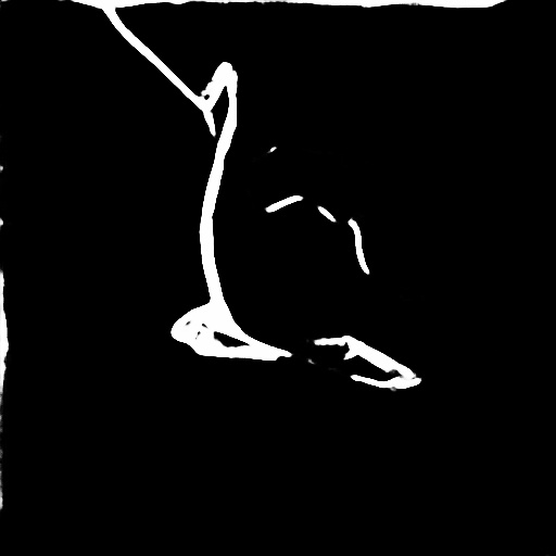
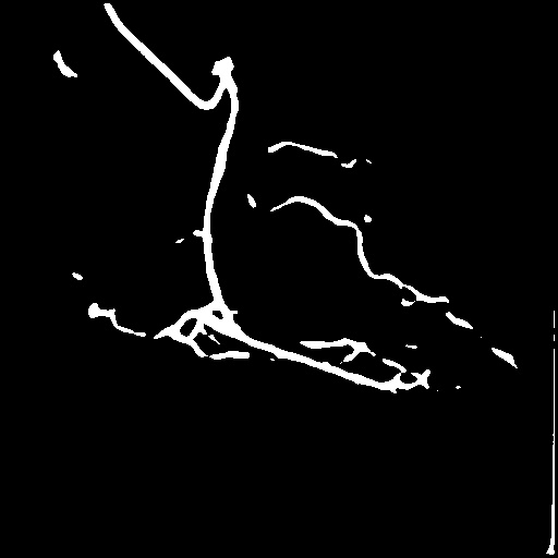
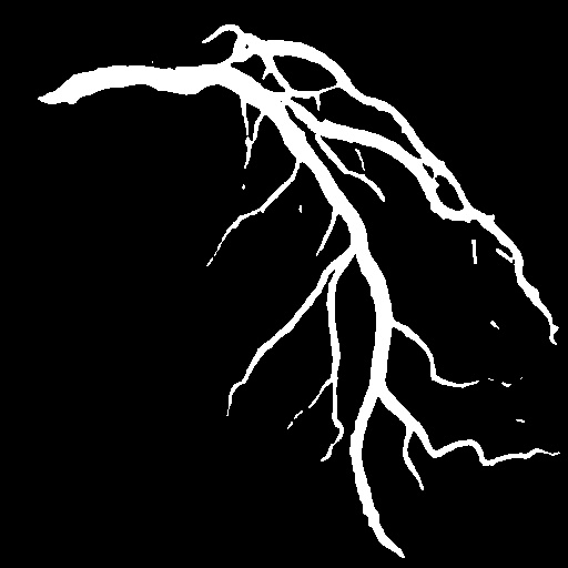
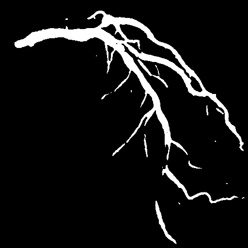
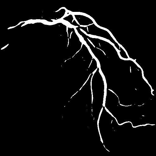
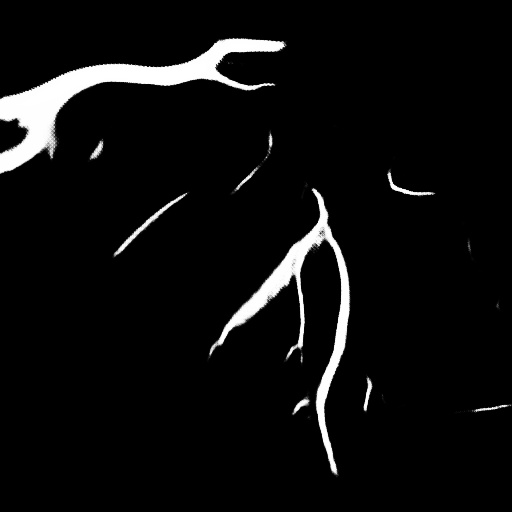
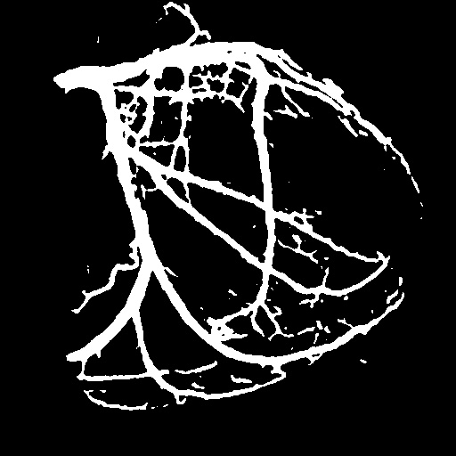
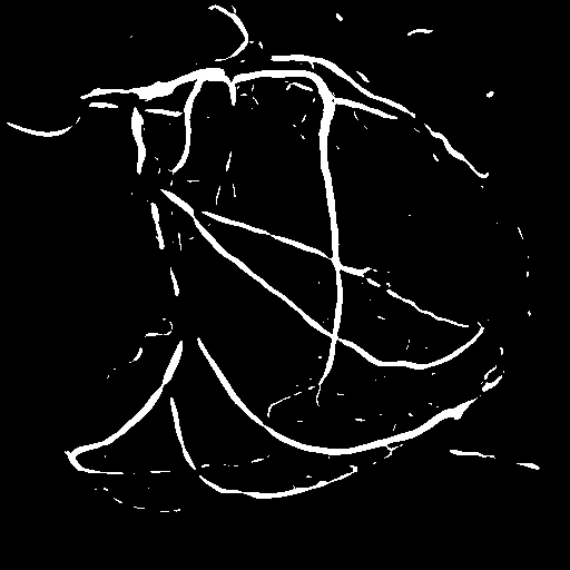
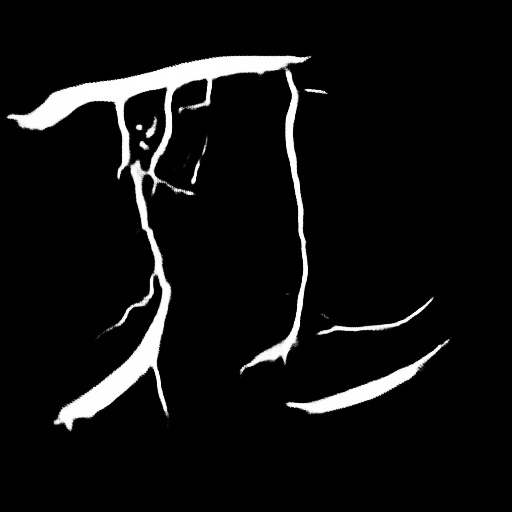
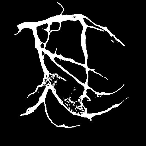
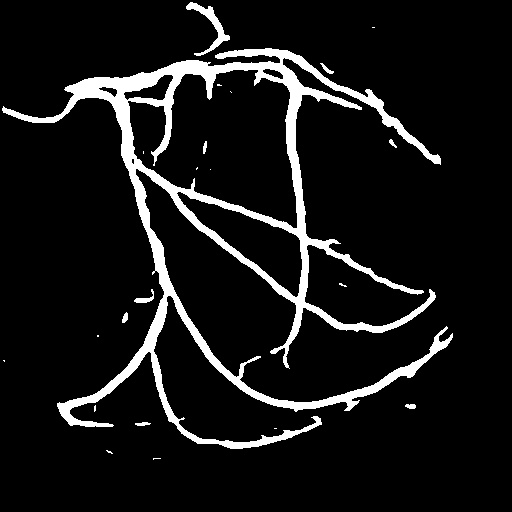
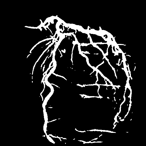
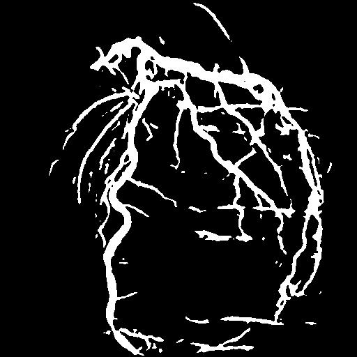
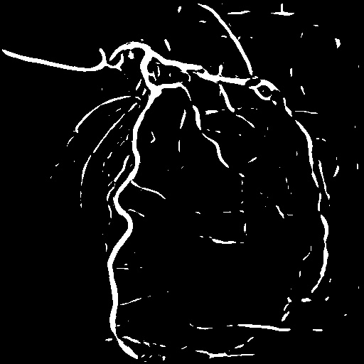
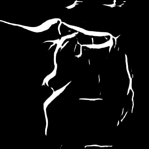
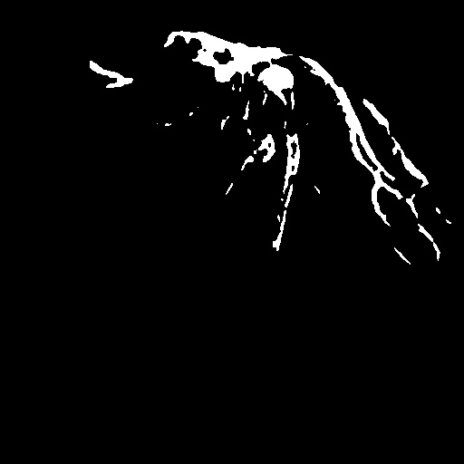
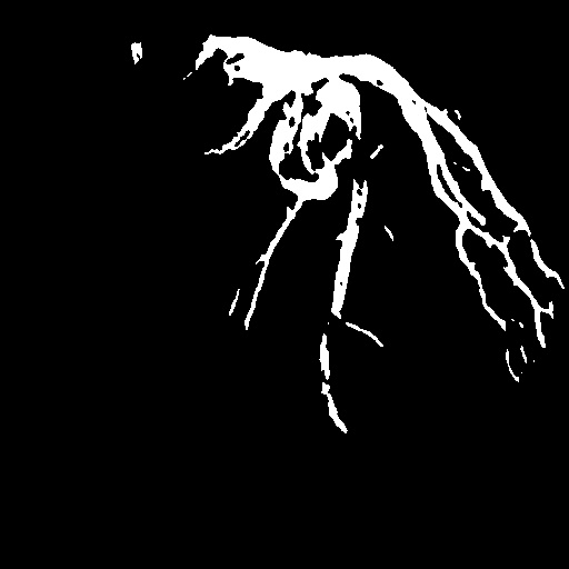
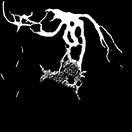
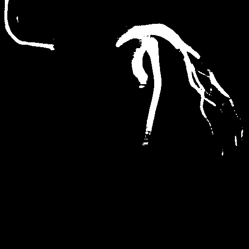
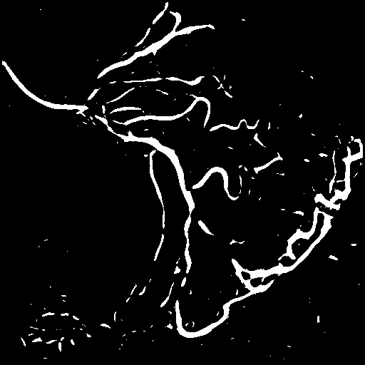
| Image | GT | Hessian | SSVS | DARL | FreeCOS | Ours |
| Traditional | Self-supervised | Unsupervised | ||||
4.4 Comparison
Table 1 reports the performance of video vessel segmentation on the XACV dataset between our proposed DeNVeR with the baseline methods. Although our method is unsupervised, for comparison with other supervised or self-supervised methods, we still partition the entire dataset into training and testing sets in an 8:2 ratio. All the results recorded in Table 1 are obtained on the testing set.
In comparison with the traditional Hessian-based filter, our method achieves a 16.9% improvement in the Jaccard Index and a 14.9% increase in the Dice Score, indicating a significant enhancement in performance while utilizing it as a prior. For self-supervised methods, we follow their tutorials to augment the training dataset and generate synthetic masks for training. Each model is trained for at least 100 epochs. The results indicate that FreeCOS [50] performs the best among them, but our approach still shows a 7.7% improvement in the Jaccard Index and a 7.3% improvement in the Dice Score compared to it. It is worth noting that, due to the sensitivity of the Hessian-based approach to the chosen threshold and its greater bias, under our intentionally selected optimal conditions, the performance of SSVS may be slightly lower than that of the Hessian-based filter.
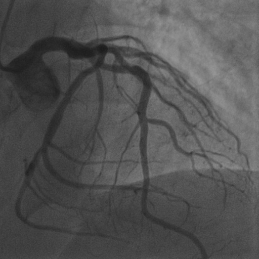
We also provide visual comparison results in Figure 6, demonstrating our vessel segmentation results are more accurate, complete, and closer to the ground truth masks. Moreover, in some sequences, our method even performs on par with supervised U-Net [48], as U-Net might face an overfitting problem with insufficient training data. Additionally, we provide visual comparisons on the CADICA [19] dataset, which is also a coronary artery X-ray video dataset but without ground truth labeling. Figure 7 demonstrates that our test-time training scheme generalizes better than existing methods. Due to the space limit, we provide more visual comparisons in the appendix.
 |
 |
 |
 |
 |
 |
 |
 |
 |
| Input frame | Without | With | Input frame | Without | With | Input frame | Without | With |
| (a) Layer separation boostrapping | (b) Hessian prior loss | (c) Parallel vessel motion loss | ||||||
| Bootstrap | Jaccard | Dice | Acc. | Sn. | Sp. | ||
| - | ✓ | ✓ | |||||
| ✓ | - | ✓ | |||||
| ✓ | ✓ | - | |||||
| ✓ | ✓ | ✓ |
4.5 Ablation Study
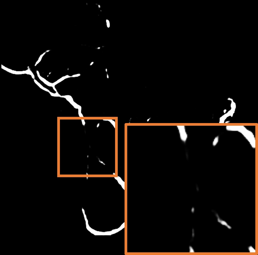
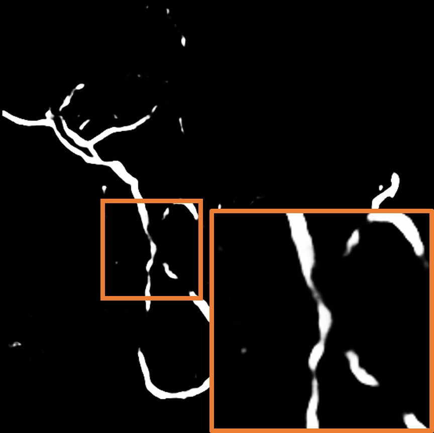
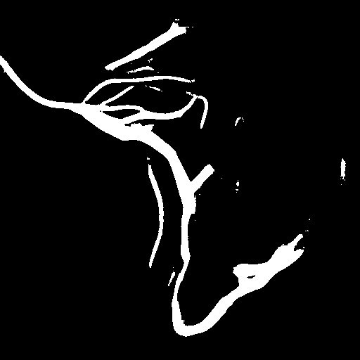
Layer separation bootstrapping.
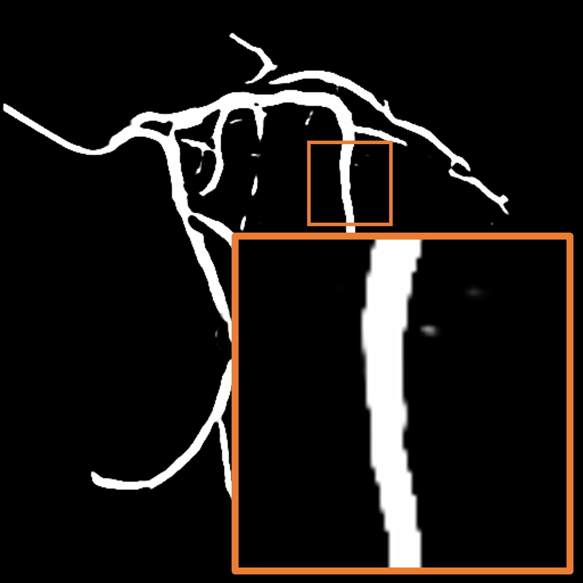
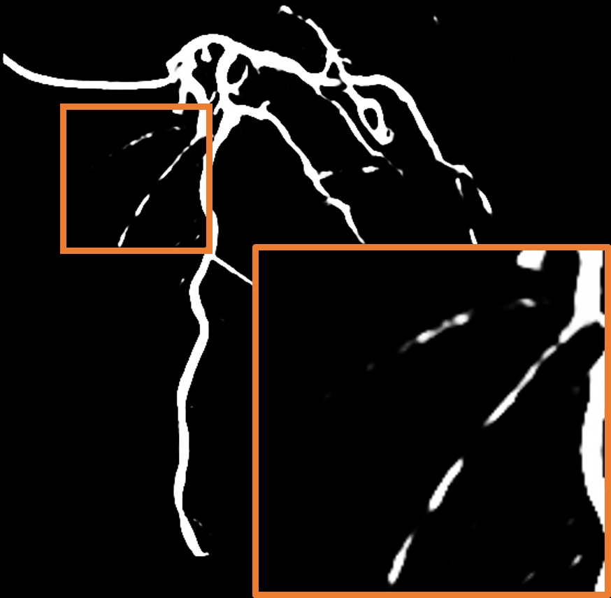
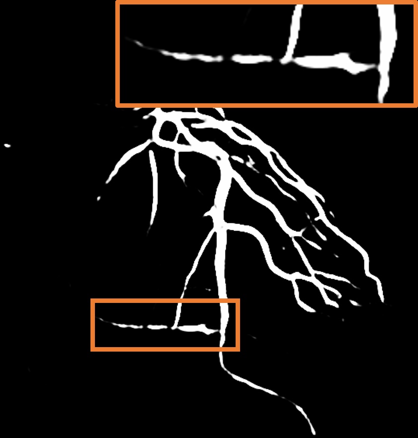
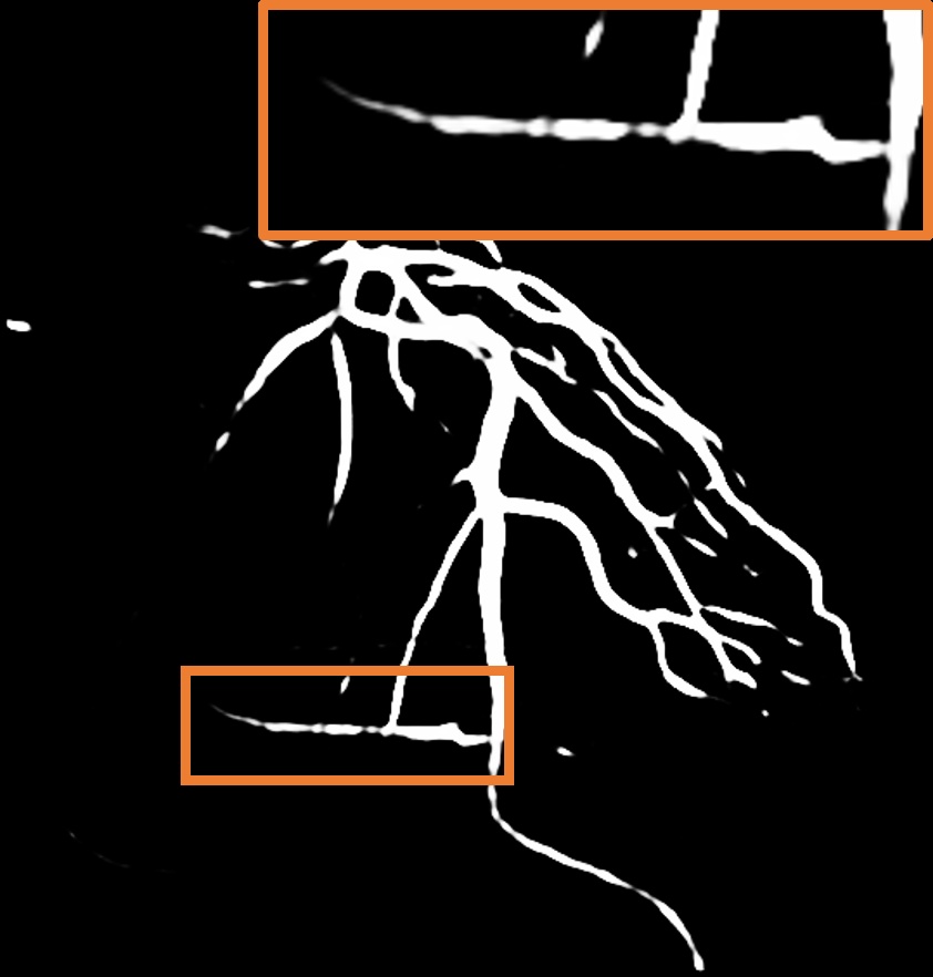
To validate the effectiveness of the layer separation bootstrapping, we train foreground and background canonical images using the same representation. The results are shown in Table 2, where optimizing both foreground and background canonical images simultaneously leads to a decrease in the Dice score by 0.0055. The comparison is shown in Figure 8 (a), where the orange area indicates the difference between without and with Layer separation bootstrapping. The bottom-right corner shows a zoom-in patch, highlighting the significant effect of the bootstrapping step.
Hessian prior loss .
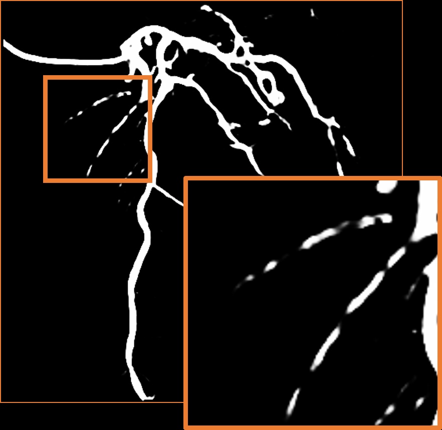
To test the effect of the Hessian prior loss, we remove the Hessian prior loss. As a result, the segmentation performance, as shown in Table 2, also decreases the Dice score by 0.0063. The comparison between the without and with is shown in Figure 8 (b), where the orange area indicates the difference between them. The zoom-in patch shows that our model predicts less noticeable vascular regions incorporating the Hessian prior loss .
Parallel vessel motion loss .
We conduct an experiment to assess the effect of the parallel vessel motion loss by removing it from the training pipeline. As shown in Table 2, the segmentation performance decreases the Dice score by 0.0047. Without this loss to enforce parallelism between blood and vessels, the segmentation results are negatively affected. In addition, the comparison between without and with is shown in Figure 8 (c). The zoom-in patch shows that the image with has clearer segmented vascular regions.
5 Conclusions
This paper introduces DeNVeR, an unsupervised test-time training framework for vessel segmentation in X-ray video data. DeNVeR utilizes optical flow and layer separation techniques to accurately segment vessels without requiring annotated datasets. Quantitative and qualitative evaluations on the XACV and CADICA datasets show that DeNVeR outperforms existing image-based self-supervised methods, offering precise delineation of vessel boundaries critical for medical diagnosis and treatment.
Limitations.
Our method uses unsupervised learning, so it is sensitive to the effects of preprocessing filters. This can cause inaccuracies in distinguishing between vascular and non-vascular tubular structures. For example, in our cases, non-vascular elements like catheters may be mistakenly identified as blood vessels. This limitation can affect the precision and reliability of our approach in clinical settings.
References
- Abdal et al. [2021] Rameen Abdal, Peihao Zhu, Niloy J Mitra, and Peter Wonka. Labels4free: Unsupervised segmentation using stylegan. In ICCV, 2021.
- Alonso et al. [2021] Inigo Alonso, Alberto Sabater, David Ferstl, Luis Montesano, and Ana C Murillo. Semi-supervised semantic segmentation with pixel-level contrastive learning from a class-wise memory bank. In ICCV, 2021.
- Bar et al. [2022] Amir Bar, Xin Wang, Vadim Kantorov, Colorado J Reed, Roei Herzig, Gal Chechik, Anna Rohrbach, Trevor Darrell, and Amir Globerson. Detreg: Unsupervised pretraining with region priors for object detection. In CVPR, 2022.
- Benaim et al. [2020] Sagie Benaim, Ariel Ephrat, Oran Lang, Inbar Mosseri, William T Freeman, Michael Rubinstein, Michal Irani, and Tali Dekel. Speednet: Learning the speediness in videos. In CVPR, 2020.
- Black and Anandan [1991] Michael J Black and Padmanabhan Anandan. Robust dynamic motion estimation over time. In CVPR, 1991.
- Brox and Malik [2010] Thomas Brox and Jitendra Malik. Object segmentation by long term analysis of point trajectories. In ECCV, 2010.
- Chen et al. [2019] Mickaël Chen, Thierry Artières, and Ludovic Denoyer. Unsupervised object segmentation by redrawing. In NeurIPS, 2019.
- Do et al. [2021] Kien Do, Truyen Tran, and Svetha Venkatesh. Clustering by maximizing mutual information across views. In ICCV, 2021.
- Dodge Jr et al. [1992] J Theodore Dodge Jr, B Greg Brown, Edward L Bolson, and Harold T Dodge. Lumen diameter of normal human coronary arteries. influence of age, sex, anatomic variation, and left ventricular hypertrophy or dilation. Circulation, 1992.
- Doersch et al. [2015] Carl Doersch, Abhinav Gupta, and Alexei A Efros. Unsupervised visual representation learning by context prediction. In ICCV, 2015.
- Fan et al. [2019] Zhun Fan, Jiajie Mo, Benzhang Qiu, Wenji Li, Guijie Zhu, Chong Li, Jianye Hu, Yibiao Rong, and Xinjian Chen. Accurate retinal vessel segmentation via octave convolution neural network. arXiv preprint arXiv:1906.12193, 2019.
- Felfelian et al. [2016] Banafsheh Felfelian, Hamid R Fazlali, Nader Karimi, S Mohamad R Soroushmehr, Shadrokh Samavi, B Nallamothu, and Kayvan Najarian. Vessel segmentation in low contrast x-ray angiogram images. In ICIP, 2016.
- Frangi et al. [1998] Alejandro F Frangi, Wiro J Niessen, Koen L Vincken, and Max A Viergever. Multiscale vessel enhancement filtering. In MICCAI, 1998.
- Gidaris et al. [2018] Spyros Gidaris, Praveer Singh, and Nikos Komodakis. Unsupervised representation learning by predicting image rotations. In ICLR, 2018.
- Götberg et al. [2017] Matthias Götberg, Evald H Christiansen, Ingibjörg J Gudmundsdottir, Lennart Sandhall, Mikael Danielewicz, Lars Jakobsen, Sven-Erik Olsson, Patrik Öhagen, Hans Olsson, Elmir Omerovic, et al. Instantaneous wave-free ratio versus fractional flow reserve to guide pci. New England Journal of Medicine, 2017.
- Isensee et al. [2018] Fabian Isensee, Jens Petersen, Andre Klein, David Zimmerer, Paul F. Jaeger, Simon Kohl, Jakob Wasserthal, Gregor Koehler, Tobias Norajitra, Sebastian Wirkert, and Klaus H. Maier-Hein. nnu-net: Self-adapting framework for u-net-based medical image segmentation, 2018.
- Iyer et al. [2023] Kritika Iyer, Brahmajee K Nallamothu, C Alberto Figueroa, and Raj R Nadakuditi. A multi-stage neural network approach for coronary 3d reconstruction from uncalibrated x-ray angiography images. 2023.
- Ji et al. [2019] Xu Ji, Joao F Henriques, and Andrea Vedaldi. Invariant information clustering for unsupervised image classification and segmentation. In ICCV, 2019.
- Jiménez-Partinen et al. [2024] Ariadna Jiménez-Partinen, Miguel A Molina-Cabello, Karl Thurnhofer-Hemsi, Esteban J Palomo, Jorge Rodríguez-Capitán, Ana I Molina-Ramos, and Manuel Jiménez-Navarro. Cadica: a new dataset for coronary artery disease detection by using invasive coronary angiography. arXiv preprint arXiv:2402.00570, 2024.
- Jojic and Frey [2001] Nebojsa Jojic and Brendan J Frey. Learning flexible sprites in video layers. In CVPR, 2001.
- Khan et al. [2020] Khan Bahadar Khan, Muhammad Shahbaz Siddique, Muhammad Ahmad, Manuel Mazzara, et al. A hybrid unsupervised approach for retinal vessel segmentation. BioMed Research International, 2020, 2020.
- Khowaja et al. [2019] Sunder Ali Khowaja, Parus Khuwaja, and Imdad Ali Ismaili. A framework for retinal vessel segmentation from fundus images using hybrid feature set and hierarchical classification. Signal, Image and Video Processing, 2019.
- Kim et al. [2023] Boah Kim, Yujin Oh, and Jong Chul Ye. Diffusion adversarial representation learning for self-supervised vessel segmentation. In ICLR, 2023.
- Kingma and Ba [2015] Diederik P Kingma and Jimmy Ba. Adam: A method for stochastic optimization. In ICLR, 2015.
- Larsson et al. [2017] Gustav Larsson, Michael Maire, and Gregory Shakhnarovich. Colorization as a proxy task for visual understanding. In CVPR, 2017.
- Law and Chung [2008] Max WK Law and Albert CS Chung. Three dimensional curvilinear structure detection using optimally oriented flux. In ECCV, 2008.
- Ledig et al. [2017] Christian Ledig, Lucas Theis, Ferenc Huszár, Jose Caballero, Andrew Cunningham, Alejandro Acosta, Andrew Aitken, Alykhan Tejani, Johannes Totz, Zehan Wang, et al. Photo-realistic single image super-resolution using a generative adversarial network. In CVPR, 2017.
- Li et al. [2021] Yunfan Li, Peng Hu, Zitao Liu, Dezhong Peng, Joey Tianyi Zhou, and Xi Peng. Contrastive clustering. In AAAI, 2021.
- Lin and Ching [2005] Chih-Yang Lin and Yu-Tai Ching. Extraction of coronary arterial tree using cine x-ray angiograms. Biomedical Engineering: Applications, Basis and Communications, 2005.
- Liu et al. [2020a] Fan Liu, Dongxiao Li, Xinyu Jin, Wenyuan Qiu, Qi Xia, and Bin Sun. Dynamic cardiac mri reconstruction using motion aligned locally low rank tensor (mallrt). Magnetic resonance imaging, 2020a.
- Liu et al. [2020b] Yu-Lun Liu, Wei-Sheng Lai, Ming-Hsuan Yang, Yung-Yu Chuang, and Jia-Bin Huang. Learning to see through obstructions. In CVPR, 2020b.
- Liu et al. [2021] Yu-Lun Liu, Wei-Sheng Lai, Ming-Hsuan Yang, Yung-Yu Chuang, and Jia-Bin Huang. Learning to see through obstructions with layered decomposition. IEEE TPAMI, 2021.
- Ma et al. [2021] Yuxin Ma, Yang Hua, Hanming Deng, Tao Song, Hao Wang, Zhengui Xue, Heng Cao, Ruhui Ma, and Haibing Guan. Self-supervised vessel segmentation via adversarial learning. In ICCV, 2021.
- Maglaveras et al. [2001] N Maglaveras, K Haris, SN Efstratiadis, J Gourassas, and G Louridas. Artery skeleton extraction using topographic and connected component labeling. In Computers in Cardiology 2001. Vol. 28 (Cat. No. 01CH37287), 2001.
- Memari et al. [2019] Nogol Memari, Abd Rahman Ramli, M Iqbal Bin Saripan, Syamsiah Mashohor, and Mehrdad Moghbel. Retinal blood vessel segmentation by using matched filtering and fuzzy c-means clustering with integrated level set method for diabetic retinopathy assessment. Journal of Medical and Biological Engineering, 2019.
- Misra and Maaten [2020] Ishan Misra and Laurens van der Maaten. Self-supervised learning of pretext-invariant representations. In CVPR, 2020.
- Misra et al. [2016] Ishan Misra, C Lawrence Zitnick, and Martial Hebert. Shuffle and learn: unsupervised learning using temporal order verification. In ECCV, 2016.
- Nam et al. [2022] Seonghyeon Nam, Marcus A Brubaker, and Michael S Brown. Neural image representations for multi-image fusion and layer separation. In ECCV, 2022.
- Nasr-Esfahani et al. [2016] Ebrahim Nasr-Esfahani, Shadrokh Samavi, Nader Karimi, SM Reza Soroushmehr, Kevin Ward, Mohammad H Jafari, Banafsheh Felfeliyan, B Nallamothu, and Kayvan Najarian. Vessel extraction in x-ray angiograms using deep learning. In EMBC, 2016.
- Noroozi and Favaro [2016] Mehdi Noroozi and Paolo Favaro. Unsupervised learning of visual representations by solving jigsaw puzzles. In ECCV, 2016.
- Ost et al. [2021] Julian Ost, Fahim Mannan, Nils Thuerey, Julian Knodt, and Felix Heide. Neural scene graphs for dynamic scenes. In CVPR, 2021.
- Park et al. [2020] Taesung Park, Alexei A Efros, Richard Zhang, and Jun-Yan Zhu. Contrastive learning for unpaired image-to-image translation. In ECCV, 2020.
- Paszke et al. [2019] Adam Paszke, Sam Gross, Francisco Massa, Adam Lerer, James Bradbury, Gregory Chanan, Trevor Killeen, Zeming Lin, Natalia Gimelshein, Luca Antiga, et al. Pytorch: An imperative style, high-performance deep learning library. In NeurIPS, 2019.
- Pathak et al. [2016] Deepak Pathak, Philipp Krahenbuhl, Jeff Donahue, Trevor Darrell, and Alexei A Efros. Context encoders: Feature learning by inpainting. In CVPR, 2016.
- Reinke et al. [2024] Annika Reinke, Minu D. Tizabi, Michael Baumgartner, Matthias Eisenmann, Doreen Heckmann-Nötzel, A. Emre Kavur, Tim Rädsch, Carole H. Sudre, Laura Acion, Michela Antonelli, Tal Arbel, Spyridon Bakas, Arriel Benis, Florian Buettner, M. Jorge Cardoso, Veronika Cheplygina, Jianxu Chen, Evangelia Christodoulou, Beth A. Cimini, Keyvan Farahani, Luciana Ferrer, Adrian Galdran, Bram van Ginneken, Ben Glocker, Patrick Godau, Daniel A. Hashimoto, Michael M. Hoffman, Merel Huisman, Fabian Isensee, Pierre Jannin, Charles E. Kahn, Dagmar Kainmueller, Bernhard Kainz, Alexandros Karargyris, Jens Kleesiek, Florian Kofler, Thijs Kooi, Annette Kopp-Schneider, Michal Kozubek, Anna Kreshuk, Tahsin Kurc, Bennett A. Landman, Geert Litjens, Amin Madani, Klaus Maier-Hein, Anne L. Martel, Erik Meijering, Bjoern Menze, Karel G. M. Moons, Henning Müller, Brennan Nichyporuk, Felix Nickel, Jens Petersen, Susanne M. Rafelski, Nasir Rajpoot, Mauricio Reyes, Michael A. Riegler, Nicola Rieke, Julio Saez-Rodriguez, Clara I. Sánchez, Shravya Shetty, Ronald M. Summers, Abdel A. Taha, Aleksei Tiulpin, Sotirios A. Tsaftaris, Ben Van Calster, Gaël Varoquaux, Ziv R. Yaniv, Paul F. Jäger, and Lena Maier-Hein. Understanding metric-related pitfalls in image analysis validation. Nature Methods, 21(2):182–194, February 2024. ISSN 1548-7105. doi: 10.1038/s41592-023-02150-0. URL http://dx.doi.org/10.1038/s41592-023-02150-0.
- Ren and Lee [2018] Zhongzheng Ren and Yong Jae Lee. Cross-domain self-supervised multi-task feature learning using synthetic imagery. In CVPR, 2018.
- Revaud et al. [2016] Jerome Revaud, Philippe Weinzaepfel, Zaid Harchaoui, and Cordelia Schmid. Deepmatching: Hierarchical deformable dense matching. IJCV, 2016.
- Ronneberger et al. [2015] Olaf Ronneberger, Philipp Fischer, and Thomas Brox. U-net: Convolutional networks for biomedical image segmentation. In MICCAI, 2015.
- Shi and Malik [1998] Jianbo Shi and Jitendra Malik. Motion segmentation and tracking using normalized cuts. In ICCV, 1998.
- Shi et al. [2023] Tianyi Shi, Xiaohuan Ding, Liang Zhang, and Xin Yang. Freecos: Self-supervised learning from fractals and unlabeled images for curvilinear object segmentation. In ICCV, 2023.
- Shit et al. [2021] Suprosanna Shit, Johannes C Paetzold, Anjany Sekuboyina, Ivan Ezhov, Alexander Unger, Andrey Zhylka, Josien PW Pluim, Ulrich Bauer, and Bjoern H Menze. cldice-a novel topology-preserving loss function for tubular structure segmentation. In CVPR, 2021.
- Soomro et al. [2019] Toufique Ahmed Soomro, Ahmed J Afifi, Ahmed Ali Shah, Shafiullah Soomro, Gulsher Ali Baloch, Lihong Zheng, Ming Yin, and Junbin Gao. Impact of image enhancement technique on cnn model for retinal blood vessels segmentation. IEEE Access, 2019.
- Stables et al. [2022] Rodney H Stables, Liam J Mullen, Mostafa Elguindy, Zoe Nicholas, Yousra H Aboul-Enien, Ian Kemp, Peter O’Kane, Alex Hobson, Thomas W Johnson, Sohail Q Khan, et al. Routine pressure wire assessment versus conventional angiography in the management of patients with coronary artery disease: the ripcord 2 trial. Circulation, 2022.
- Teed and Deng [2020] Zachary Teed and Jia Deng. Raft: Recurrent all-pairs field transforms for optical flow. In ECCV, 2020.
- Toth et al. [2014] Gabor Toth, Michalis Hamilos, Stylianos Pyxaras, Fabio Mangiacapra, Olivier Nelis, Frederic De Vroey, Luigi Di Serafino, Olivier Muller, Carlos Van Mieghem, Eric Wyffels, et al. Evolving concepts of angiogram: fractional flow reserve discordances in 4000 coronary stenoses. European heart journal, 2014.
- Ulyanov et al. [2018] Dmitry Ulyanov, Andrea Vedaldi, and Victor Lempitsky. Deep image prior. In CVPR, 2018.
- Wang and Chung [2020] Jierong Wang and Albert CS Chung. Higher-order flux with spherical harmonics transform for vascular analysis. In MICCAI, 2020.
- Wang and Adelson [1994] John YA Wang and Edward H Adelson. Representing moving images with layers. IEEE TIP, 1994.
- Wang et al. [2022] Xuehui Wang, Kai Zhao, Ruixin Zhang, Shouhong Ding, Yan Wang, and Wei Shen. Contrastmask: Contrastive learning to segment every thing. In CVPR, 2022.
- Wu et al. [2021] Haiyan Wu, Yanyun Qu, Shaohui Lin, Jian Zhou, Ruizhi Qiao, Zhizhong Zhang, Yuan Xie, and Lizhuang Ma. Contrastive learning for compact single image dehazing. In CVPR, 2021.
- Xie et al. [2021] Enze Xie, Jian Ding, Wenhai Wang, Xiaohang Zhan, Hang Xu, Peize Sun, Zhenguo Li, and Ping Luo. Detco: Unsupervised contrastive learning for object detection. In ICCV, 2021.
- Xu et al. [2019] Dejing Xu, Jun Xiao, Zhou Zhao, Jian Shao, Di Xie, and Yueting Zhuang. Self-supervised spatiotemporal learning via video clip order prediction. In CVPR, 2019.
- Yang et al. [2018] Siyuan Yang, Jian Yang, Yachen Wang, Qi Yang, Danni Ai, and Yongtian Wang. Automatic coronary artery segmentation in x-ray angiograms by multiple convolutional neural networks. In Proceedings of the 3rd international conference on multimedia and image processing, 2018.
- Yang et al. [2019a] Su Yang, Jihoon Kweon, Jae-Hyung Roh, Jae-Hwan Lee, Heejun Kang, Lae-Jeong Park, Dong Jun Kim, Hyeonkyeong Yang, Jaehee Hur, Do-Yoon Kang, et al. Deep learning segmentation of major vessels in x-ray coronary angiography. Scientific reports, 2019a.
- Yang et al. [2019b] Yanchao Yang, Antonio Loquercio, Davide Scaramuzza, and Stefano Soatto. Unsupervised moving object detection via contextual information separation. In CVPR, 2019b.
- Ye et al. [2022] Vickie Ye, Zhengqi Li, Richard Tucker, Angjoo Kanazawa, and Noah Snavely. Deformable sprites for unsupervised video decomposition. In CVPR, 2022.
- Zhong et al. [2021] Yuanyi Zhong, Bodi Yuan, Hong Wu, Zhiqiang Yuan, Jian Peng, and Yu-Xiong Wang. Pixel contrastive-consistent semi-supervised semantic segmentation. In ICCV, 2021.
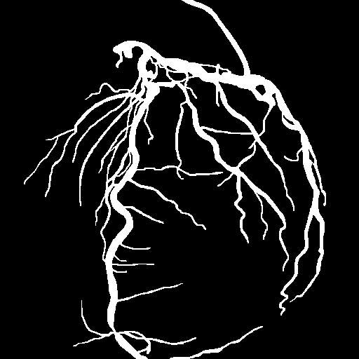
Appendix A Appendix
A.1 Additional Visualization Results
Figure 9 and Figure 10 demonstrate a comprehensive comparison where we consider supervised learning [16] as the upper bound for the vessel segmentation task, as well as all baseline methods mentioned in the main paper. For the supervised learning approach, both image-based and video-based inputs were considered. The image-based input utilized only the annotated image, while the video-based input involved using the annotated image along with two preceding and two subsequent frames, totaling five frames, as input.
The results show that although supervised learning theoretically offers the best performance, our method achieves close to those of supervised learning methods without ground truth. Additionally, we found that using five consecutive images as input for nn-UNet [16] were only slightly better than using a single image as input. In contrast, our method exhibits significant improvement compared to both the traditional Hessian-based filter and self-supervised methods, demonstrating that the robust performance of our approach is not solely attributed to the increase in input images.
We showcase some examples at the following URL: https://colab.research.google.com/drive/1IYGiJECwAaoLPq7KGHQE_dvtrdHz9fUA?authuser=2&hl=zh-tw#scrollTo=n1ppvOhqbRkV
 |
 |
 |
 |
 |
| Image | Ground truth | U-Net (image) | U-Net (video) | Hessian |
| Supervised (upper bound) | ||||
 |
 |
 |
 |
|
| SSVS | DARL | FreeCOS | Ours | |
| Self-supervised | Unsupervised | |||
 |
 |
 |
 |
 |
| Image | Ground truth | U-Net (image) | U-Net (video) | Hessian |
| Supervised (upper bound) | ||||
 |
 |
 |
 |
|
| SSVS | DARL | FreeCOS | Ours | |
| Self-supervised | Unsupervised | |||
 |
 |
 |
 |
 |
| Image | Ground truth | U-Net (image) | U-Net (video) | Hessian |
| Supervised (upper bound) | ||||
 |
 |
 |
 |
|
| SSVS | DARL | FreeCOS | Ours | |
| Self-supervised | Unsupervised | |||
 |
 |
 |
 |
 |
| Image | Ground truth | U-Net (image) | U-Net (video) | Hessian |
| Supervised (upper bound) | ||||
 |
 |
 |
 |
|
| SSVS | DARL | FreeCOS | Ours | |
| Self-supervised | Unsupervised | |||
 |
 |
 |
 |
 |
| Image | Ground truth | U-Net (image) | U-Net (video) | Hessian |
| Supervised (upper bound) | ||||
 |
 |
 |
 |
|
| SSVS | DARL | FreeCOS | Ours | |
| Self-supervised | Unsupervised | |||
 |
 |
 |
 |
 |
| Image | Ground truth | U-Net (image) | U-Net (video) | Hessian |
| Supervised (upper bound) | ||||
 |
 |
 |
 |
|
| SSVS | DARL | FreeCOS | Ours | |
| Self-supervised | Unsupervised | |||
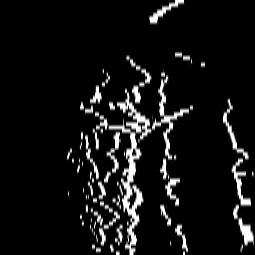
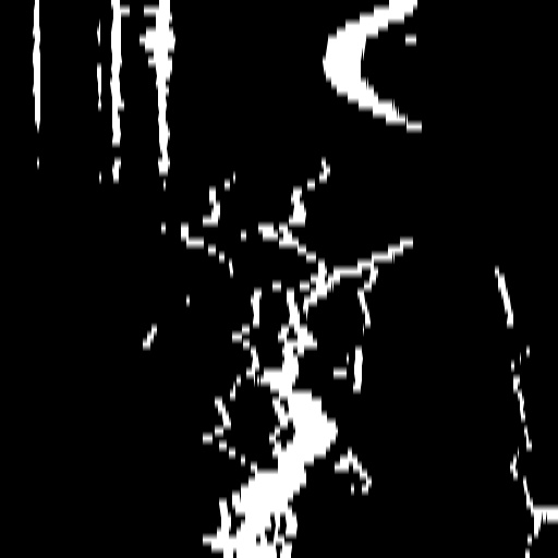
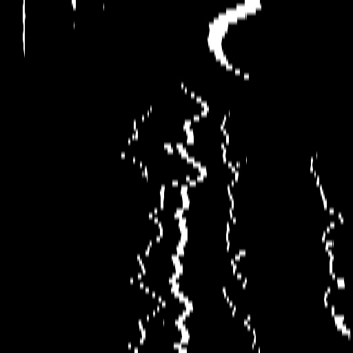
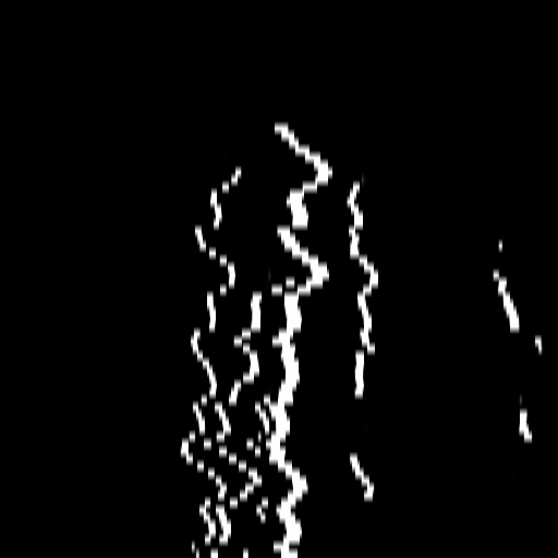
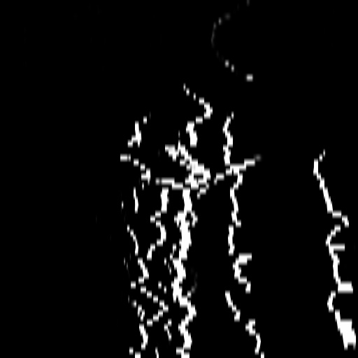
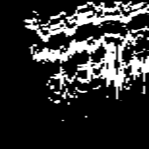
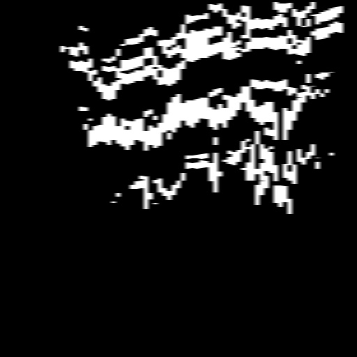
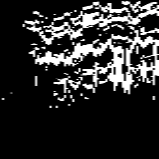
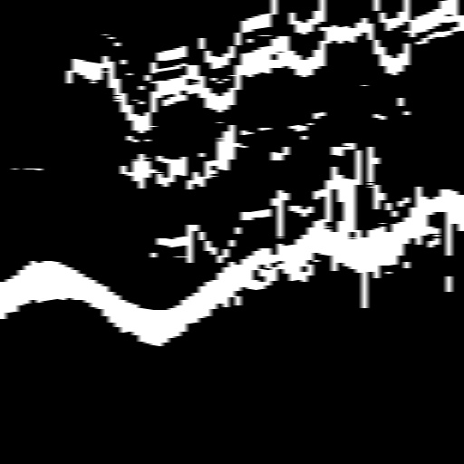
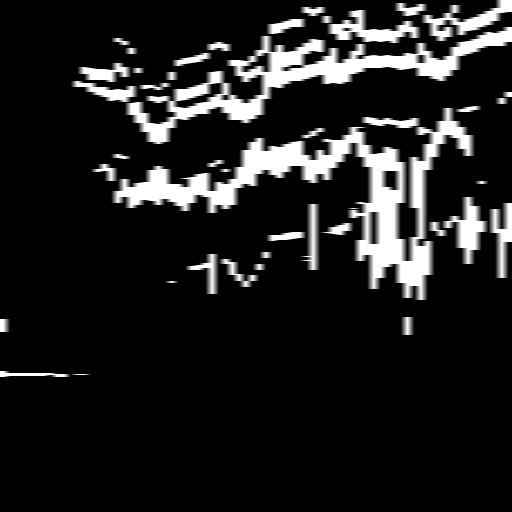
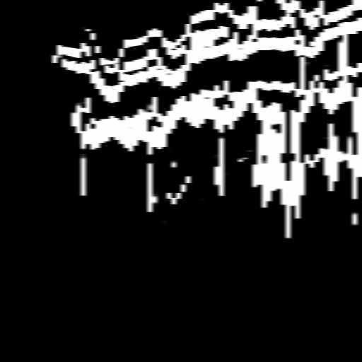
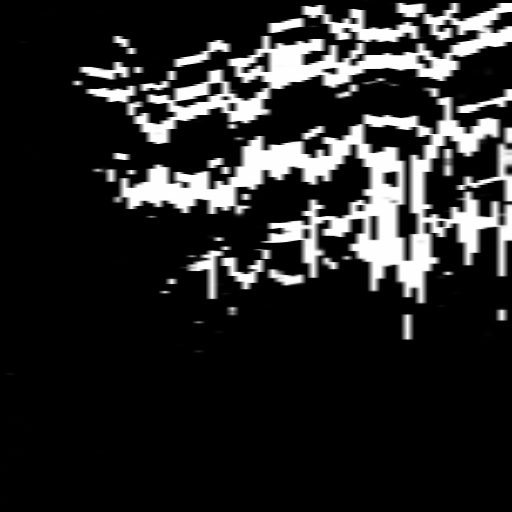
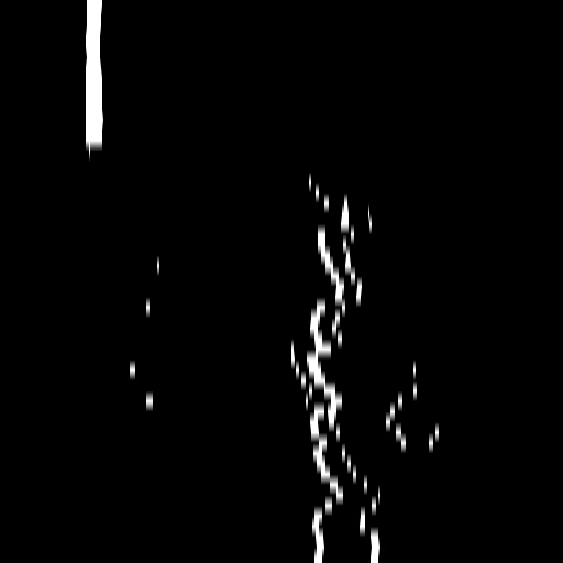
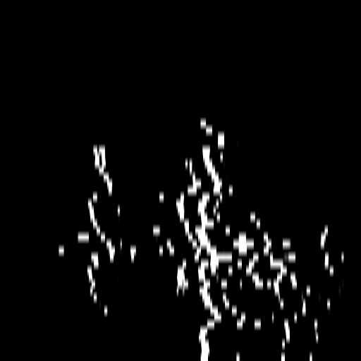
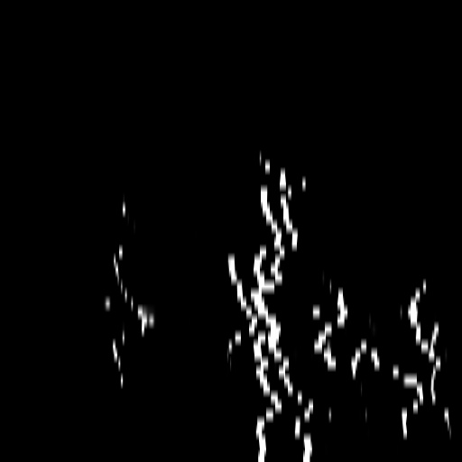
A.2 Temporal Coherency
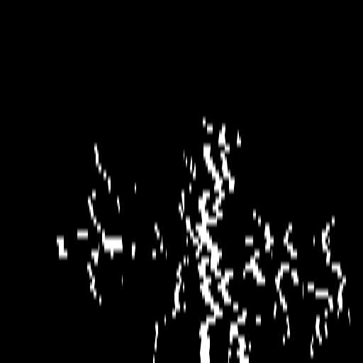
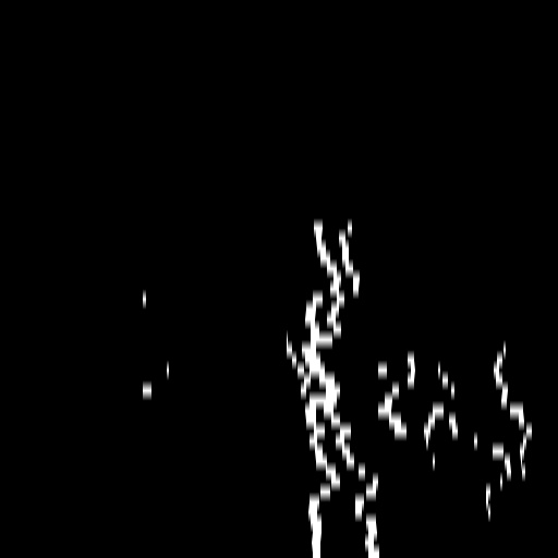
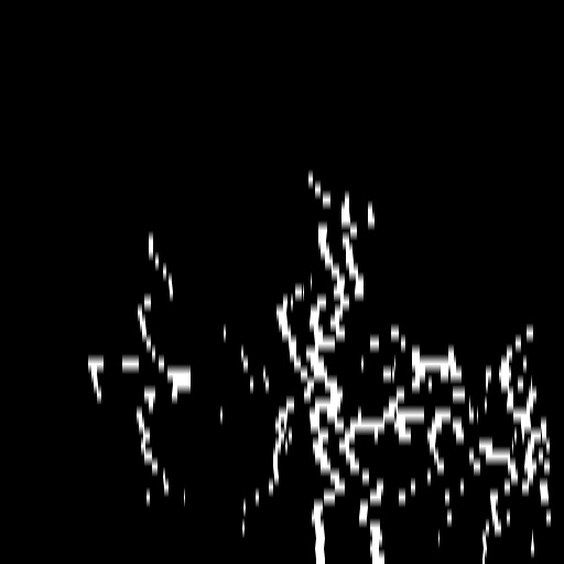
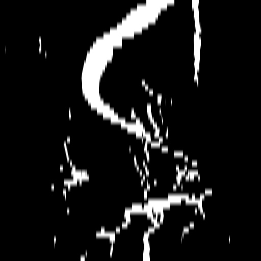
Our method takes an entire X-ray video as input, thus producing segmentation results with better temporal coherency. Temporal coherency is essential for making medical diagnoses, especially when dealing with blood flow in vessels. Therefore, we conduct visual comparisons between our method and other compared methods by slicing horizontally or vertically and stacking the segmentation results. The results in Figure 11 show our method strikes a better balance between segmentation accuracy and temporal coherency. While other baseline methods either produce false segmentation results or do not maintain consistent prediction along the temporal dimension.
 |
 |
 |
 |
 |
 |
 |
 |
 |
 |
 |
 |
 |
 |
 |
 |
 |
 |
 |
 |
 |
 |
 |
 |
 |
 |
 |
 |
 |
 |
 |
 |
 |
 |
 |
 |
| Frame & Temporal slice | U-Net (image) | U-Net (video) | Hessian | SSVS | DARL | FreeCOS | Ours | |
| Supervised (upper bound) | Traditional | Self-supervised | Unsupervised | |||||