55email: apooria@student.ubc.ca
Vision-Language Synthetic Data Enhances Echocardiography Downstream Tasks
Abstract
High-quality, large-scale data is essential for robust deep learning models in medical applications, particularly ultrasound image analysis. Diffusion models facilitate high-fidelity medical image generation, reducing the costs associated with acquiring and annotating new images. This paper utilizes recent vision-language models to produce diverse and realistic synthetic echocardiography image data, preserving key features of the original images guided by textual and semantic label maps. Specifically, we investigate three potential avenues: unconditional generation, generation guided by text, and a hybrid approach incorporating both textual and semantic supervision. We show that the rich contextual information present in the synthesized data potentially enhances the accuracy and interpretability of downstream tasks, such as echocardiography segmentation and classification with improved metrics and faster convergence. Our implementation with checkpoints, prompts, and the created synthetic dataset will be publicly available at GitHub.
Keywords:
Vision-Language Models Image Synthesis Diffusion Models Echocardiography1 Introduction
Cardiovascular diseases (CVDs) pose a growing threat to global health, emerging as prominent contributors to mortality rates worldwide [7]. Owing to its widespread availability, cost-effectiveness, non-invasive nature, and real-time capabilities, echocardiography (echo) stands as the most commonly employed medical imaging approach for heart evaluation. Manual data collection, difficulty in interpreting noisy data, and low sensitivity and specificity of clinical assessment continue to be the major obstacles associated with echo imaging. While deep learning has emerged as a powerful tool in the modern medical imaging stack, the limited availability of echo data impedes the full extent of potential advancements facilitated by it. To mitigate the aforementioned challenges, employing synthetic echo data for deep learning-based analysis presents a promising avenue, leading to precise assessment of anatomical and functional structures [15].
Since their inception, generative model architectures have garnered immense interest in medical imaging [21, 11]. The pioneering generative adversarial networks (GANs) [6] have proven versatile in generating high-quality medical samples rapidly. However, GANs are potentially known for poor coverage, mode collapse, and training instability which hinders accurate anatomical reproduction with significant transformations of guide images, leading to the seen anatomy collapse. Recently, diffusion models [9] have proven to be less prone to the drawbacks of GANs [4] leading to a surge of contributions in the medical domain [11] surpassing GAN models. Specifically, diffusion models have shown the capacity to produce high-quality samples with precise and detailed characteristics, which is crucial in medical image analysis where the generated samples must be both visually authentic and medically relevant [16]. These models perform a forward diffusion phase where input data is incrementally perturbed with Gaussian noise, followed by learning to reverse this process to extract noise-free data from noisy samples. Owing to their ability to learn complex data distributions, diffusion models excel in recognizing sharp and detailed features, particularly effective in medical images [12, 5]. Despite being feasibly designed, pioneering works of these generative models have the limitation of lack of conditioning, necessitating the need to train separate models for different tasks, which becomes time-consuming and resource-intensive [29].
In this work, we aim to explore the ability of diffusion-based models to create synthetic echo data by prompting relevant medical keywords or concepts of interest. Specifically, we propose a novel framework leveraging the vision-language entanglement of pre-trained text/label-map conditional models that could provide an intuitive mechanism to capture the highly structured and spatially correlated nature of echo images. Moreover, we investigate the generated synthetic images in their ability to enhance the performance of downstream medical segmentation/classification tasks by generating reliable and compositionally diverse synthetic images.
1.1 Related work and our contribution
Diffusion Models:
Diffusion models are a formidable force in modern deep learning
stack, breaking the long-time dominance of GANs showing potential in a variety of domains [4, 11, 14].
There has been such a surge of methods proposed recently to improve diffusion models. These range from exploiting pre-trained text-to-image diffusion models for more complex or fine-grained control of synthesis results to mitigating their computational burden. Specifically, Stable Diffusion (SD) [19] applies the diffusion process over a lower dimensional latent space, instead of using the actual pixel space to reduce the memory and computational time. The ControlNet model [27] introduces a parallel network alongside the UNet [20] architecture of SD serving as a plug-in controlling pre-trained large diffusion models using additional conditions including edge maps, segmentation maps, key points, and more.
Diffusion Models in Medical Applications. Leveraging the progress in computer vision, the medical imaging domain has observed a growing interest in diffusion models [11]. Their wealth of research has been dedicated to applications including anomaly
detection [10], synthetic image generation [16], and segmentation [26], just to name a few. Diffusion models have risen to prominence in the context of ultrasound images for various applications [22, 24].
Echo Synthesis via Diffusion Models. Noticeable works have been done in echo image/video synthesis using diffusion models [23, 15]. Stojanovski
et al. [23] used a Semantic Diffusion Model (SDM) [25] guided by semantic label maps as conditions to generate synthetic images. Synthetic Boost [1] uses synthetic datasets from diffusion models to enhance vision-language models for echo segmentation. Methods for generating echo image sequences have
also been proposed. Specifically, [18] proposes a video diffusion model that synthesizes video data from single frames by conditioning on interpretable clinical parameters.
Our Contributions: What previous papers have in common is the lack of exploiting rich semantic and domain-specific guidance to synthesize echo images. Our contributions include: ➊ For the first time, we investigate leveraging the joint representation of anatomical semantic label map and text modalities to guide the echo generation process to incorporate rich contextual information. ➋ We explore the representational capacity of large vision-language models when coupled with fine-grained control, aiming to accurately capture the diverse compositional complexity inherent in ultrasound images. ➌ We incorporate our synthesized data on two downstream tasks (segmentation and classification) to prove the fidelity and conceptual correctness of our generations. ➍ We demonstrate that our synthesis method not only generalizes well to real datasets, achieving perceptual realism but also outperforms other counterparts in various generation and downstream task evaluation metrics.
2 Methods
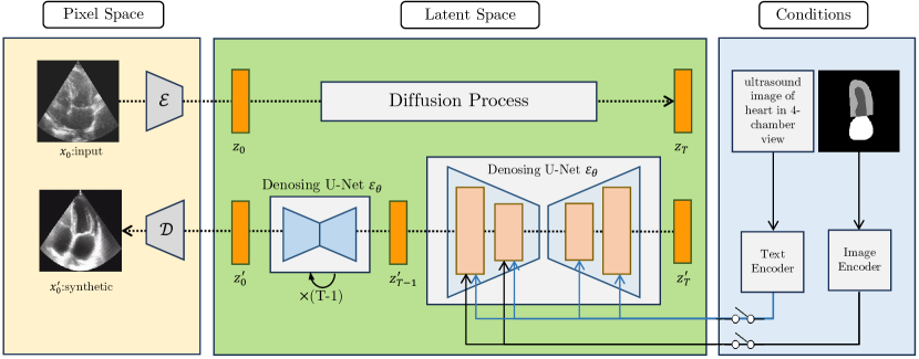
In our proposed network, illustrated in Figure 1, taking an input image with spatial dimensions and , and channels. We first compress the images into a lower-dimensional latent representation using the encoder of a variational autoencoder (VAE), such that [19]. The generative process in the latent space is modeled by a diffusion process, trained to invert the diffusion from a noise distribution to the original data distribution. Based on the auxiliary branches incorporating additional conditioning signals, we consider three possible scenarios in our generation process. Lastly, the decoder maps processed latent representations back to the pixel space.
2.1 Unconditional Image Generation
For the first setting, we utilized a simple Denoising Diffusion Probabilistic Model (DDPM) [9] where the encoder and decoder are identity functions and no guidance signals are applied. The forward process, adding noise over time, is expressed as . Solving this equation yields , where , , and . The reverse process, a denoising process, is formulated with a conditional probability , where and are neural network parameterized functions predicting mean and variance. The model is trained by empirically, parameterizing to predict and using the loss function at step .
2.2 Text-Guided Image Generation
To distill in-domain knowledge in the generation process and improve the representation capabilities, we condition our synthesis on text prompts in the second scenario. Specifically, our pipeline is based on the Stable Diffusion (SD) [19] model that offers a computationally efficient approach by operating in a latent space. The feature vector generated from the encoder is concatenated with the CLIP encoding [17] of the text prompt which is then combined into the denoising process using the cross-attention model [3]. The generative process in this latent space is modeled by a diffusion process, which is learned to reverse the diffusion from a noise distribution back to the data distribution. The training objective for our model is defined as:
| (1) |
where is the latent representation at time step , is the denoising UNet with cross-attention layers, is the conditioning text prompt, and is the text tokenizer. and are jointly optimized during the training process. The conditional generation of echo images is performed by guiding the reverse diffusion process with a text condition as elaborated in subsection 3.1.
2.3 Text + Segmentation Guided Image Generation
In addition to the text prompt, we use a semantic label map as part of the guidance signal to further improve the generation capacity. Here, the ControlNet [27] model is utilized to support additional input conditions providing greater flexibility and customization of the condition signals that allow for simultaneous structure-controlled and subject-controlled image synthesis. ControlNet model is a plug-and-play model that duplicates an SD branch to gather hierarchical features from the provided condition while keeping the initial SD branch frozen to maintain its generation capability. The trainable and fixed neural network components are linked via zero-convolution layers, where the trainable features are processed as input, and their output is combined with the frozen features. This way, we are capable of inserting, combining, and modifying imaging appearances through text prompts and corresponding segmentation maps, featuring highly detailed image correlations of the corresponding medical concepts.
3 Results
Training and Inference Settings. Our proposed technique was developed using PyTorch and Diffusers libraries and executed on four NVIDIA V100 16GB GPUs. A batch size of 1 (per device) and an Adam optimizer with a learning rate of were utilized during the training process, which was carried out for 120000 iterations. We employed a DDPM scheduler with 1000 denoising steps, and for quicker inference, a UniPC multi-step scheduler [28] with 50 steps.
Dataset and Evaluation Metrics. For our study, we used the CAMUS echocardiography dataset [13], featuring 2D apical views of both two-chamber (2CH) and four-chamber (4CH) perspectives from 500 patients across end-diastole (ED) and end-systole (ES) phases. The initial dataset randomly selected images from 50 patients for the test split, without segmentation labels, while the remaining 450 patients were assigned to the training split. Following the baseline work [23], we allocated the first 50 patients for validation and utilized the remaining 400 patients for training, leading to a total of 1600/200 images for train/validation respectively. We used Fréchet Inception Distance (FID) [8] and Kernel Inception Distance (KID) [2] to evaluate the quality of image synthesis. FID measures the distance between feature vectors of real and generated images, while KID quantifies the similarity between probability distributions of real and generated images’ features. For the downstream tasks, namely segmentation, and classification, we opt for well-established metrics as shown in Table 2 and 3(b).
3.1 Image Synthesis
Prompt Engineering. We examined two distinct strategies for prompt engineering applied to text-only and text+segmentation models: employing textual and abstract prompts. Textual prompts consist of straightforward natural language directives, such as “ultrasound image of the heart in 4-chamber view,” which proved effective with text+segmentation models due to the additional guidance from segmentation maps. However, for text-only models, we hypothesized that natural language terms might confuse the fine-tuning process, given that these models are initially trained on vast collections of non-medical images. To address this, we introduced an abstract training approach for our text-only models. This method involved using prompts structured like “{ultrasound image} displays the {heart} in a {two-chamber} view during the {ed} phase,” where each phrase within braces was replaced by a randomly generated string, aiming to minimize potential misunderstandings inherent to natural language processing in these models.
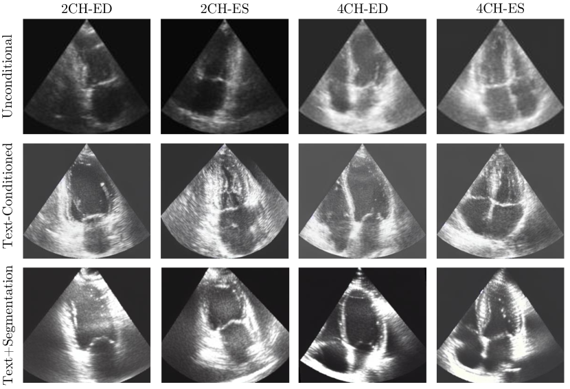
Generation Results. Table 1 presents a comparison of our setting proposals with the baseline SOTA method [23] using the FID and KID metrics across 4 possible view/cycles. The text+segmentation-based method has the lowest mean FID of 1.4902 across all views/cycles, which surpasses baseline SOTA by a clear margin of 1.6330 showing anatomical realism
and a high fidelity and diversity of the generated medical images. Moreover, this model outperforms unguided/text-guided counterparts in the KID metric achieving a significant performance boost.
FID Mean KID 2CH-ED 2CH-ES 4CH-ED 4CH-ES 2CH-ED 2CH-ES 4CH-ED 4CH-ES Unconditional 4.7712 4.5490 5.0679 4.1077 1.4619 2.5799 2.5452 2.0171 Text-Conditioned 1.4499 2.3445 1.7494 2.8399 1.2570 2.4584 1.5466 2.6263 SDM [23] 1.8188 1.7528 4.4704 4.4508 2.8221 2.9019 5.3975 5.4414 Text+Segmentation 1.3957 1.6251 1.6080 1.3322 1.4022 1.6186 1.6246 1.3322
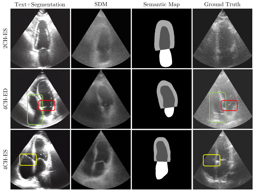
Figure 2 illustrates a qualitative result of our methods. Generally, our visuals guided by both text and segmentation maps exhibit higher fidelity and closely resemble real echo images. Overall, we observe that our model’s generated images have high overlap with both the input image and corresponding segmentation map, suggesting that our model closely adhered to the input guidance during image generation, significantly outperforming the generated images produced by SDM [23]. Specifically, the 2CH view focuses exclusively on the heart’s left ventricle (LV, upper chamber) and left atrium (LA, lower chamber), in contrast to the 4CH view, which additionally includes the RV and RA. The segmentation maps are limited to the anatomical features surrounding the LV endocardium (LV-endo), LV epicardium (LV-epi), and LA, lacking any data on the right chambers. Consequently, models, notably the SDM [23], tend to show enhanced performance for 2CH views as they are not required to predict the right chambers. Nevertheless, in the case of 4CH views, the SDM models lack directives for generating the right chambers, leading to subpar performance in their depiction. As illustrated in the second row of Figure 2, our text+segmentation model demonstrates superior accuracy in predicting the right chambers (indicated by green boxes) as the prompts explicitly specify the chamber count. Additionally, the ground truth in the third row reveals a visible tricuspid valve between the RV and RA, accurately predicted by our text+segmentation model (highlighted with yellow boxes). Moreover, our model accurately represents the mitral valve’s behavior, which remains closed during the ED phase and opens during the ES phase of the cardiac cycle, as shown in the outputs for the second and third rows (marked with red boxes).
Downstream Tasks. We evaluate the effect of the synthesized data obtained from our models on downstream tasks of echo image segmentation using UNet architecture [20] and linear probing for the classification of ED/ES classes.
Data Dice HD ASD Mean LV-endo LV-epi LA Mean LV-endo LV-epi LA Mean LV-endo LV-epi LA Real+200% 0.8685 0.8931 0.9200 0.7924 11.57 8.91 10.18 15.61 5.38 3.61 3.80 6.96 Real+100% 0.8759 0.8995 0.9244 0.8038 12.65 9.55 10.54 17.85 5.66 3.87 3.73 7.58 Real+50% 0.8721 0.8950 0.9248 0.7966 11.93 9.08 9.63 17.09 5.46 3.64 3.63 7.30 Real 0.8700 0.8902 0.9206 0.7991 15.02 9.48 10.96 24.62 6.15 3.79 3.97 8.33 SDM [23] 0.8576 0.8661 0.9006 0.8061 13.83 12.87 11.76 16.84 5.52 4.65 4.61 6.43
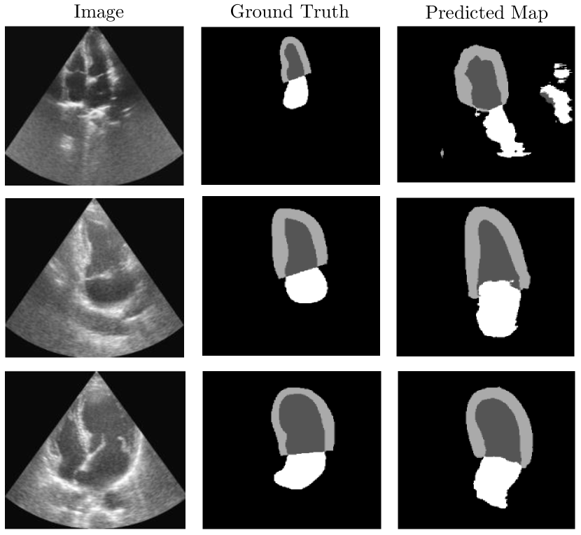
We investigated multiple data augmentation methods, including only real images (Real), a combination of 1600 real with 800 synthetic images (Real+50%), 1600 real with 1600 synthetic images (Real+100%), and 1600 real with 3200 synthetic images (Real+200%). These synthetic images were created using our text+segmentation model. For validation, we used 50 held-out patients, following [23] (It is noteworthy to mention that [23] developed their segmentation model using 8000 synthetic images.). Table 2 and 3(b) present the comparative effectiveness of our generated data in diverse scenarios. Specifically, Table 2 shows that our synthetic data distinctly impact the performance of different models, resulting in additional performance gains when utilizing data generated with comprehensive guidance compared to other competitors, as evidenced by various evaluation metrics. This also aligns with the visual comparison of echo segmentation in 3(a) which indicates that our text+segmentation guided model can produce echo images with better overall realism and a tendency to adhere to anatomical input constraints. Lastly, the classification results shown in 3(b) highlight the value of synthetic data, demonstrating that image-level details (text prompts) outperform those from full guidance (text+segmentation).
![[Uncaptioned image]](/html/2403.19880/assets/x3.png) |
| Methods & dataset | Metrics | |||
|---|---|---|---|---|
| ACC | PR | RC | F1 | |
| ResNet18 (Real data only) | 0.8400 | 0.8400 | 0.8400 | 0.8399 |
| ResNet18 (Unconditional) | 0.8350 | 0.8366 | 0.8350 | 0.8347 |
| ResNet18 (Text-Conditioned) | 0.8700 | 0.8773 | 0.8700 | 0.8693 |
| ResNet18 (SDM [23]) | 0.8300 | 0.8311 | 0.8300 | 0.8298 |
| ResNet18 (Text+Segmentation) | 0.8650 | 0.8659 | 0.8650 | 0.8640 |
| VGG16 (Real data only) | 0.7450 | 0.7471 | 0.7450 | 0.7445 |
| VGG16 (Uniconditional) | 0.7500 | 0.7549 | 7500 | 0.7487 |
| VGG16 (Text-Conditioned) | 0.7850 | 0.7850 | 0.7850 | 0.7849 |
| VGG16 (SDM [23]) | 0.7600 | 0.7687 | 0.7600 | 0.7580 |
| VGG16 (Text+Segmentation) | 0.7750 | 0.7763 | 0.7750 | 0.7747 |
4 Conclusion
Generating echocardiograms poses unique challenges compared to standard computer vision tasks due to the inherent noise in ultrasound images. In this paper, we demonstrate the first attempt to synthesize echo images using a diffusion-based model with both semantic segmentation map and text supervision which is an optimal way to guide models in generating high-fidelity yet diverse images. We prove this hypothesis about generation through extensive experiments with different prompting scenarios, along with two downstream tasks showcasing the effectiveness of the synthesized data.
Acknowledgment
We acknowledge the support of the Natural Sciences and Engineering Research Council of Canada (NSERC), [funding reference number RGPIN-2023-03575]. Cette recherche a été financée par le Conseil de recherches en sciences naturelles et en génie du Canada (CRSNG), [numéro de référence RGPIN-2023-03575].
References
- [1] Adhikari, R., Dhakal, M., Thapaliya, S., Poudel, K., Bhandari, P., Khanal, B.: Synthetic boost: Leveraging synthetic data for enhanced vision-language segmentation in echocardiography. In: International Workshop on Advances in Simplifying Medical Ultrasound. pp. 89–99. Springer (2023)
- [2] Bińkowski, M., Sutherland, D.J., Arbel, M., Gretton, A.: Demystifying mmd gans. arXiv preprint arXiv:1801.01401 (2018)
- [3] Chen, C.F.R., Fan, Q., Panda, R.: Crossvit: Cross-attention multi-scale vision transformer for image classification. In: Proceedings of the IEEE/CVF international conference on computer vision. pp. 357–366 (2021)
- [4] Dhariwal, P., Nichol, A.: Diffusion models beat gans on image synthesis. Advances in neural information processing systems 34, 8780–8794 (2021)
- [5] Fiaz, M., Heidari, M., Anwer, R.M., Cholakkal, H.: Sa2-net: Scale-aware attention network for microscopic image segmentation (2022)
- [6] Goodfellow, I., Pouget-Abadie, J., Mirza, M., Xu, B., Warde-Farley, D., Ozair, S., Courville, A., Bengio, Y.: Generative adversarial nets. Advances in neural information processing systems 27 (2014)
- [7] Heidenreich, P.A., Trogdon, J.G., Khavjou, O.A., Butler, J., Dracup, K., Ezekowitz, M.D., Finkelstein, E.A., Hong, Y., Johnston, S.C., Khera, A., et al.: Forecasting the future of cardiovascular disease in the united states: a policy statement from the american heart association. Circulation 123(8), 933–944 (2011)
- [8] Heusel, M., Ramsauer, H., Unterthiner, T., Nessler, B., Hochreiter, S.: Gans trained by a two time-scale update rule converge to a local nash equilibrium. Advances in neural information processing systems 30 (2017)
- [9] Ho, J., Jain, A., Abbeel, P.: Denoising diffusion probabilistic models. Advances in neural information processing systems 33, 6840–6851 (2020)
- [10] Iqbal, H., Khalid, U., Chen, C., Hua, J.: Unsupervised anomaly detection in medical images using masked diffusion model. In: International Workshop on Machine Learning in Medical Imaging. pp. 372–381. Springer (2023)
- [11] Kazerouni, A., Aghdam, E.K., Heidari, M., Azad, R., Fayyaz, M., Hacihaliloglu, I., Merhof, D.: Diffusion models for medical image analysis: A comprehensive survey. arXiv preprint arXiv:2211.07804 (2022)
- [12] Khader, F., Mueller-Franzes, G., Arasteh, S.T., Han, T., Haarburger, C., Schulze-Hagen, M., Schad, P., Engelhardt, S., Baessler, B., Foersch, S., et al.: Medical diffusion–denoising diffusion probabilistic models for 3d medical image generation. arXiv preprint arXiv:2211.03364 (2022)
- [13] Leclerc, S., Smistad, E., Pedrosa, J., Østvik, A., Cervenansky, F., Espinosa, F., Espeland, T., Berg, E.A.R., Jodoin, P.M., Grenier, T., et al.: Deep learning for segmentation using an open large-scale dataset in 2d echocardiography. IEEE transactions on medical imaging 38(9), 2198–2210 (2019)
- [14] Lin, L., Li, Z., Li, R., Li, X., Gao, J.: Diffusion models for time-series applications: a survey. Frontiers of Information Technology & Electronic Engineering pp. 1–23 (2023)
- [15] Olive Pellicer, A., Yadav, A.K.S., Bhagtani, K., Xiang, Z., Pizlo, Z., Gradus-Pizlo, I., Delp, E.J.: Synthetic echocardiograms generation using diffusion models. bioRxiv pp. 2023–11 (2023)
- [16] Peng, W., Adeli, E., Bosschieter, T., Park, S.H., Zhao, Q., Pohl, K.M.: Generating realistic brain mris via a conditional diffusion probabilistic model. In: International Conference on Medical Image Computing and Computer-Assisted Intervention. pp. 14–24. Springer (2023)
- [17] Radford, A., Kim, J.W., Hallacy, C., Ramesh, A., Goh, G., Agarwal, S., Sastry, G., Askell, A., Mishkin, P., Clark, J., et al.: Learning transferable visual models from natural language supervision. In: International conference on machine learning. pp. 8748–8763. PMLR (2021)
- [18] Reynaud, H., Qiao, M., Dombrowski, M., Day, T., Razavi, R., Gomez, A., Leeson, P., Kainz, B.: Feature-conditioned cascaded video diffusion models for precise echocardiogram synthesis. arXiv preprint arXiv:2303.12644 (2023)
- [19] Rombach, R., Blattmann, A., Lorenz, D., Esser, P., Ommer, B.: High-resolution image synthesis with latent diffusion models. In: Proceedings of the IEEE/CVF conference on computer vision and pattern recognition. pp. 10684–10695 (2022)
- [20] Ronneberger, O., Fischer, P., Brox, T.: U-net: Convolutional networks for biomedical image segmentation. In: Medical Image Computing and Computer-Assisted Intervention–MICCAI 2015: 18th International Conference, Munich, Germany, October 5-9, 2015, Proceedings, Part III 18. pp. 234–241. Springer (2015)
- [21] Skandarani, Y., Jodoin, P.M., Lalande, A.: Gans for medical image synthesis: An empirical study. Journal of Imaging 9(3), 69 (2023)
- [22] Stevens, T.S., Meral, F.C., Yu, J., Apostolakis, I.Z., Robert, J.L., Van Sloun, R.J.: Dehazing ultrasound using diffusion models. IEEE Transactions on Medical Imaging (2024)
- [23] Stojanovski, D., Hermida, U., Lamata, P., Beqiri, A., Gomez, A.: Echo from noise: synthetic ultrasound image generation using diffusion models for real image segmentation. International Workshop on Advances in Simplifying Medical Ultrasound, MICCAI-ASMUS (2023)
- [24] Tang, F., Ding, J., Wang, L., Xian, M., Ning, C.: Multi-level global context cross consistency model for semi-supervised ultrasound image segmentation with diffusion model. arXiv preprint arXiv:2305.09447 (2023)
- [25] Wang, W., Bao, J., Zhou, W., Chen, D., Chen, D., Yuan, L., Li, H.: Semantic image synthesis via diffusion models. arXiv preprint arXiv:2207.00050 (2022)
- [26] Wolleb, J., Sandkühler, R., Bieder, F., Valmaggia, P., Cattin, P.C.: Diffusion models for implicit image segmentation ensembles. In: International Conference on Medical Imaging with Deep Learning. pp. 1336–1348. PMLR (2022)
- [27] Zhang, L., Rao, A., Agrawala, M.: Adding conditional control to text-to-image diffusion models. In: Proceedings of the IEEE/CVF International Conference on Computer Vision. pp. 3836–3847 (2023)
- [28] Zhao, W., Bai, L., Rao, Y., Zhou, J., Lu, J.: Unipc: A unified predictor-corrector framework for fast sampling of diffusion models. Advances in Neural Information Processing Systems 36 (2024)
- [29] Zhou, M., Khalvati, F.: Conditional generation of 3d brain tumor regions via vqgan and temporal-agnostic masked transformer. In: Medical Imaging with Deep Learning (2024)
Appendix