Ensembling and Test Augmentation for Covid-19 Detection and Covid-19 Domain Adaptation from 3D CT-Scans
Junia, UMR 8520, CNRS, Centrale Lille, Univerity of Polytechnique Hauts-de-France, 59000 Lille, France
faresbougourzi@gmail.com; fares.bougourzi@junia.com
\And
Junia, UMR 8520, CNRS, Centrale Lille, Univerity of
Polytechnique Hauts-de-France, 59000 Lille, France
feryal.windal@junia.com
\And
Centre d’ Enseignement, de Recherche et
d’Innovation Systèmes Numériques, Lille, France
halim.benhabiles@imt-nord-europe.fr
\And
University of the Basque Country UPV/EHU,
San Sebastian, SPAIN; IKERBASQUE, Basque
Foundation for Science, Bilbao, SPAIN
fadi.dornaika@ehu.eus
\And
IEMN UMR CNRS 8520, Université
Polytechnique Hauts de France, UPHF
Abdelmalik.Taleb-Ahmed@uphf.fr
Abstract
Since the emergence of Covid-19 in late 2019, medical image analysis using artificial intelligence (AI) has emerged as a crucial research area, particularly with the utility of CT-scan imaging for disease diagnosis. This paper contributes to the 4th COV19D competition, focusing on Covid-19 Detection and Covid-19 Domain Adaptation Challenges. Our approach centers on lung segmentation and Covid-19 infection segmentation employing the recent CNN-based segmentation architecture PDAtt-Unet, which simultaneously segments lung regions and infections. Departing from traditional methods, we concatenate the input slice (grayscale) with segmented lung and infection, generating three input channels akin to color channels. Additionally, we employ three 3D CNN backbones—Customized Hybrid-DeCoVNet, along with pretrained 3D-Resnet-18 and 3D-Resnet-50 models—to train Covid-19 recognition for both challenges. Furthermore, we explore ensemble approaches and testing augmentation to enhance performance. Comparison with baseline results underscores the substantial efficiency of our approach, with a significant margin in terms of F1-score (14%). This study advances the field by presenting a comprehensive methodology for accurate Covid-19 detection and adaptation, leveraging cutting-edge AI techniques in medical image analysis.
Keywords Covid-19 Deep Leaning CNNs Recognition
1 Introduction
Since the onset of the Covid-19 pandemic in late 2019, Reverse Transcription-Polymerase Chain Reaction (RT-PCR) has been widely established as the primary method for Covid-19 detection. Nevertheless, this testing modality presents several drawbacks, including limited availability of RT-PCR kits, lengthy procedures, and a notable incidence of false negative results [1]. Consequently, medical imaging techniques such as X-rays and CT-scans have gained prominence as complementary tools for Covid-19 detection [2, 3]. CT-scans not only serve in identifying Covid-19 infections but also play a crucial role in monitoring patients’ conditions and predicting disease severity [3, 4].
In recent years, Deep Learning methodologies have risen to prominence in computer vision tasks, showcasing remarkable performance gains over conventional techniques [5, 6, 7]. However, one of the primary challenges associated with Deep Learning, particularly in the realm of Convolutional Neural Networks (CNNs), lies in the necessity for extensive labeled datasets, a resource often scarce in medical domains [3, 8]. Furthermore, the majority of existing CNN architectures are tailored for processing static images, which proves inadequate in capturing the intricacies inherent in medical imaging data, especially for the volumetric scans [4]. On the other hand, domain adaptation is one of the most challenging aspects encountered in medical imaging, owing to the high variability of data from one center to another due to the variety of recording settings and scanners. Machine learning techniques used in computer-aided medical image analysis usually suffer from the domain shift problem caused by different distributions between source/reference data and target data [9].
In our paper, we present an approach to address both Covid-19 detection and domain adaptation challenges on the 4th COV19D competition. Our method revolves around lung segmentation and Covid-19 infection segmentation using the PDAtt-Unet CNN-based segmentation architecture, which concurrently segments lung regions and infections. Departing from traditional methods, we integrate the input slice with segmented lung and infection, creating three input channels akin to color channels. We utilize three 3D CNN backbones— Customized Hybrid-DeCoVNet, pretrained 3D-Resnet-18, and 3D-Resnet-50 models—to train Covid-19 recognition for both challenges. Additionally, we explore ensemble approaches and testing augmentation to enhance performance. Our main contributions are:
-
•
We adopted a Customized Hybrid-DeCoVNet architecture for both Covid-19 Detection and Covid-19 Domain Adaptation Challenges. This architecture incorporates the concatenation of the original slice, the segmented lung, and the segmented Covid-19 infection as the three input channels.
-
•
In addition to our proposed Customized Hybrid-DeCoVNet architecture, we leveraged two pretrained 3D-CNNs: 3D-Resnet-18 and 3D-Resnet-50.
-
•
We explored ensemble approaches and testing augmentation techniques to enhance the robustness and performance of our method.
-
•
Our approach demonstrated a substantial improvement in efficiency compared to baseline results, with a significant margin in F1-score (14%).
-
•
We have made our codes and pretrained models publicly available in 111https://github.com/faresbougourzi/4th-COV19D-Competition. ( Last accessed on March, 17th 2024).
2 Our Approaches
Our approach is tailored to capitalize on region of interest segmentation, specifically lung segmentation, and infection segmentation alongside input slices from CT scans. The objective is to develop a model proficient in discerning COVID-19 cases from non-COVID-19 cases. We evaluate three baseline architectures: Customized Hybrid-DeCoVNet [10], 3D-ResNet-18, and 3D-ResNet-50 [11].
2.1 Data Preprocessing
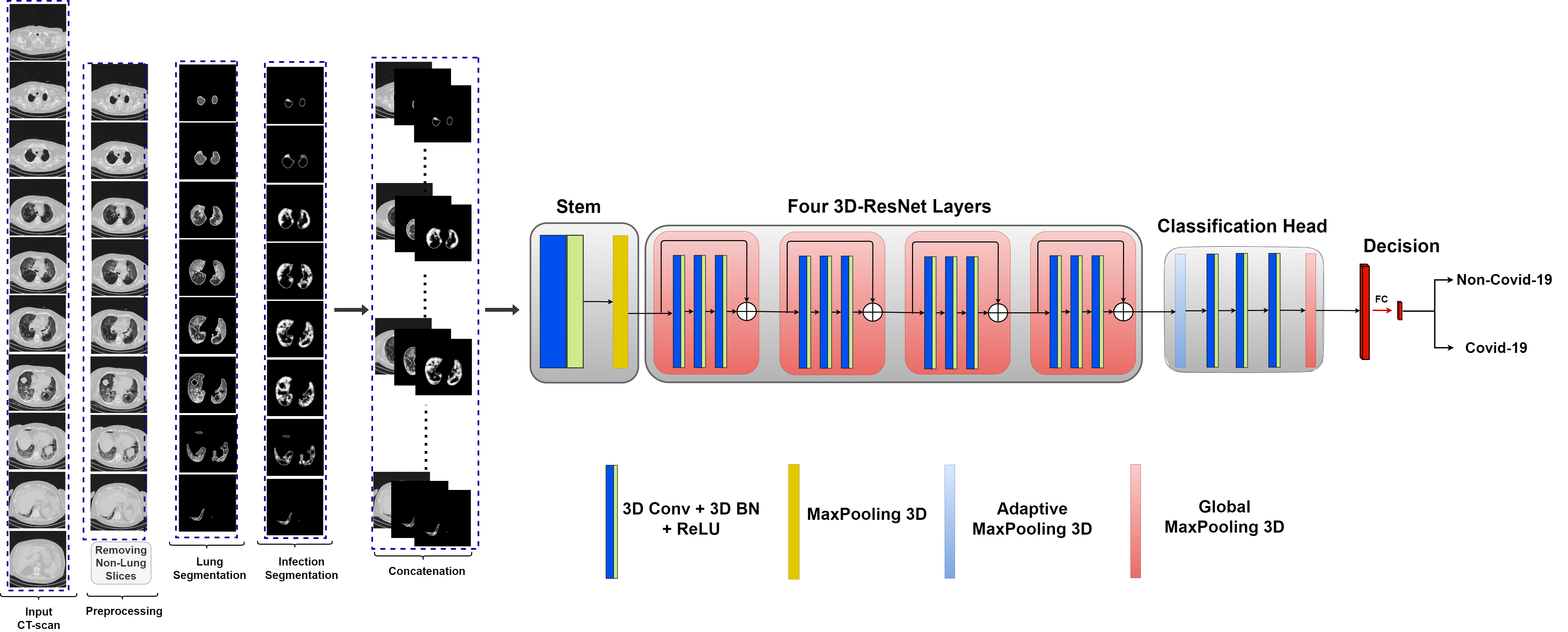
The objective of this phase is to eliminate slices that do not exhibit any lung structures and to identify lung features in the remaining slices. Following our previous approach in the 2nd COV19D competition and 3rd COV19D competition [12, 10], ResneXt-50 [13] is used to filter the CT slices that does not show lung regions, to concentrate only on the slices that may have infection.
2.2 Customized Hybrid-DeCoVNet
In this challenge, we adopted our proposed Customized Hybrid-DeCoVNet which were proposed for Covid-19 severity prediction, to perform Covid-19 recognition in this challenge. The first modification is by considering the input slice, their region of interest segmentation and the infection segmentation as input, these three images are concatenated to form 3 channels. For segmenting the lung and the infection we used the PDAtt-Unet [8] for segmenting the infection and the lung simultaneously. PDAtt-Unet is trained using three datasets Segmentation dataset nr. 2 [14], COVID-19 CT segmentation [14] and CC-CCII [15], each dataset is divided into 70%-30% as training and validation splits, then PDAtt-Unet is trained on the ensemble of 70% of the three datasets and evaluated on the ensemble of the their 30%.
As illustrated in Figure 1, Customized Hybrid-DeCoVNet comprises of four components. First, the three images depicting the input slice, the segmented lung and segmented infection are merged into a three-channel image. This is performed for every slice of the input CT-scans, then all of these merged 3 channels images are concatenated. For a CT-scan of slice this will produce . Since the number of slices is different from one CT-scan to another, S is resized into a fixed size of . This resulting volume is fed into the Stem block, which is a 3D convolutional layer with a kernel of size (7, 7, 5) for height, width, and depth, respectively. The Stem block transforms the two input channels into 16 channels and is followed by Batch Normalization Layer (BN) and ReLU activation function. The second block of Customized Hybrid-DeCoVNet consists of four 3D-Resnet layers, which expand the channels to 64, 128, 256, and 512, respectively. The Classification Head comprises of 3D Adaptive MaxPooling, three 3D convolutional layers, and 3D Global MaxPooling. The output of the Classification Head is flattened into a single-channel deep feature map and fed into the Decision Head, which consists of one FC layer that has two outputs (Non-Covid-19 and Covid-19). Our proposed architecture is designed to enhance the performance of Covid-19 Prediction. It should be noted that Customized Hybrid-DeCoVNet does not have any pretrained weight in contrast with 3D-Resnet-18 and 3D-Resnet-50.
2.3 3D-Resnet-18 and 3D-Resnet-50
In addition to our proposed Customized Hybrid-DeCoVNet, we evaluated the use of pretrained 3D CNN architectures. To this end, we used the pretrained 3D-Resnet-18 and 3D-Resnet-50 from [11]. These pretrained models were trained for action recognition from 3D-video. For 3D-Resnet-18, the pretrained weights was trained on the ensemble of Kinetics-700 and Moments in Time datasets. While, 3D-Resnet-50 was trained on the ensemble of Kinetics-700, Moments in Time, STAIR-Actions datasets. To adopt these models for Covid-19 recognition, we changed the decision layer to give 2 output which corresponds to Non-Covid-19 and Covid-19 classes.
3 Experiments and Results
3.1 The COV19-CT-DB Database
In this competition, the COVID19-CT-Database (COV19-CT-DB) [16, 17, 18, 19, 20, 21, 22, 23] is used for two sub challenges, which are Covid-19 Detection Challenge and Covid-19 Domain Adaptation Challenge. In Covid-19 Detection Challenge, many CT scans have been aggregated, each one of which has been manually annotated in terms of Covid-19 and non-Covid-19 categories. The resulting dataset is split into training, validation and test partitions. The provided training and validation partitions for developing the approach are summarized in Table 1.
In the second challenge, Covid-19 Domain Adaptation Challenge, CT scans have been aggregated from various hospitals and medical centres. Each CT scan has been manually annotated with respect to Covid-19 and non-Covid-19 categories. The resulting dataset is split into training, validation and test partitions. Participants will be provided with a training set that consists of: i) the annotated data of the 1st Challenge which are aggregated from some hospitals and medical centres (case A); ii) a small number of annotated data and a larger number of non-annotated data (case B), all of which are aggregated from other hospitals and medical centres and their distribution is different from that of case A. Participants will be provided with a validation set that consists of a small number of annotated data of case B.
| Sub-Competition | Train | Validation | ||
|---|---|---|---|---|
| Covid-19 | Non-Covid-19 | Covid-19 | Non-Covid-19 | |
| Covid-19 Detection Challenge | 703 | 655 | 170 | 156 |
| Covid-19 Domain Adaptation Challenge | 120 | 120 | 56 | 113 |
3.2 Experimental Setup
We utilized the Pytorch Library and four NVIDIA GPU Device GeForce TITAN RTX 16 GB for Deep Learning training and testing. The batch size of 16 CT-scan volumes was used to train the Customized Hybrid-DeCoVNet and 3D-Resnet-18 architectures for 80 epochs. While, a batch size of 8 is used to train 3D-Resnet-50 for 40 epochs. Warm up Cosine learning rate Schedule is adopted with initial learning rate of 0.0001. The following data augmentations are used for training and testing augmentation approach: random rotation with an angle between -40∘ to 40∘, vertical and horizontal flipping with a probability of 20% for each, Multiplicative Noise, Random Brightness, Random Brightness Contrast, Random Contrast, and Random Grid Shuffle .
3.3 Results
3.3.1 Results of the first evaluation scenario
In this part, we used the training data of Covid-19 Detection Challenge (first challenge) and the validation data to train and save the best model on the validation data after each epoch, this two splits will be denoted as Train1 and Val1. We also used the training and validation data of the Covid-19 Domain Adaptation Challenge (the second challenge) to evaluate the performance of our approach in unseen data, these two splits will be denoted as Train2 and Val2. Table 2 summarizes the obtained results. From these results, it is noticed that the performance on the Train2 and Val2 splits decreased compared with the results on Val1, this is due to the change of data domain. However, the drop in results in not too big, this shows that our approach can achieve a good result. On the other hand, the ensembling approach achieves better performance on Train2 compared with the single architectures.
| Architecture | Val1 | Train2 | Val2 | |||
| Accuracy | F1-score | Accuracy | F1-score | Accuracy | F1-score | |
| Customized Hybrid-DeCoVNet | 92.33 | 92.33 | 83.75 | 83.72 | 82.58 | 82.09 |
| 3D-Resnet-18 | 91.41 | 91.41 | 82.08 | 81.88 | 83.70 | 82.59 |
| 3D-Resnet-50 | 91.41 | 91.40 | 84.58 | 84.58 | 83.70 | 82.59 |
| Ensemble | 91.10 | 91.10 | 85 | 84.93 | 83.70 | 82.59 |
3.3.2 Results of the second evaluation scenario
In the second evaluation scenario, we combined the training data of Covid-19 Detection and Covid-19 Domain Adaptation challenges (Train1+Train2) in order to compare the performance of the three backbones in the scenario where the training data is augmented. The obtained results are summarized in Table 3, in which, Val1 and Val2 correspond to the validation of the first and the second challenge, respectively (correspond to the same splits used in Table 2). By comparing the results of Tables 2 and 3, it is noticed that augmenting the training data improve the performance of the three backbones, especially 3D-Resnet-18.
Table 4 depicts the results of using testing augmentation, in which, each CT-scan is augmented ten times and the CT-scan prediction corresponds to the average probabilities of the prediction of the ten augmentations. Compared with the results of Table 3, using testing augmentation further improves the performance.
| Architecture | Val1 | Val2 | ||
| Accuracy | F1-score | Accuracy | F1-score | |
| Customized Hybrid-DeCoVNet | 92.33 | 92.33 | 83.14 | 80.64 |
| 3D-Resnet-18 | 92.33 | 92.32 | 87.07 | 85.60 |
| 3D-Resnet-50 | 92.63 | 92.60 | 84.26 | 82.92 |
| Architecture | Val1 | Val2 | ||
| Accuracy | F1-score | Accuracy | F1-score | |
| Customized Hybrid-DeCoVNet | 91.41 | 91.40 | 84.83 | 83.23 |
| 3D-Resnet-18 | 92.33 | 92.33 | 88.76 | 87.52 |
| 3D-Resnet-50 | 92.33 | 92.33 | 85.39 | 84.34 |
| Ensemble | 92.33 | 92.33 | 88.20 | 87.14 |
3.4 Comparison with the Baseline
Table 5, depicts the comparison with the baseline results from [24]. The comparison of our approach and the baseline results shows that our approach achieved better performance on both challenges. In more details, our approach achieved better performance than the baseline approach by 14.33% in terms of F1-score for Covid-19 Detection Challenge. Similarly for Covid-19 Domain Adaptation Challenge, our approach achieved better performance than the baseline by 14.52% in terms of F1-score.
| Architecture | sub-challenge 1 | sub-challenge 2 | ||
| Accuracy | F1-score | Accuracy | F1-score | |
| Baseline | - | 78 | - | 73 |
| Customized Hybrid-DeCoVNet | 91.41 | 91.40 | 84.83 | 83.23 |
| 3D-Resnet-18 | 92.33 | 92.33 | 88.76 | 87.52 |
| 3D-Resnet-50 | 92.33 | 92.33 | 85.39 | 84.34 |
| Ensemble | 92.33 | 92.33 | 88.20 | 87.14 |
4 Conclusion
In this paper, we introduced a new approach for addressing the Covid-19 Detection and Covid-19 Domain Adaptation Challenges. Our approach primarily leveraged lung segmentation and Covid-19 infection segmentation through the utilization of state-of-the-art CNN-based segmentation architecture, namely PDAtt-Unet. This architecture enables simultaneous segmentation of lung regions and infections. Rather than feeding individual input slices to the training network, we concatenated the input slice (grayscale) with the segmented lung and infection, resulting in three input channels akin to color channels.
Moreover, we employed three distinct 3D CNN backbones to train Covid-19 recognition for both challenges: Customized Hybrid-DeCoVNet, as well as pretrained 3D-Resnet-19 and 3D-Resnet-50 models. To further enhance performance, we investigated ensemble approaches and testing augmentation techniques. Comparative analysis against baseline results demonstrates the significant efficiency of our proposed approach, exhibiting a substantial margin in terms of F1-score (14%).
Our findings underscore the effectiveness of our methodology in addressing the complexities of Covid-19 detection and domain adaptation challenges. By integrating cutting-edge segmentation architectures and leveraging ensemble strategies, our approach demonstrates promising advancements in accurately identifying Covid-19 infections, thus contributing to the ongoing efforts in combatting Covid-19 pandemic and the future ones.
References
- [1] Ying-Hui Jin, Lin Cai, Zhen-Shun Cheng, and others., “A rapid advice guideline for the diagnosis and treatment of 2019 novel coronavirus (2019-nCoV) infected pneumonia (standard version),” Military Medical Research, vol. 7, no. 1, pp. 4, Feb. 2020.
- [2] Fares Bougourzi, Riccardo Contino, Cosimo Distante, and Abdelmalik Taleb-Ahmed, “Recognition of COVID-19 from CT Scans Using Two-Stage Deep-Learning-Based Approach: CNR-IEMN,” Sensors, vol. 21, no. 17, pp. 5878, 2021.
- [3] Fares Bougourzi, Fadi Dornaika, Amir Nakib, and Abdelmalik Taleb-Ahmed, “Emb-trattunet: a novel edge loss function and transformer-CNN architecture for multi-classes pneumonia infection segmentation in low annotation regimes,” Artificial Intelligence Review, vol. 57, no. 4, pp. 90, Mar. 2024.
- [4] Fares Bougourzi, Riccardo Contino, Cosimo Distante, and Abdelmalik Taleb-Ahmed, “CNR-IEMN: A Deep Learning based approach to recognise COVID-19 from CT-scan,” in ICASSP 2021-2021 IEEE International Conference on Acoustics, Speech and Signal Processing (ICASSP). IEEE, 2021, pp. 8568–8572.
- [5] F. Bougourzi, F. Dornaika, K. Mokrani, A. Taleb-Ahmed, and Y. Ruichek, “Fusing Transformed Deep and Shallow features (FTDS) for image-based facial expression recognition,” Expert Systems with Applications, vol. 156, pp. 113459, Oct. 2020.
- [6] F. Bougourzi, F. Dornaika, and A. Taleb-Ahmed, “Deep Learning Based Face Beauty Prediction via Dynamic Robust Losses and Ensemble Regression,” Knowledge-Based Systems, vol. 242, pp. 108246, Apr. 2022.
- [7] Fares Bougourzi, Fadi Dornaika, Nagore Barrena, Cosimo Distante, and Abdelmalik Taleb-Ahmed, “Cnn based facial aesthetics analysis through dynamic robust losses and ensemble regression,” Applied Intelligence, vol. 53, no. 9, pp. 10825–10842, 2023.
- [8] Fares Bougourzi, Cosimo Distante, Fadi Dornaika, and Abdelmalik Taleb-Ahmed, “Pdatt-unet: Pyramid dual-decoder attention unet for covid-19 infection segmentation from ct-scans,” Medical Image Analysis, p. 102797, 2023.
- [9] Hao Guan and Mingxia Liu, “Domain adaptation for medical image analysis: a survey,” IEEE Transactions on Biomedical Engineering, vol. 69, no. 3, pp. 1173–1185, 2021.
- [10] Fares Bougourzi, Fadi Dornaika, Amir Nakib, Cosimo Distante, and Abdelmalik Taleb-Ahmed, “Deep-covid-sev: an ensemble 2d and 3d cnn-based approach for covid-19 severity prediction from 3d ct-scans,” in 2023 IEEE International Conference on Acoustics, Speech, and Signal Processing Workshops (ICASSPW). IEEE, 2023, pp. 1–5.
- [11] Kensho Hara, Hirokatsu Kataoka, and Yutaka Satoh, “Can spatiotemporal 3d cnns retrace the history of 2d cnns and imagenet?,” in Proceedings of the IEEE conference on Computer Vision and Pattern Recognition, 2018, pp. 6546–6555.
- [12] Fares Bougourzi, Cosimo Distante, Fadi Dornaika, and Abdelmalik Taleb-Ahmed, “CNR-IEMN-CD and CNR-IEMN-CSD Approaches for Covid-19 Detection and Covid-19 Severity Detection from 3D CT-scans,” in Computer Vision–ECCV 2022 Workshops: Tel Aviv, Israel, October 23–27, 2022, Proceedings, Part VII. Springer, 2023, pp. 593–604.
- [13] Saining Xie, Ross Girshick, Piotr Dollár, Zhuowen Tu, and Kaiming He, “Aggregated residual transformations for deep neural networks,” 2017.
- [14] RADIOLOGISTS, “COVID-19 CT-scans segmentation datasets, available at: http://medicalsegmentation.com/covid19/,” 2019, Last visited: 18-08-2021.
- [15] Xiaohong Liu, Kai Wang, Ke Wang, Ting Chen, Kang Zhang, and Guangyu Wang, “Kiseg: a three-stage segmentation framework for multi-level acceleration of chest ct scans from covid-19 patients,” in International Conference on Medical Image Computing and Computer-Assisted Intervention. Springer, 2020, pp. 25–34.
- [16] Dimitrios Kollias, Anastasios Arsenos, and Stefanos Kollias, “Ai-enabled analysis of 3-d ct scans for diagnosis of covid-19 & its severity,” in 2023 IEEE International Conference on Acoustics, Speech, and Signal Processing Workshops (ICASSPW). IEEE, 2023, p. 1–5.
- [17] Anastasios Arsenos, Andjoli Davidhi, Dimitrios Kollias, Panos Prassopoulos, and Stefanos Kollias, “Data-driven covid-19 detection through medical imaging,” in 2023 IEEE International Conference on Acoustics, Speech, and Signal Processing Workshops (ICASSPW). IEEE, 2023, p. 1–5.
- [18] Dimitrios Kollias, Anastasios Arsenos, and Stefanos Kollias, “A deep neural architecture for harmonizing 3-d input data analysis and decision making in medical imaging,” Neurocomputing, vol. 542, pp. 126244, 2023.
- [19] Dimitrios Kollias, Anastasios Arsenos, and Stefanos Kollias, “Ai-mia: Covid-19 detection & severity analysis through medical imaging,” arXiv preprint arXiv:2206.04732, 2022.
- [20] Anastasios Arsenos, Dimitrios Kollias, and Stefanos Kollias, “A large imaging database and novel deep neural architecture for covid-19 diagnosis,” in 2022 IEEE 14th Image, Video, and Multidimensional Signal Processing Workshop (IVMSP). IEEE, 2022, p. 1–5.
- [21] Dimitrios Kollias, Anastasios Arsenos, Levon Soukissian, and Stefanos Kollias, “Mia-cov19d: Covid-19 detection through 3-d chest ct image analysis,” in Proceedings of the IEEE/CVF International Conference on Computer Vision, 2021, p. 537–544.
- [22] Dimitrios Kollias, N Bouas, Y Vlaxos, V Brillakis, M Seferis, Ilianna Kollia, Levon Sukissian, James Wingate, and S Kollias, “Deep transparent prediction through latent representation analysis,” arXiv preprint arXiv:2009.07044, 2020.
- [23] Dimitris Kollias, Y Vlaxos, M Seferis, Ilianna Kollia, Levon Sukissian, James Wingate, and Stefanos D Kollias, “Transparent adaptation in deep medical image diagnosis.,” in TAILOR, 2020, p. 251–267.
- [24] Dimitrios Kollias, Anastasios Arsenos, and Stefanos Kollias, “Domain adaptation, explainability & fairness in ai for medical image analysis: Diagnosis of covid-19 based on 3-d chest ct-scans,” arXiv preprint arXiv:2403.02192, 2024.