Gating single-molecule fluorescence with electrons
Abstract
Tip-enhanced photoluminescence (TEPL) measurements are performed with sub-nanometer spatial resolution on individual molecules decoupled from a metallic substrate by a thin NaCl layer. TEPL spectra reveal progressive fluorescence quenching with decreasing tip-molecule distance when electrons tunneling from the tip of a scanning tunneling microscope are injected at resonance with the molecular states. Rate equations based on a many-body model reveal that the luminescence quenching is due to a progressive population inversion between the ground neutral (S0) and the ground charge () states of the molecule occurring when the current is raised. We demonstrate that both the bias voltage and the atomic-scale lateral position of the tip can be used to gate the molecular emission. Our approach can in principle be applied to any molecular system, providing unprecedented control over the fluorescence of a single molecule.
Control over the optical properties of individual molecules can be achieved by modifying their chemical structure or influencing their local environment. In that respect, the ability to switch the molecule from a bright to a dark state is one of the most striking modifications of these characteristics. Such a mechanism is employed in, for example, superresolution microscopy to localize individual emittersFolling2008 ; Heilemann2009 ; Vogelsang2009 and holds the potential to be applied in sensorsBrasselet2000 or even molecular quantum devicesToninelli2021 to gate the properties of single-photon emittersYu2022 . Usually, switching the chromophore to a dark state relies on a transition to a long-lived and non-emissive triplet state or on modifying the redox state of the molecule by attaching positive or negative charges to prevent the formation of charge-neutral excited statesBrasselet2000 ; Ji2015a ; Vogelsang2009 ; Dickson1997 . The latter process can be induced on demand by an electric stimulus, as is the case in macroscopic electrochromic materialsJi2015a ; Gu2022 . However, gating the photoluminescence of a targeted molecule by the controlled injection of individual charges remains a thought experiment.
Scanning tunneling microscopy (STM) allows the controlled charge transfer to single molecules with angstrom-scale precisionRepp2005 ; Nazin2005a ; Swart2011 ; Fernandez-Torrente2012 ; Hauptmann2013 ; Cochrane2018 ; Kaiser2023a . At the same time, the field enhancement in the junction between the metallic sample and the atomically sharp metallic tip, a geometry denoted as plasmonic picocavityBenz2016 ; Roslawska2022 , enables probing electrically-driven STM-induced luminescence (STML)Qiu2003 ; Imada2016 ; Zhang2016 ; Doppagne2017 ; Luo2019 ; Cao2021 ; Doppagne2018 ; Rai2020 ; Dolezal2021a ; Dolezal2021b ; Dolezal2022 ; Jiang2023 ; Hung2023 ; Roslawska2022 ; Rai2023 ; Kaiser2024 or optically-driven tip-enhanced photoluminescence (TEPL)Yang2020 ; Imada2021 ; Imai-Imada2022 ; Roslawska2023 ; Dolezal2024 of individual molecules adsorbed on ultrathin insulating films with nearly atomic precision. STML has, in a few cases, been used to address the fluorescence of charged species, however, the electrical excitation renders it impossible to separate luminescence and charge transport from each other and to control them separatelyDoppagne2018 ; Rai2020 ; Dolezal2021a ; Dolezal2021b ; Dolezal2022 ; Jiang2023 ; Hung2023 ; Rai2023 ; Kaiser2024 .
Here, we show that we can deliberately manipulate the photoluminescence yield from a single molecule located in the double barrier tunneling junction of an STM, up to nearly full luminescence quenching, by resonant charge transport through the molecule. We achieve this by adjusting the tip-molecule distance, the applied bias voltage, and the in-plane position of the tip with respect to the molecule. This versatile control of the fluorescence yield of a single molecule with sub-nm precision is explained using a straightforward many-body description of our system.
The experiments were performed with a low-temperature (6 K) ultrahigh vacuum STM (Unisoku USM1400) equipped with two adjustable lenses used to focus the beam of a laser to and collect the luminescence from the junction of the STM (more details in Supplementary Materials). We used an Ag(111) substrate that was cleaned by successive argon-ion sputtering and annealing cycles. NaCl was thermally sublimed onto Ag(111) kept at room temperature and then mildly annealed to obtain two to four-monolayer thick NaCl films. The molecules were thermally sublimed onto the cold (6 K) NaCl/Ag(111) substrate. We used electrochemically etched Ag tips, that were sputtered and annealed in the ultrahigh vacuum. The plasmonic response of the tips was optimized by voltage pulses and gentle indentations into the Ag(111) substrate.
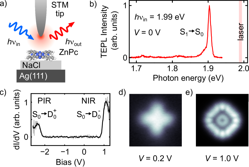
Fig. 1a shows a scheme of the experiment. A zinc phthalocyanine (ZnPc) molecule deposited on 4 monolayer (ML) thick NaCl is optically excited by a tunable laser source focused on the tip-sample junction. Within the junction, the electromagnetic field is strongly amplified between the tip apex and the metallic substrate acting as a plasmonic picocavity. This leads to increased absorption and a strong enhancement of radiative transitions (i.e., Purcell effect) so that the TEPL signal of individual molecules can be detected in the far fieldYang2020 ; Imada2021 ; Imai-Imada2022 ; Roslawska2023 ; Dolezal2024 . A typical single-molecule TEPL spectrum obtained with an incident photon energy of 1.99 eV is shown in Fig. 1b. It reveals a prominent emission line at an energy 1.91 eV, and low-energy vibronic peaks. Both stem from the S1 S0 transition of neutral ZnPcMurray2011 ; Zhang2016 . Fig. 1c shows a differential conductance (dd) spectrum of ZnPc/4 ML NaCl/Ag(111) with two distinct features at = -2.1 V and = 0.8 V, corresponding to the positive and negative ion resonance (PIR and NIR), respectivelyRepp2005 . For voltages between the PIR and the NIR (referred to as in-gap conditions), the charge transport through the double-barrier tunneling junction is non-resonant and only perturbed by the presence of the molecule Grewal2023 . In contrast, at the PIR and NIR, the charge transport through the junction is dominated by a two-step process where the molecule is first charged by tunneling from the tip and then discharged by tunneling to the sample. Thus, for resonant conditions, the molecule repetitively switches between a neutral and a charged configuration. Fig. 1d and Fig. 1e show STM images of ZnPc recorded in these two transport regimes, namely for in-gap ( = 0.2 V) and at NIR ( = 1 V), respectively, that is Fig. 1e shows the density of the lowest unoccupied molecular orbital (LUMO). Note that the image in Fig. 1d was recorded in constant current mode for a molecule adsorbed on 3 ML NaCl, other data shown in the main manuscript are recorded on 4 ML.
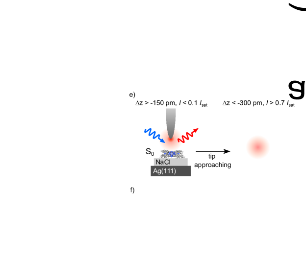
In the following, we investigate how charge transport in the two regimes influences the optical properties of the neutral molecule. Fig. 2a shows a series of TEPL spectra recorded in-gap ( = 0.5 V) for different relative tip-molecule distances . This parameter is defined relatively to an initial tip-molecule separation (z = 0) at a setpoint of = 1 V and = 5 pA. Upon approaching the tip to the molecule (decreasing ), the peak in TEPL increases in intensity, broadens from 6 meV to more than 10 meV (full width at half maximum), as extracted from Lorentzian fits, and shifts by 6 meV towards smaller energies at very small distances. This is in line with previous experimentsRoslawska2022 , which assigned a similar peak broadening and increase in emission intensity to an increased coupling between the molecules’ transition dipole moments and the picocavity plasmons upon reduction of the relative tip-molecule distance. The observed red-shift can be attributed to a combination of Lamb and Stark effects. The same dependence, but recorded at the NIR ( = 1 V), is shown in Fig. 2b. Here, as well, we observe a broadening of the peak in TEPL for decreasing . For z -150 pm, however, the photoluminescence intensity decreases and is almost entirely suppressed for z -400 pm.
This gradual reduction of the emission yield with decreasing , which is only observed at resonance, suggests a quenching mechanism related to the transient charging of the molecule. To confirm this, we recorded the tunnel current simultaneously with the integrated TEPL intensity of the emission line (Fig. 2c, d). Note that, for = 0.5 V and , the current is too low to be measured. At pm, where recorded at resonance follows the expected exponential dependence (red squares in Fig. 2d), the TEPL intensity increases similarly for both bias voltages. Upon approaching the molecule ( -150 pm), the curve measured at resonance deviates from the exponential dependence, while the TEPL intensity slowly decreases. This behaviour contrasts with the TEPL intensity recorded in-gap that increases monotonously with the tip approach (gray-scale circles in Fig. 2c). Eventually, at very small relative tip-molecule distances and for resonant tunneling conditions (blue squares in Fig. 2c, d), the current saturates ( 400 pA), and the TEPL intensity is nearly fully quenched.
The saturation in the tunnel current is a direct consequence of the charge transport mechanism that takes place in the double-barrier tunneling junction of our experimentSteurer2014 ; Kaiser2023a . At NIR, the molecule is successively (i) charged negatively by an electron tunneling through the vacuum barrier from the tip to the molecule, and then (ii) discharged by a second tunneling event taking place through the NaCl layer. The overall tunnel current is composed of these two consecutive events and is limited by the process with the lower tunneling probability, i.e., the slower process. While the tunneling probability through the vacuum gap varies with , the tunneling probability through the NaCl is merely determined by the thickness of the NaCl layer and thus remains constant as the tip approaches. At large relative tip-molecule distances, tunneling through the vacuum is, therefore, the rate-limiting process ( -150 pm, Fig. 2e, f), and the tunneling current increases exponentially with decreasing . At smaller relative tip-molecule distances ( -150 pm) the progressive saturation of the current indicates that the discharging process through the NaCl becomes the rate-limiting step. This affects the relative populations of the neutral and charged states of the molecule, as indicated in the many-body diagrams in Fig. 2f, h: At large relative tip-molecule distances ( -150 pm), the charging rate indicated by the dark grey arrow is low and the molecule spends most of the time in the neutral state . Optical excitation of the S0 S1 transition by incoming photons and the emission from the reverse S1 S0 transition (Fig. 2f) is thus possible. For decreasing , the population of the negatively charged state increases at the expense of the population, up to a predominant occupation of in the saturation regime (Fig. 2g, h). Since the D S1 transition cannot be induced by a photon, which carries a net zero charge, the formation of a neutral exciton is gradually hindered and the TEPL signal vanishes. Thus, the controllable transition to a configuration where the charged doublet state is occupied most of the time, instead of the neural singlet, is responsible for the quenching of the luminescence, akin to the process taking place in electrochromic systems. A similar effect is expected to take place at PIR when the D population is promoted. At this condition, however, electrical excitation of S1 via D leads to a strong STML signal at the same photon energyDolezal2019 ; Dolezal2021a ; Hung2023 , precluding the observation of the photoluminescence quenching.
To quantify the mechanism behind the photoluminescence quenching, we develop a model encompassing the many-body transitions between the relevant states in our system, , and , illustrated in Fig. 2f, h (see Supplementary Materials for more details). The total tunnel current through the double-barrier tunneling junction can be expressed using the tunneling rates through vacuum, ( transition), and NaCl, ( transition), respectivelyKaiser2023a :
| (1) |
with the elementary charge , the tunneling rate from the tip for = 0, the decay constant and the lifetime . The steady-state ground state population is then given by:
| (2) |
Fitting recorded for V in Fig. 2d (solid line) with the expression in Eq. (1) yields 400 ps, in line with recent reports on decoupled emitters Kaiser2023a ; Roslawska2018 . Once the tunneling rate from the tip becomes comparable to the tunneling rate from the substrate, which in our system is observed for -150 pm, begins to substantially deviate from 1 (dashed line in Fig. 2c).
Next we analyse the and transitions that are responsible for the observed photoluminescence. The total light intensity can be written as , where is the excitation rate. In agreement with recent literatureYang2020 ; Imai-Imada2022 , we find that () is well-described by an exponential function, which reflects the strongly increased plasmonic field when the tip approaches the molecule. Therefore
| (3) |
with being the excitation rate for = 0 and the decay constant. Fitting the data in Fig. 2c with Eq. (3) and = 1 for = 0.5 V, that is assuming very short excited state lifetime Roslawska2022 ; Dolezal2024 and thus 0, we obtain a very good agreement with the experiment. For fitting the curve recorded at = 1 V, we use the same values of the parameters ( and ) but described by Eq. (2). Again, our model is in excellent agreement with the experimental data. Comparing the = 1 V and curves in Fig. 2c, we find that the quenching effect onsets already for 0.8, when -150 pm.
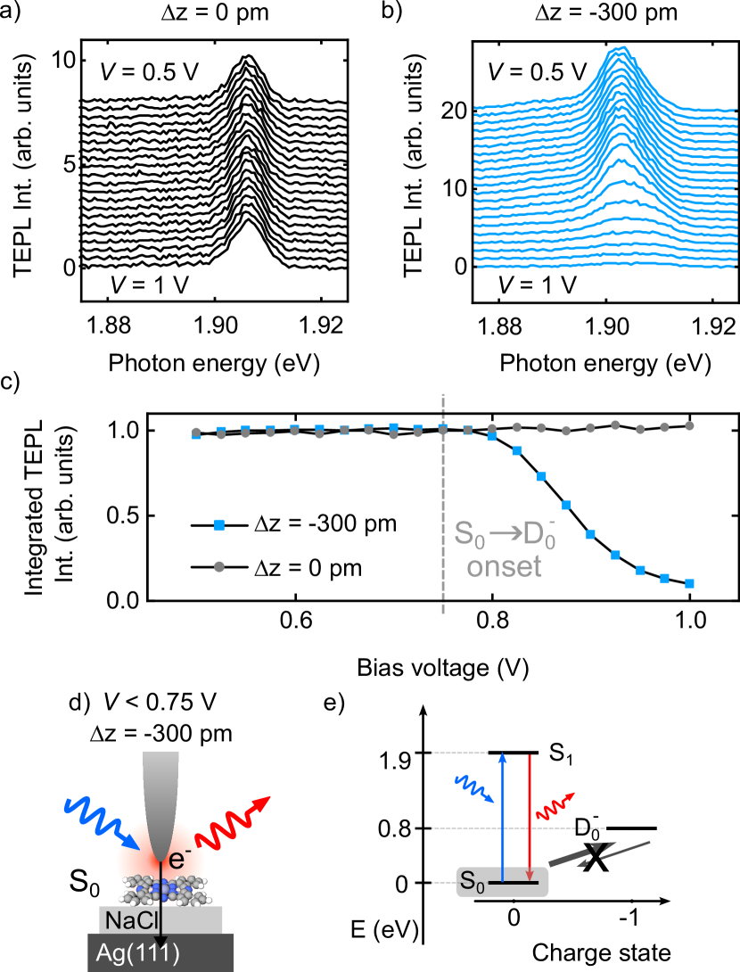
Since the ability to excite a molecule with incoming photons depends on the relative population of , we can conveniently gate the TEPL yield by the applied bias voltage. Fig. 3a and Fig. 3b show constant-height TEPL spectra as a function of the applied bias voltage for large ( pm) and small ( pm) relative tip-molecule distances, respectively. At pm (Fig. 3a), the TEPL spectra remain unaffected by the change in bias voltage from 0.5 V to 1 V. In contrast, at small relative tip-molecule distances (Fig. 3b), increasing the bias from 0.5 V to 1 V results in gradual quenching of the TEPL signal. The integrated intensity as a function of the bias voltage for these two distances is compared in Fig. 3c. At large relative tip-molecule distances (gray circles), the TEPL signal intensity remains constant. For small distances (blue squares), we find that the TEPL intensity begins decreasing at V, a value that agrees very well with the onset of the NIR (Fig. 1d). At large distances, the tunneling probability between the tip and molecule is low, and thus the relative population of S0 remains high regardless of the applied voltage. This is also true at small distances to the molecule for low bias voltages, as the D state cannot be accessed. Hence, in both conditions, the TEPL signal remains intense (Fig. 3d, e). In contrast, at small distances and at bias voltages that are resonant with the NIR of the molecule, the charged state is efficiently populated, and the luminescence is quenched. Overall, this shows that one can gate the photoluminescence of a single molecule by tuning the voltage applied across the junction and the distance to one of the electrodes.
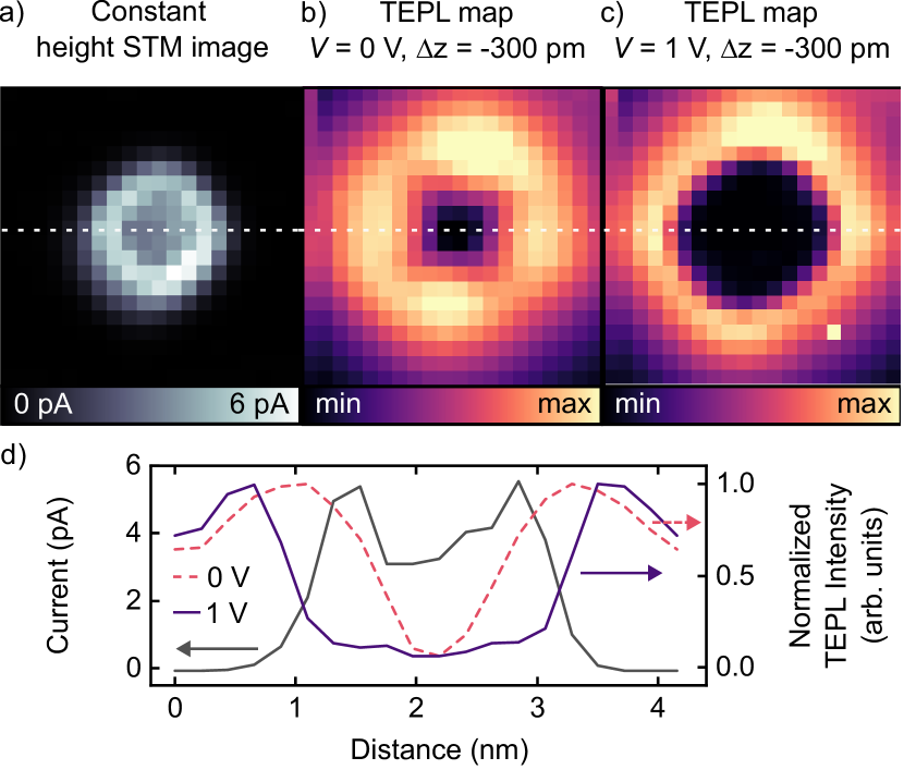
The local character of this TEPL gating is demonstrated by the TEPL intensity dependence on the tip position. Fig. 4a shows the spatial extent of the electronic orbital at V (LUMO). Fig. 4b, c show maps of the TEPL intensity recorded atop the molecule in-gap (b) and at NIR (c) at a constant height of pm. The TEPL map recorded at = 0 V reveals a radially symmetrical pattern and a low TEPL intensity in the middle of the molecule. This expected spatial dependence originates from the coupling of the picocavity plasmon to the doubly-degenerate transition dipoles of S1 for ZnPc. The low intensity in the center arises due to the symmetry of the system which leads to a vanishing coupling between the picocavity field and the molecular transition densityNeuman2018 ; Yang2020 . At resonant tunneling conditions ( = 1 V, Fig. 4c), the TEPL map reveals a much broader dark center compared to the map recorded at = 0 V. This dark region matches well the spatial extent of the LUMO in the constant height current map in Fig. 4a. In contrast, in regions where resonant tunneling through the molecule is not possible (at tip positions exceeding the lateral extent of the LUMO) the intensity pattern of Fig. 4b is recovered. This demonstrates that, for = 1 V and pm, as soon as direct tunneling into the ZnPc is possible, we can deliberately quench the TEPL yield by controllably decreasing the relative population of . This is further illustrated in the cross-section in Fig. 4d where the onset of the orbital (increase of the current) coincides with the onset of TEPL quenching. The occupancy in the current saturation regime (Fig. 2d) is entirely governed by the substrate-mediated discharging rate and is therefore independent of the tip position. Overall, this photoluminescence quenching strategy can be generalized to other molecules as we demonstrate in the Supplementary Materials for a free-base phthalocyanine, H2Pc.
In conclusion, we have demonstrated that we can gate the photoluminescence of a single molecule with sub-nm precision.
We use the tip of an STM simultaneously as an atomically precise electrode, which allows us to modify the charge state of a molecule, and as an optical antenna to probe the chromophore’s photoluminescence. By selectively injecting individual charges into the molecule we can controllably and gradually shift the time-averaged population of the molecule from its neutral ground state to a transient charge state from which the neutral exciton cannot be formed. Our approach can be used, for example, to selectively alter the properties of individual chromophores in a molecular layer or assembly. This can influence for example coherent dipole-dipole couplingZhang2016 ; Doppagne2017 ; Luo2019 or energy transfer mechanismsImada2016 ; Cao2021 , thus modifying the collective optical responses of the system.
We thank Tomáš Neuman and Stéphane Berciaud for fruitful discussions and Virginie Speisser and Halit Sumar for technical support. This project has received funding from the European Research Council (ERC) under the European Union’s Horizon 2020 research and innovation program (grant agreement No 771850). This work of the Interdisciplinary Thematic Institute QMat, as part of the ITI 2021 2028 program of the University of Strasbourg, CNRS and Inserm, was supported by IdEx Unistra (ANR 10 IDEX 0002), and by SFRI STRAT’US project (ANR 20 SFRI 0012) and EUR QMAT ANR-17-EURE-0024 under the framework of the French Investments for the Future Program. K. K. acknowledges funding from the SNSF under the Postdoc.Mobility grant agreement No 206912. A. R. acknowledges funding from the Emmy Noether Programme of the Deutsche Forschungsgemeinschaft (DFG, German Research Foundation) - 534367924.
References
- (1) Fölling, J. et al. Fluorescence nanoscopy by ground-state depletion and single-molecule return. Nat. Methods 5, 943–945 (2008).
- (2) Heilemann, M., van de Linde, S., Mukherjee, A. & Sauer, M. Super-Resolution Imaging with Small Organic Fluorophores. Angew. Chem. Int. Ed. 48, 6903–6908 (2009).
- (3) Vogelsang, J., Cordes, T., Forthmann, C., Steinhauer, C. & Tinnefeld, P. Controlling the fluorescence of ordinary oxazine dyes for single-molecule switching and superresolution microscopy. Proc. Natl. Acad. Sci. 106, 8107–8112 (2009).
- (4) Brasselet, S. & Moerner, W. Fluorescence Behavior of Single-Molecule pH-Sensors. Single Mol. 1, 17–23 (2000).
- (5) Toninelli, C. et al. Single organic molecules for photonic quantum technologies. Nat. Mater. 20, 1615–1628 (2021).
- (6) Yu, M., Yim, D., Seo, H. & Lee, J. Electrical charge control of h-BN single photon sources. 2D Mater. 9, 035020 (2022).
- (7) Ji, Z., Doorn, S. K. & Sykora, M. Electrochromic Graphene Molecules. ACS Nano 9, 4043–4049 (2015).
- (8) Dickson, R. M., Cubitt, A. B., Tsien, R. Y. & Moerner, W. E. On/off blinking and switching behaviour of single molecules of green fluorescent protein. Nature 388, 355–358 (1997).
- (9) Gu, C., Jia, A.-B., Zhang, Y.-M. & Zhang, S. X.-A. Emerging electrochromic materials and devices for future displays. Chem. Rev. 122, 14679–14721 (2022).
- (10) Repp, J., Meyer, G., Stojković, S. M., Gourdon, A. & Joachim, C. Molecules on Insulating Films: Scanning-Tunneling Microscopy Imaging of Individual Molecular Orbitals. Phys. Rev. Lett. 94, 026803 (2005).
- (11) Nazin, G. V., Wu, S. W. & Ho, W. Tunneling rates in electron transport through double-barrier molecular junctions in a scanning tunneling microscope. Proc. Natl. Acad. Sci. 102, 8832–8837 (2005).
- (12) Swart, I., Sonnleitner, T. & Repp, J. Charge State Control of Molecules Reveals Modification of the Tunneling Barrier with Intramolecular Contrast. Nano Lett. 11, 1580–1584 (2011).
- (13) Fernández-Torrente, I., Kreikemeyer-Lorenzo, D., Stróżecka, A., Franke, K. J. & Pascual, J. I. Gating the Charge State of Single Molecules by Local Electric Fields. Phys. Rev. Lett. 108, 036801 (2012).
- (14) Hauptmann, N., Hamann, C., Tang, H. & Berndt, R. Switching and charging of a ruthenium dye on Ag(111). Phys. Chem. Chem. Phys. 15, 10326–10330 (2013).
- (15) Cochrane, K. A. et al. Molecularly resolved electronic landscapes of differing acceptor-donor interface geometries. J. Phys. Chem. C 122, 8437–8444 (2018).
- (16) Kaiser, K., Lieske, L.-A., Repp, J. & Gross, L. Charge-state lifetimes of single molecules on few monolayers of NaCl. Nat. Commun. 14, 4988 (2023).
- (17) Benz, F. et al. Single-molecule optomechanics in “picocavities”. Science 354, 726–729 (2016).
- (18) Rosławska, A. et al. Mapping Lamb, Stark, and Purcell Effects at a Chromophore-Picocavity Junction with Hyper-Resolved Fluorescence Microscopy. Phys. Rev. X 12, 011012 (2022).
- (19) Qiu, X. H., Nazin, G. V. & Ho, W. Vibrationally Resolved Fluorescence Excited with Submolecular Precision. Science 299, 542–546 (2003).
- (20) Imada, H. et al. Real-space investigation of energy transfer in heterogeneous molecular dimers. Nature 538, 364–367 (2016).
- (21) Zhang, Y. et al. Visualizing coherent intermolecular dipole–dipole coupling in real space. Nature 531, 623–627 (2016).
- (22) Doppagne, B. et al. Vibronic Spectroscopy with Submolecular Resolution from STM-Induced Electroluminescence. Phys. Rev. Lett. 118, 127401 (2017).
- (23) Luo, Y. et al. Electrically Driven Single-Photon Superradiance from Molecular Chains in a Plasmonic Nanocavity. Phys. Rev. Lett. 122, 233901 (2019).
- (24) Cao, S. et al. Energy funnelling within multichromophore architectures monitored with subnanometre resolution. Nat. Chem. 13, 766–770 (2021).
- (25) Doppagne, B. et al. Electrofluorochromism at the single-molecule level. Science 361, 251–255 (2018).
- (26) Rai, V. et al. Boosting Light Emission from Single Hydrogen Phthalocyanine Molecules by Charging. Nano Lett. 20, 7600–7605 (2020).
- (27) Doležal, J., Canola, S., Merino, P. & Švec, M. Exciton-Trion Conversion Dynamics in a Single Molecule. ACS Nano 15, 7694–7699 (2021).
- (28) Doležal, J. et al. Real Space Visualization of Entangled Excitonic States in Charged Molecular Assemblies. ACS Nano (2021).
- (29) Doležal, J. et al. Evidence of exciton-libron coupling in chirally adsorbed single molecules. Nat. Commun. 13, 6008 (2022).
- (30) Jiang, S. et al. Many-Body Description of STM-Induced Fluorescence of Charged Molecules. Phys. Rev. Lett. 130, 126202 (2023).
- (31) Hung, T.-C. et al. Bipolar single-molecule electroluminescence and electrofluorochromism. Phys. Rev. Res. 5, 033027 (2023).
- (32) Rai, V. et al. Activating Electroluminescence of Charged Naphthalene Diimide Complexes Directly Adsorbed on a Metal Substrate. Phys. Rev. Lett. 130, 036201 (2023).
- (33) Kaiser, K. et al. Electrically driven cascaded photon-emission in a single molecule. arXiv:2402.17536 (2024).
- (34) Yang, B. et al. Sub-nanometre resolution in single-molecule photoluminescence imaging. Nat. Photonics 14, 693–699 (2020).
- (35) Imada, H. et al. Single-molecule laser nanospectroscopy with micro–electron volt energy resolution. Science 373, 95–98 (2021).
- (36) Imai-Imada, M. et al. Orbital-resolved visualization of single-molecule photocurrent channels. Nature 603, 829–834 (2022).
- (37) Rosławska, A. et al. Submolecular-scale control of phototautomerization. Nat. Nanotechnol. (2024).
- (38) Doležal, J., Sagwal, A., de Campos Ferreira, R. C. & Švec, M. Single-Molecule Time-Resolved Spectroscopy in a Tunable STM Nanocavity. Nano Lett. 24, 1629–1634 (2024).
- (39) Murray, C. et al. Visible luminescence spectroscopy of free-base and zinc phthalocyanines isolated in cryogenic matrices. Phys. Chem. Chem. Phys. 13, 17543–17554 (2011).
- (40) Grewal, A., Leon, C. C., Kuhnke, K., Kern, K. & Gunnarsson, O. Character of Electronic States in the Transport Gap of Molecules on Surfaces. ACS Nano 17, 13176–13184 (2023).
- (41) Steurer, W., Gross, L. & Meyer, G. Local thickness determination of thin insulator films via localized states. Appl. Phys. Lett. 104, 231606 (2014).
- (42) Doležal, J. et al. Charge Carrier Injection Electroluminescence with CO-Functionalized Tips on Single Molecular Emitters. Nano Lett. 19, 8605–8611 (2019).
- (43) Rosławska, A. et al. Single Charge and Exciton Dynamics Probed by Molecular-Scale-Induced Electroluminescence. Nano Lett. 18, 4001–4007 (2018).
- (44) Neuman, T., Esteban, R., Casanova, D., García-Vidal, F. J. & Aizpurua, J. Coupling of Molecular Emitters and Plasmonic Cavities beyond the Point-Dipole Approximation. Nano Lett. 18, 2358–2364 (2018).