Low-coercive-field ferroelectric hafnia with mobile domain walls
Abstract
The high coercive field () of hafnia-based ferroelectrics presents a major obstacle to their applications. The ferroelectric switching mechanisms in hafnia that dictate , especially those related to nucleation-and-growth at the domain wall (DW), have remained elusive. Through deep-learning-assisted multiscale simulations, we determine the finite-temperature thermodynamics and switching mechanisms for diverse types of 180∘ DWs, revealing a complex, stress-sensitive mobility landscape. The propagation velocities for mobile DW types under various thermal conditions can be characterized with a single creep equation, featuring a creep exponent of 2. This unconventional critical exponent results from the nucleation of a half-unit-cell-thin, elliptically-shaped critical nucleus. Our multiscale approach not only reproduces the experimental thickness () scaling, , but also predicts that of HfO2 can be engineered to 0.1 MV/cm, even lower than perovskite ferroelectrics. The theoretical lower bound of afforded by ferroelectric hafnia offers opportunities to realize power-efficient, high-fidelity ferroelectric nanoelectronics.
Ferroelectric hafnia is emerging as a viable candidate for incorporating ferroelectric capabilities into semiconductor technologies for its nanoscale ferroelectricity and proven compatibility with silicon [1, 2]. The phase of HfO2 is widely acknowledged as the ferroelectric phase in thin films [3, 4, 5, 6]. The discovery of switchable spontaneous polarization in this fluorite-structured binary oxide has significantly advanced our understanding of ferroelectric physics, an area historically focused on perovskite oxides. For example, recent investigations have uncovered several unique aspects of hafnia-based ferroelectrics, including its metastable nature compared to the bulk monoclinic phase [5], the presence of flat phonon bands [7], and intertwined polar-nonpolar structural polymorphism with oxygen vacancy diffusion [8, 9].
However, the widespread applications of ferroelectric hafnia-based devices are mainly hindered by reliability issues, especially their limited endurance [10]. Unlike devices based on perovskite ferroelectrics like Pb(Zr,Ti)O3 and BiFeO3, which can be made close to fatigue-free [11], hafnia-based devices, depending on their architectures, typically exhibit poor endurance, with switching cycles ranging between and [12, 13, 14, 15]. This falls short of the required cycles for embedded random access memory. Such a detrimental issue is attributed to the high coercive field (, the electric field needed to switch the polarization) of hafnia-based ferroelectrics (typically 1 MV/cm in polycrystalline thin films and 2 MV/cm in epitaxial thin films) [16, 17, 18]. Repeated applications of high electric fields near the breakdown strength to reverse the polarization are expected to accelerate breakdown, causing rapid performance deterioration. Reducing without sacrificing nanoscale ferroelectricity of hafnia has become a pressing challenge for the development of fast, durable, and energy-efficient nanoelectronics.
The atomic-level switching mechanisms affecting in ferroelectric hafnia remain elusive, due to the multiple competing pathways and the complex variety of domain walls (DWs), which are interfaces separating differently polarized domains. Previous studies using zero-Kelvin density functional theory (DFT) have focused on polarization switching at the unit-cell level [3, 19] and the behavior of specific types of DWs [7, 20, 21, 22]. These studies overlooked the thermally activated nucleation-and-growth process, a crucial aspect for accurate predictions [23]. Here, utilizing a deep neural network-based force field trained exclusively on first-principles data of hafnia [24, 25], we employ large-scale molecular dynamics (MD) simulations to investigate the finite-temperature thermodynamics and kinetics of all types of 180∘ DWs, identified through comprehensive symmetry analysis. Computational details are provided in Supplementary Sect. I and an online notebook [26] is developed to share details regarding the machine-learning force field.
Our MD simulations unveil, without a priori assumptions, a stress-tunable, spontaneous transformation of DW type at room temperatures, which plays a critical role in the “divide-and-conquer”-like mechanism underpins the low switching fields for two types of DWs.
We quantify the temperature- and field-dependence of propagation velocities for these walls in HfO2, revealing a universal creep exponent of and the underlying nucleation-and-growth mechanisms characterized by a half-unit-cell-thin, elliptically-shaped critical nucleus. A Landau–Ginzburg–Devonshire (LGD) analytical model incorporating this insight is developed, reproducing the experimentally observed thickness () scaling of in thin films, . Importantly, we demonstrate that the intrinsic of HfO2 determined by mobile DWs is, unexpectedly, on par with Pb(Ti,Zr)O3, and can be as low as 0.1 MV/cm in thin films.
Classification of 180∘ DWs based on symmetry
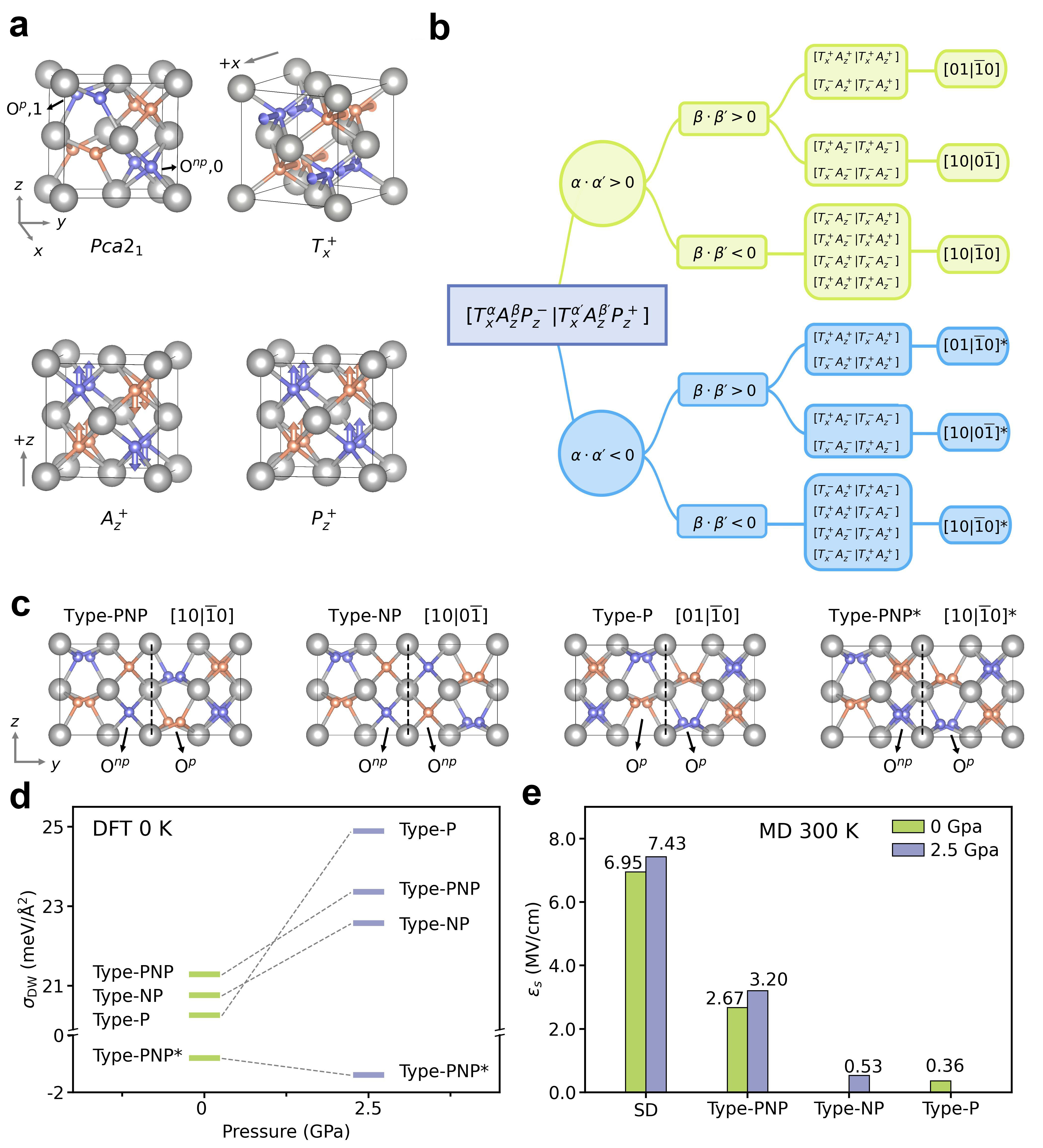
We start with a symmetry analysis to classify all possible 180∘ DWs, interfaces separating domains with opposite polarization. The unit cell of HfO2 has a nonpolar layer of fourfold-coordinated oxygen ions (Onp) and a polar layer of threefold-coordinated oxygen ions (Op), and these layers are ordered alternately (Fig. 1a). Application of Euclidean transformations within the unit cell results in eight different representations (Fig. S7). This feature, refereed to as conditional chirality [27], leads to a rich spectrum of 180∘ DWs. We introduce a mode vector with (, , =) denoting the sign of the mode amplitude, to label the unit-cell configuration, where the tetragonal mode (), the antipolar mode (), and the polar mode () are the three main modes bridging the cubic phase and polar orthorhombic phase with polarization aligned along the axis () [28]. The and mode are characterized by antiparallel -displacements and -displacements of neighboring oxygen ions (Fig. 1a), respectively.
A general 180∘ wall can be expressed as ], allowing for 16 configurations (Fig. S8). As illustrated in Fig. 1b, these configurations can be further classified based on the sign of and . In cases where , the -displacement pattern of oxygen atoms across the DW remains unchanged, whereas indicates a sign reversal of the mode. For , the oxygen atoms at the wall are of the same type (either Op or Onp), while a negative sign indicates Op and Onp atoms are adjacent. Symmetry analysis reveals six unique 180∘ DWs, which are labeled as , , and , along with their counterparts having the sign of the mode reversed across the wall, denoted as , , and . Here, “1”(“”) labels the Op atom displaced along (), and “0” represents the Onp atom (Fig. 1c). For clarity in the following discussion, , , and walls are also named as type-P, type-NP, and type-PNP, based on the type of oxygen atoms at the wall; their counterparts with reversed mode are type-P∗, type-NP∗, and type-PNP∗.
As shown in Fig. 1d, our DFT calculations of DW energy () reveal that the type-PNP∗ wall, resembling the antiferroelectric-like phase, possesses the lowest energy (), followed by type-P, type-NP, and type-PNP walls at 0 GPa.
In contrast, the other two -sign-reversed DWs, type-P∗ and type-NP∗, are highly unstable. Interestingly, applying a compressive hydrostatic pressure of 2.5 GPa substantially destabilizes type-P compared to type-NP and type-PNP walls.
It is noted that is an expected consequence of HfO2 being lower in energy than HfO2. Previously, this intriguing negative DW energy led to the proposal that each polar layer in HfO2 is strongly localized, allowing for individual switching and scale-free ferroelectricity [7]. Many subsequent studies focused on the motion of a type-PNP∗ wall [20, 22], despite its substantial energy barrier of eV [21].
We will demonstrate with MD simulations that the polarization switching in HfO2 does not involve the type-PNP∗ wall as it is immobile.
Spontaneous transformation of DW types at room temperatures
An important discovery from our MD simulations, contrasting with earlier findings from DFT modeling, is the spontaneous change in DW type at room temperatures. At 300 K and 0 GPa, the type-NP wall is unstable, automatically transforming into the type-P wall through OnpOp (). This change can be intuitively expressed as , where the wall shifts laterally by half a lattice constant along the -axis, . At a higher pressure of 2.5 GPa, a reverse transition of type-Ptype-NP occurs spontaneously via , and can be represented as . The high-energy type-PNP wall remains stable at 300 K under both stress conditions. As we will elaborate below, this thermally-induced spontaneous transition between Onp and Op atoms at the interface is pivotal for the dynamics of type-P and type-NP walls.
We then gauge DW mobility at 300 K by determining the lowest electric field strength () that triggers DW propagation within 1 nanosecond in MD simulations, with results summarized in Fig. 1e. Our simulations reveal that the type-PNP∗ wall is essentially immobile, showing no movement under a substantial of 7 MV/cm. Rather, the adjacent domain undergoes polarization reversal, since this field strength is sufficient to drive single-domain (SD) switching within 1 nanosecond. This aligns with the high DFT-calculated energy barrier of 1.3 eV for moving a type-PNP∗ wall, compared to a considerably lower barrier of 0.22 eV for homogeneous SD switching [19]. The resistance of a type-PNP∗ wall to stems from the inability of a -axis field to flip mode that features antipolar -displacements of oxygen atoms.
For -sign-conserved DWs, a wall with a lower value mobilize more readily under , similar to the trend observed in perovskite ferroelectrics [23]. Notably, the type-P wall at 0 GPa exhibits a minimal of 0.36 MV/cm. At 2.5 GPa, the type-NP wall has the lowest of 0.53 MV/cm. Our following discussions will focus on the most mobile type-P and type-NP walls.
Mechanisms and kinetics of domain wall motions from MD simulations
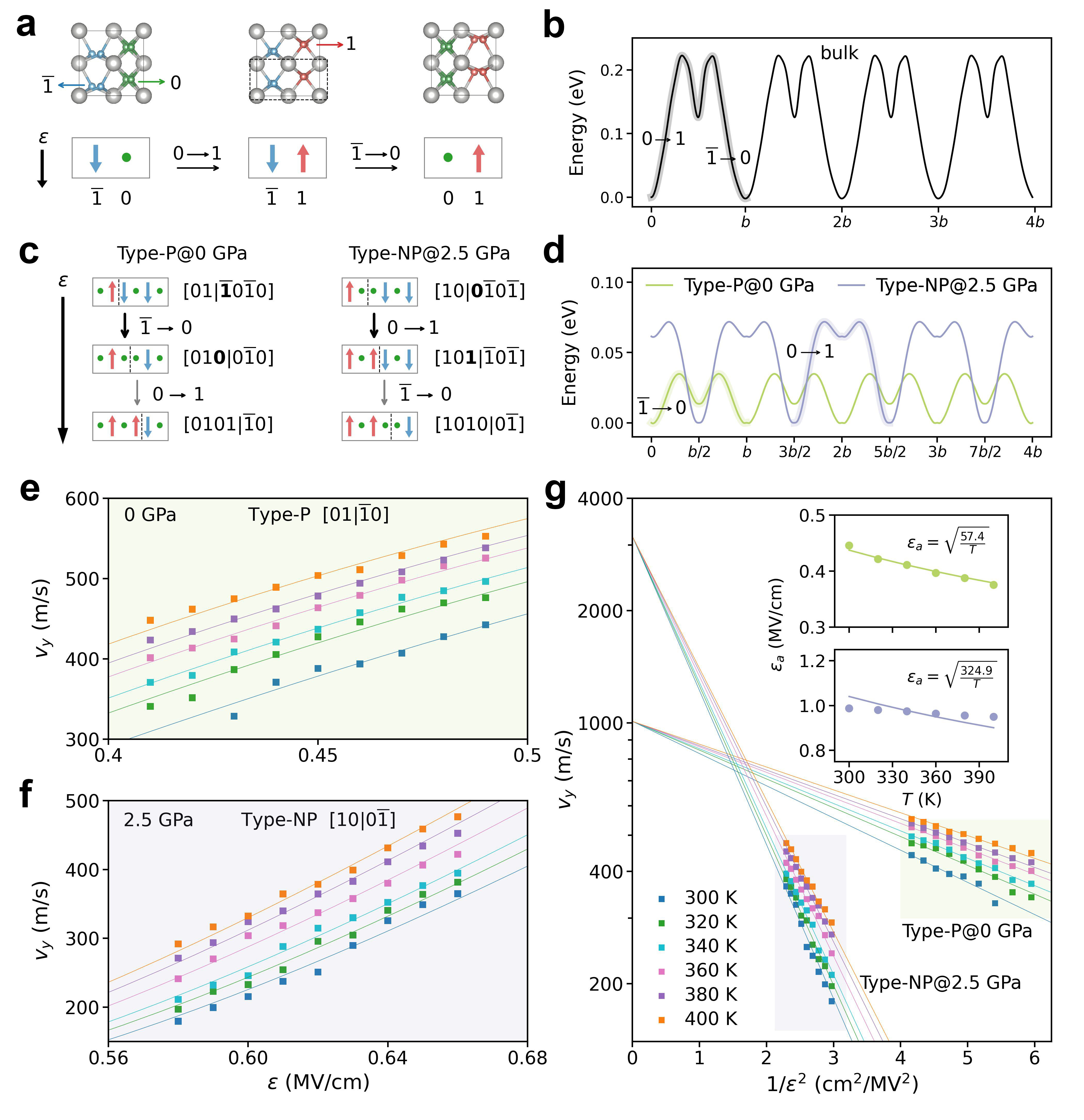
Figure 2a and c compare the SD switching to DW motion mechanisms extracted from finite-field MD simulations at 300 K. At 0 GPa, a type-P wall initially transforms into a type-NP wall that serves as a bridging state. Conversely, at 2.5 GPa, a type-NP wall progresses via a transient type-P wall. A common feature is the lateral one-unit-cell translation being accomplished through two half-unit-cell hops (), with the initial hop kinetically determinant. This is markedly different from perovskite ferroelectrics, where DWs advance by one unit cell at a time [29, 23]. Such a “first-hop-limiting” mechanism naturally results from the thermally-induced spontaneous change in DW types. Specifically, as shown in Fig. 2c, under a downward , a type-P wall transitions through to a type-NP wall. This type is thermodynamically unstable at 0.0 GPa, causing a spontaneous hop of . At 2.5 GPa, the type-NP wall moves via , forming an unstable type-P wall that then shifts another via . These findings also imply that the rate-limiting step can be manipulated by mechanical boundary conditions, being either at 0 GPa or under higher pressure.
The unusual mechanisms of DW motions in HfO2 revealed by MD are corroborated by DFT-based nudged elastic band (NEB) calculations. As plotted in Fig. 2d, the minimum energy pathways (MEPs) at both pressures display clear two-hop characteristics: a larger barrier followed by a shallower one. The MEP for SD switching (Fig. 2b) also involves successive and hops, but the first hop is associated with a larger barrier. We propose that the presence of DWs decouples the movements of Op and Onp atoms, rendering only one hop kinetically significant. This “divide-and-conquer”-like mechanism explains the lower switching fields for type-P and type-NP walls.
The velocities () for type-P and type-NP walls at 0 and 2.5 GPa, respectively, are quantified using MD simulations across various temperatures () and field strengths. For both stress conditions, as shown in Fig. 2e-f, the -dependence of follows a creep process [30, 31]:
| (1) |
where is the DW velocity under an infinite field, is Boltzmann’s constant, and and represent the characteristic energy barrier and electric field at zero Kelvin, respectively; is the creep exponent, which depends on the dimensionality of the interface and the universality class of the disorder landscape pinning the interface [32]. Equation (1) can be reformulated as:
| (2) |
where corresponds to the famous Merz’s law [33, 34], and is the -dependent activation field. By comparing Equations (1) and (2), is given as with . Remarkably, all velocity data fits well with . This is evidenced by the linear relationship between and and an accurate representation of ’s -dependence as (Fig. 2g and insets). A creep exponent of 2 is higher than the well-known value of for 1D magnetic domain walls in ultrathin magnetic films [32] and for 2D ferroelectric domain walls in typical perovskite ferroelectrics [29, 23].
Nucleation-and-growth at domain walls
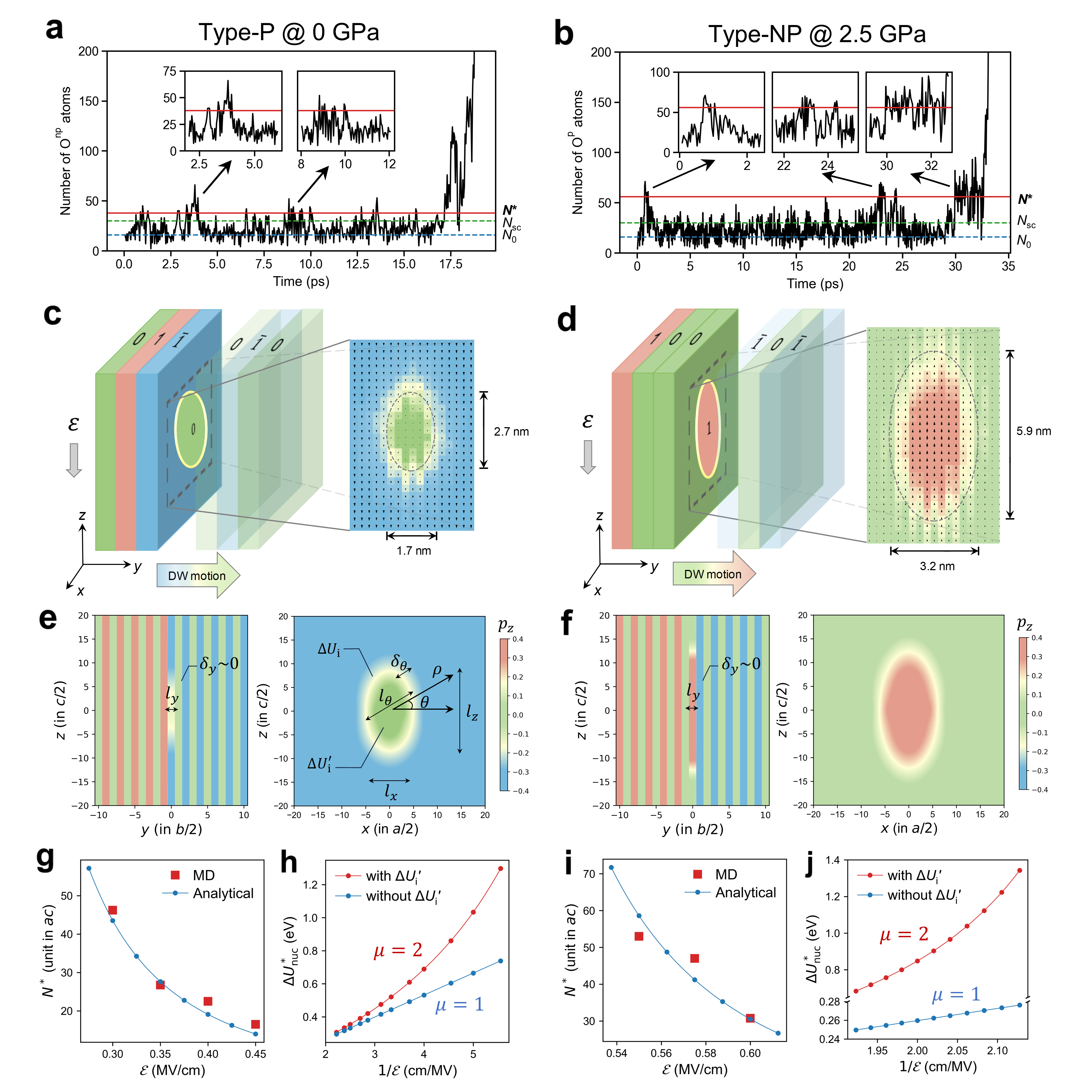
We now develop an analytical model based on classical nucleation theory and Landau–Ginzburg–Devonshire (LGD) theory to explain the origin of for both types of DWs. The DW motion can be understood as the formation of a quasi-two-dimensional nucleus with polarization aligned with at the wall, followed by the growth of the nucleus. The energy of the critical nucleus gives the nucleation barrier () which can be further related to in equation (2) via the Avrami theory of transformation kinetics. We use a revised persistent-embryo method (PEM) [35] based on MD simulations to determine the size () and shape of the critical nucleus at the DW (see Supplementary Sect. I). Specifically, a two-dimensional (2D) embryo with switched oxygen atoms is initially embedded in the unswitched DW, and a tunable harmonic potential is applied to each oxygen atoms in the embryo to prevent back-switching. The potential is gradually weakened and completely removed when the nucleus size () surpasses a subcritical threshold ( and ). The plateau of the curve then corresponds to the period over which the nucleus size fluctuates around .
Figure 3a displays the curve for the type-P wall at 0.0 GPa, 300 K, and MV/cm, revealing multiple plateaus, from which the average of is determined to be 38. The rate-limiting step of involves a half-unit-cell-thin critical nucleus in the plane consisting of Onp atoms. This nucleus is elliptical, measuring nm in width and nm in height (Fig. 3c). For the type-NP wall at 2.5 GPa and 300 K under 0.55 MV/cm, the curve in Fig. 3b yields a half-unit-cell-thin, elliptically-shaped critical nucleus of Op atoms, and the dimensions are nm and nm.
Having determined the shape of the critical nucleus, we can now proceed to develop a LGD nucleation model. For an elliptical nucleus of size , its boundary within the plane is conveniently described with polar coordinates and . Here, is given by . In terms of the local displacement of oxygen atom () along the polar axis, the atomistic profile for a type-P or type-NP wall containing a nucleus can be represented by a single generalized equation:
| (3) |
where and is the diffuseness parameter; with being an integer derived from and taking for a nucleus of Onp atoms and for a nucleus of Op atoms; is the ground-state value for the local displacement of Op. The profiles in the and planes generated by equation (3) are shown in Fig. 3e and 3f for type-P and type-NP walls, respectively. In the presence of , the energy of a wall with a nucleus relative to the wall without any nucleus can be expressed as:
| (4) |
with
| (5) |
and
| (6) | ||||
Here, is the electric field-polarization coupling term and is the energy penalty for creating new interfaces due to nucleation; is a scaling constant that relates oxygen displacement to local polarization; is the atomistic profile for a nucleus-free wall. Taking advantage of the 2D nature of the nucleus (, ) revealed from MD simulations, we can derive (see details in Supplementary Sect. IV) that , and is given as:
| (7) | ||||
We note that results from the change in DW type during the formation of a half-unit-cell-thin nucleus, where is proportional to the DW energy difference between type-P and type-NP walls, . This energy penalty is distinctive to DWs in HfO2. In contrast, nucleation at DWs in perovskite ferroelectrics such as PbTiO3 does not involve a change in the DW type that separates differently polarized domains (see Fig. S10). The term quantifies the energy penalty arising from polarization changes and their gradient at the nucleus boundary, where is the -dependent gradient coefficient along , and scales with the energy barrier of DW motion.
For a given , the energy of a 2D nucleus with the shape and size specified by depends on just a few parameters: , , , , and . All these parameters can be estimated straightforwardly with low costs. This enables the facile determination of and across a range of field strengths. As demonstrated in Fig. 3g and 3i, for both wall types, the values of predicted by the LGD model show remarkable agreement with the results obtained from MD-based PEM, confirming the validity and accuracy of the nucleation model as universally applicable to both type-P and type-NP walls. Interestingly, we find that scales with the (Fig. 3h and j), indicating the nucleation rate can be reformulated as:
| (8) |
where is a pre-exponential factor and is the effective activation field for nucleation.
This further corroborates the finding that the creep exponent is in equation (2).
A series of model calculations are performed using a nucleation model without to assess the impact of this unique energy term on the field-dependence of . We observe that omitting recovers a linear relationship between and . These results demonstrate is the atomistic origin for .
Intrinsic coercive fields from multiscale simulations
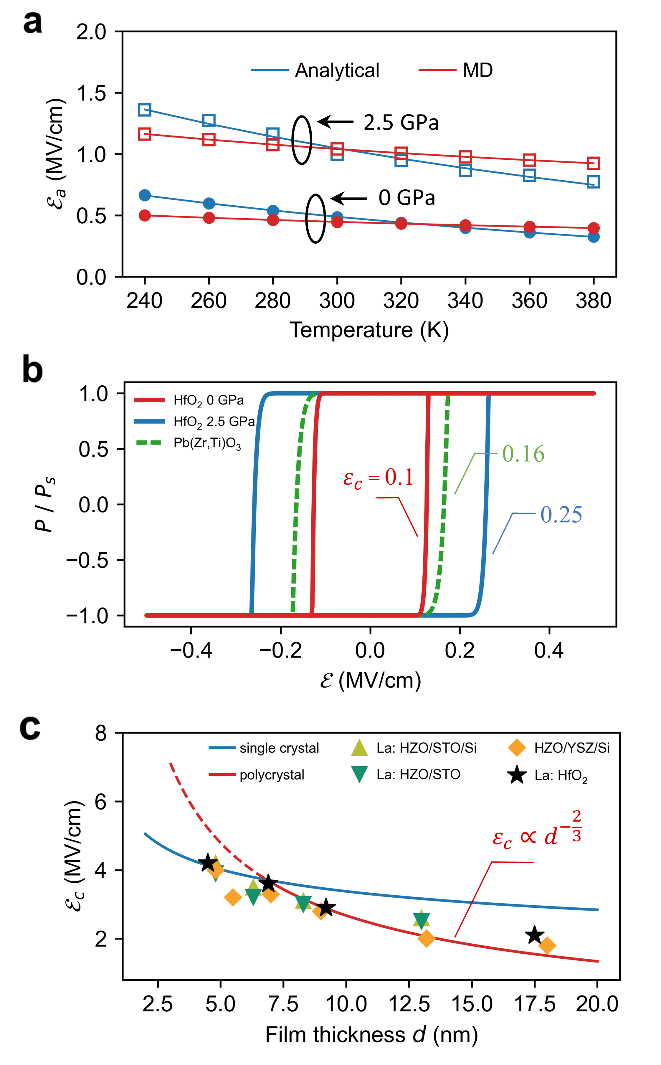
Based on Avrami’s theory of transformation kinetics, we derive the relationship between and for DW motions with as , where represents the dimensionality and is a scaling parameter mapping , associated with a half-unit-cell hop, to the effective barrier encountered by a DW moving one unit cell at a time (see Supplementary Sect. V). With and , we compute values using the LGD model at various temperatures, and the analytical values for two types of DWs agree with MD results (Fig. 4a). Utilizing the LGD model’s efficient prediction capability, we can conveniently predict the values of by simulating the polarization-electric (-) hysteresis loops resulting from DW motions (see Supplementary Sect. VI). As depicted in Fig. 4b, the - loop for type-P walls in HfO2 at 0 GPa yields 0.1 MV/cm, even lower than that of 0.2 MV/cm due to 180∘ DWs in Pb(Zr,Ti)O3 thin films [23].
The analysis above suggests that the intrinsic coercive fields of hafnia-based ferroelectrics could be quite low. Yet, why do they often exhibit much larger values experimentally [16, 17, 18]? We propose that this is due to a “thermodynamic-kinetic” dilemma unique to 180∘ DWs in HfO2. The most thermodynamically stable 180∘ DW is the -like type-PNP∗ wall, which is immobile; whereas the type-P wall is the most mobile at room temperatures but its higher thermodynamic energy makes its formation in thin films rare. Therefore, the magnitude of in hafnia-based thin films is likely determined by the SD switching process, which can also be predicted by our LGD nucleation model.
We first consider a single-crystal, single-domain HfO2 thin film with a thickness and a (111) orientation that is commonly observed in experiments.
Assuming that is the minimum electric field required to extend the critical nucleus length to , we determine the intrinsic values as a function of , plotted as a blue line in Fig. 4c. Notably, the theoretical is 4 MV/cm for nm, an excellent agreement with experimental values for La-doped HfO2 [18] and Hf0.5Zr0.5O2 films [36, 37] in the thin thickness limit where they are predominantly composed of ferroelectric phases. Furthermore, we consider the decreasing volume fraction of the polar phase with increasing thickness in polycrystalline thin films, by introducing a thickness-dependent into the nucleation model (see Supplementary Sect. VII).
Despite this simple treatment, our analytical values (red line in Fig. 4c) not only reproduce experimental results across various film thickness, but also the scaling relationship, , in epitaxial polycrystalline thin films [18, 36, 37].
Conclusions
In conclusion, we have developed a multiscale approach to elucidate the pivotal role of DW dynamics in dictating the coercive fields of ferroelectric HfO2. Our finite-field MD simulations indicate that the -like DW, though extensively studied, actually plays a rather limited role in ferroelectric switching, and we have identified two mobile DW types.
A thermally-induced spontaneous transformation of DW types is responsible for the unusual “first-hop-limiting” mechanism in the movements of mobile walls at room temperatures. The formation of a half-unit-cell-thin elliptical nucleus is identified, which supports the unconventional creep exponent of .
The LGD analytical model, incorporating atomistic insights of nucleation,
not only reproduces the experimentally observed thickness scaling of coercive fields in hafnia-based thin films but also uncovers an unexpectedly low intrinsic coercive field for HfO2 due to mobile walls, rivaling that of perovskite ferroelectrics. Our results offer a deeper understanding of the fundamental mechanisms governing ferroelectric switching in this promising material, highlighting the potential for achieving ultralow coercive fields in hafnia-based ferroelectrics through domain wall engineering.
Acknowledgments J.W., J.Y., and S.L. acknowledge the supports from National Key R&D Program of China (2021YFA1202100), National Natural Science Foundation of China (12074319, 12361141821), and Westlake Education Foundation. The computational resource is provided by Westlake HPC Center.
Author Contributions S.L. conceived and led the project. J.W. developed the force field, performed MD simulations, and analyzed the MD data. J.Y. identified the critical nucleus using PEM with assistance from Y.S., derived the LGD analytical model, and predicted the coercive fields. All authors contributed to the discussion and the manuscript preparation.
Competing Interests The authors declare no competing financial or non-financial interests.
References
- Böscke et al. [2011] T. S. Böscke, J. Müller, D. Bräuhaus, U. Schröder, and U. Böttger, Ferroelectricity in hafnium oxide thin films, Appl. Phys. Lett. 99, 102903 (2011).
- Schroeder et al. [2022] U. Schroeder, M. H. Park, T. Mikolajick, and C. S. Hwang, The fundamentals and applications of ferroelectric HfO2, Nat. Rev. Mater. 7, 653–669 (2022).
- Huan et al. [2014] T. D. Huan, V. Sharma, G. A. Rossetti, and R. Ramprasad, Pathways towards ferroelectricity in hafnia, Phys. Rev. B 90, 064111 (2014).
- Sang et al. [2015] X. Sang, E. D. Grimley, T. Schenk, U. Schroeder, and J. M. LeBeau, On the structural origins of ferroelectricity in HfO2 thin films, Appl. Phys. Lett. 106, 162905 (2015).
- Materlik et al. [2015] R. Materlik, C. Künneth, and A. Kersch, The origin of ferroelectricity in Hf1-xZrxO2: A computational investigation and a surface energy model, J. Appl. Phys. 117, 134109 (2015).
- Park et al. [2015] M. H. Park, Y. H. Lee, H. J. Kim, Y. J. Kim, T. Moon, K. D. Kim, J. Müller, A. Kersch, U. Schroeder, T. Mikolajick, and C. S. Hwang, Ferroelectricity and antiferroelectricity of doped thin HfO2-based films, Adv. Mater. 27, 1811 (2015).
- Lee et al. [2020] H.-J. Lee, M. Lee, K. Lee, J. Jo, H. Yang, Y. Kim, S. C. Chae, U. Waghmare, and J. H. Lee, Scale-free ferroelectricity induced by flat phonon bands in HfO2, Science 369, 1343 (2020).
- Nukala et al. [2021] P. Nukala, M. Ahmadi, Y. Wei, S. de Graaf, E. Stylianidis, T. Chakrabortty, S. Matzen, H. W. Zandbergen, A. Björling, D. Mannix, D. Carbone, B. Kooi, and B. Noheda, Reversible oxygen migration and phase transitions in hafnia-based ferroelectric devices, Science 372, 630 (2021).
- Ma and Liu [2023] L.-Y. Ma and S. Liu, Structural polymorphism kinetics promoted by charged oxygen vacancies in HfO2, Phys. Rev. Lett. 130, 096801 (2023).
- Silva et al. [2023] J. P. B. Silva, R. Alcala, U. E. Avci, N. Barrett, L. Bégon-Lours, M. Borg, S. Byun, S.-C. Chang, S.-W. Cheong, D.-H. Choe, J. Coignus, V. Deshpande, A. Dimoulas, C. Dubourdieu, I. Fina, H. Funakubo, L. Grenouillet, A. Gruverman, J. Heo, M. Hoffmann, H. A. Hsain, F.-T. Huang, C. S. Hwang, J. Íñiguez, J. L. Jones, I. V. Karpov, A. Kersch, T. Kwon, S. Lancaster, M. Lederer, Y. Lee, P. D. Lomenzo, L. W. Martin, S. Martin, S. Migita, T. Mikolajick, B. Noheda, M. H. Park, K. M. Rabe, S. Salahuddin, F. Sánchez, K. Seidel, T. Shimizu, T. Shiraishi, S. Slesazeck, A. Toriumi, H. Uchida, B. Vilquin, X. Xu, K. H. Ye, and U. Schroeder, Roadmap on ferroelectric hafnia- and zirconia-based materials and devices, APL Mater. 11, 089201 (2023).
- de Araujo et al. [1995] C. A.-P. de Araujo, J. Cuchiaro, L. McMillan, M. Scott, and J. Scott, Fatigue-free ferroelectric capacitors with platinum electrodes, Nature 374, 627 (1995).
- Park et al. [2020] J. Y. Park, K. Yang, D. H. Lee, S. H. Kim, Y. Lee, P. R. S. Reddy, J. L. Jones, and M. H. Park, A perspective on semiconductor devices based on fluorite-structured ferroelectrics from the materials–device integration perspective, J. Appl. Phys. 128, 240904 (2020).
- Pešić et al. [2016] M. Pešić, F. P. G. Fengler, L. Larcher, A. Padovani, T. Schenk, E. D. Grimley, X. Sang, J. M. LeBeau, S. Slesazeck, U. Schroeder, and T. Mikolajick, Physical mechanisms behind the field-cycling behavior of HfO2-based ferroelectric capacitors, Adv. Funct. Mater. 26, 4601 (2016).
- Yurchuk et al. [2014] E. Yurchuk, J. Müller, J. Paul, T. Schlösser, D. Martin, R. Hoffmann, S. Müeller, S. Slesazeck, U. Schröeder, R. Boschke, R. van Bentum, and T. Mikolajick, Impact of scaling on the performance of HfO2-based ferroelectric field effect transistors, IEEE Trans. Electron Devices 61, 3699 (2014).
- Ambriz-Vargas et al. [2017] F. Ambriz-Vargas, G. Kolhatkar, M. Broyer, A. Hadj-Youssef, R. Nouar, A. Sarkissian, R. Thomas, C. Gomez-Yáñez, M. A. Gauthier, and A. Ruediger, A complementary metal oxide semiconductor process-compatible ferroelectric tunnel junction, ACS Appl. Mater. Interfaces 9, 13262 (2017).
- Mimura et al. [2018] T. Mimura, T. Shimizu, H. Uchida, O. Sakata, and H. Funakubo, Thickness-dependent crystal structure and electric properties of epitaxial ferroelectric Y2O3-HfO2 films, Appl. Phys. Lett. 113, 102901 (2018).
- Wei et al. [2018] Y. Wei, P. Nukala, M. Salverda, S. Matzen, H. J. Zhao, J. Momand, A. S. Everhardt, G. Agnus, G. R. Blake, P. Lecoeur, B. J. Kooi, J. Íñiguez, B. Dkhil, and B. Noheda, A rhombohedral ferroelectric phase in epitaxially strained Hf0.5Zr0.5O2 thin films, Nat. Mater. 17, 1095–1100 (2018).
- Song et al. [2021] T. Song, R. Bachelet, G. Saint-Girons, N. Dix, I. Fina, and F. Sánchez, Thickness effect on the ferroelectric properties of La-doped HfO2 epitaxial films down to 4.5 nm, J. Mater. Chem. C 9, 12224 (2021).
- Ma et al. [2023] L. Ma, J. Wu, T. Zhu, Y. Huang, Q. Lu, and S. Liu, Ultrahigh oxygen ion mobility in ferroelectric hafnia, Phys. Rev. Lett. 131, 256801 (2023).
- Ding et al. [2020] W. Ding, Y. Zhang, L. Tao, Q. Yang, and Y. Zhou, The atomic-scale domain wall structure and motion in HfO2-based ferroelectrics: A first-principle study, Acta Mater. 196, 556 (2020).
- Choe et al. [2021] D.-H. Choe, S. Kim, T. Moon, S. Jo, H. Bae, S.-G. Nam, Y. S. Lee, and J. Heo, Unexpectedly low barrier of ferroelectric switching in HfO2 via topological domain walls, Mater. Today 50, 8 (2021).
- Wu et al. [2023] Y. Wu, Y. Zhang, J. Jiang, L. Jiang, M. Tang, Y. Zhou, M. Liao, Q. Yang, and E. Y. Tsymbal, Unconventional polarization-switching mechanism in (Hf, Zr)O2 ferroelectrics and its implications, Phys. Rev. Lett. 131, 226802 (2023).
- Liu et al. [2016] S. Liu, I. Grinberg, and A. M. Rappe, Intrinsic ferroelectric switching from first principles, Nature 534, 360 (2016).
- Zhang et al. [2018] L. Zhang, J. Han, H. Wang, R. Car, and W. E, Deep potential molecular dynamics: A scalable model with the accuracy of quantum mechanics, Phys. Rev. Lett. 120, 143001 (2018).
- Wu et al. [2021] J. Wu, Y. Zhang, L. Zhang, and S. Liu, Deep learning of accurate force field of ferroelectric HfO2, Phys. Rev. B 103, 024108 (2021).
- [26] Low-coercive-field ferroelectric hafnia with ultrafast domain walls, https://nb.bohrium.dp.tech/user/update/52795761357.
- Zhao et al. [2022] G.-D. Zhao, X. Liu, W. Ren, X. Zhu, and S. Yu, Symmetry of ferroelectric switching and domain walls in hafnium dioxide, Phys. Rev. B 106, 064104 (2022).
- Zhou et al. [2022] S. Zhou, J. Zhang, and A. M. Rappe, Strain-induced antipolar phase in hafnia stabilizes robust thin-film ferroelectricity, Sci. Adv. 8, eadd5953 (2022).
- Shin et al. [2007] Y. H. Shin, I. Grinberg, I. W. Chen, and A. M. Rappe, Nucleation and growth mechanism of ferroelectric domain-wall motion, Nature 449, 881 (2007).
- Tybell et al. [2002] T. Tybell, P. Paruch, T. Giamarchi, and J. Triscone, Domain wall creep in epitaxial ferroelectric Pb(Zr0.2Ti0.8)O3 thin films, Phys. Rev. Lett. 89, 097601 (2002).
- Jo et al. [2009] J. Jo, S. Yang, T. Kim, H. Lee, J. Yoon, S. Park, Y. Jo, M. Jung, and T. W. Noh, Nonlinear dynamics of domain-wall propagation in epitaxial ferroelectric thin film, Phys. Rev. Lett. 102, 045701 (2009).
- Lemerle et al. [1998] S. Lemerle, J. Ferré, C. Chappert, V. Mathet, T. Giamarchi, and P. Le Doussal, Domain wall creep in an ising ultrathin magnetic film, Phys. Rev. Lett. 80, 849 (1998).
- Merz [1954] W. J. Merz, Domain formation and domain wall motions in ferroelectric BaTiO3 single crystals, Phys. Rev. 95, 690 (1954).
- Miller and Weinreich [1960] R. C. Miller and G. Weinreich, Mechanism for the sidewise motion of 180° domain walls in barium titanate, Phys. Rev. 117, 1460 (1960).
- Sun et al. [2018] Y. Sun, H. Song, F. Zhang, L. Yang, Z. Ye, M. I. Mendelev, C.-Z. Wang, and K.-M. Ho, Overcoming the time limitation in molecular dynamics simulation of crystal nucleation: A persistent-embryo approach, Phys. Rev. Lett. 120, 085703 (2018).
- Song et al. [2020] T. Song, R. Bachelet, G. Saint-Girons, R. Solanas, I. Fina, and F. Sánchez, Epitaxial ferroelectric La-doped Hf0.5Zr0.5O2 thin films, ACS Appl. Electron. Mater. 2, 3221 (2020).
- Lyu et al. [2019] J. Lyu, I. Fina, J. Fontcuberta, and F. Sanchez, Epitaxial integration on Si (001) of ferroelectric Hf0.5Zr0.5O2 capacitors with high retention and endurance, ACS Appl. Mater. Interfaces 11, 6224 (2019).