Decomposing Vision-based LLM Predictions
for Auto-Evaluation with GPT-4
Abstract
The volume of CT exams being done in the world has been rising every year, which has led to radiologist burn-out. Large Language Models (LLMs) have the potential to reduce their burden, but their adoption in the clinic depends on radiologist trust, and easy evaluation of generated content. Presently, many automated methods are available to evaluate the reports generated for chest radiographs, but such an approach is not available for CT presently. In this paper, we propose a novel evaluation framework to judge the capabilities of vision-language LLMs in generating accurate summaries of CT-based abnormalities. We input CT slices containing an abnormality (e.g., lesion) to a vision-based LLM (GPT-4V, LLaVA-Med, and RadFM), to generate a free-text summary of the predicted characteristics of the abnormality. Next, a GPT-4 model decomposed the summary into specific aspects (body part, location, type, and attributes), automatically evaluated the characteristics against the ground-truth, and generated a score for each aspect based on its clinical relevance and factual accuracy. These scores were then contrasted against those obtained from a clinician, and a high correlation ( 85%, p .001) was observed. Although GPT-4V outperformed other models in our evaluation, it still requires overall improvement. Our evaluation method offers valuable insights into the specific areas that need the most enhancement, guiding future development in this field.
Keywords:
Computer Tomography Deep Learning Automatic Evaluation Large Language Models GPT-4.1 Introduction
In current clinical practice, a radiologist communicates the results of an imaging exam for a patient to their referring doctor through a signed report. While reading the patient exam, the radiologist uses Speech Recognition Software (SRS) that converts dictated speech into text. SRS is widely used and has significantly reduced the report turn-around time. However, any errors resulting from the dictation have to be corrected by the radiologists themselves, and persistent communication errors can negatively impact the interpretation of patient diagnoses and lead to medical malpractice suits [1]. These errors are most common for cross-sectional imaging [2], such as CT and MR, and the volume of these exams has steadily increased each year [3]. This has led to a 54-72% radiologist burn-out rate [4] where they are under increased pressure to deal with a substantially higher number of patients while maintaining a high level of accuracy.
To reduce their burden, a myriad number of transformer-based approaches have been proposed to generate radiology reports in one shot [5, 6]. However, chest radiographs (CXR) have been the singular focus of these works with scant effort devoted to other modalities, such as CT [7]. The development of such a method for CT presents unique challenges, attributable to the intrinsic 3D nature of CT data, computational complexity, and extensive reporting detail necessary for clinical applications. However, there have been recent advances with Large Language Models (LLMs), like GPT-4 [8], and vision-based LLMs, such as GPT-4 Vision (GPT-4V), LLaVA-Med [9], and Radiology Foundation Model (RadFM) [10]. These multimodal models have demonstrated their capabilities across several tasks, such as passing medical exams, medical note-taking and diagnosing diseases [11, 12, 13]. Therefore, they have immense potential to pre-fill the “findings” section of a radiology report with relevant information that a radiologist can quickly review [14].
Despite these advances, crucial factors determining their clinical use involve: (1) radiologist trust, and (2) easy interpretation and evaluation of the generated content. Current evaluation metrics, including Natural Language Generation (NLG) and Clinical Efficacy (CE) metrics, are notoriously limited [15, 14, 16, 17] when it comes to capturing the semantic richness and clinical relevance necessary for radiology reports. Additionally, they lack the explanatory power that is required for clinical use. For CXR images, a variety of automated methods validating the clinical accuracy of reports have been established [18, 19, 15]. But, an equivalent automated system for the validation of clinical accuracy in CT is notably absent. These limitations have prompted the exploration of LLM-based evaluation methods that promise a nuanced assessment aligned with human judgment [20, 16] and emphasize accuracy and relevance of generated content.
In this paper, we present a novel evaluation framework that judges the ability of a vision-based LLM in generating diagnostically accurate findings, such that they can be pre-filled into the radiology report. CT slices containing an abnormality (e.g., lesion) were input to a vision-based LLM (e.g., GPT-4V), and it generated a free-text summary of the predicted characteristics of the abnormality. Next, a language-centric GPT-4 model decomposed the summary into specific aspects (body part, location, type, and attributes), automatically evaluated the characteristics against the ground-truth annotations, and generated a score for each aspect based on its clinical relevance and factual accuracy. These scores were then contrasted against those obtained from a clinician, and a high correlation ( 85%, p .001) was observed. Our approach is unique in that it combines the expertise of radiologists with the Chain-of-Thought (COT) [21] reasoning from LLMs to evaluate the characteristics of abnormalities predicted by vision-based LLMs.
Contributions: (1) The proposed auto-evaluation framework is a novel approach that decomposes the characteristics of a CT-based abnormal finding generated by a vision-based LLM into specific aspects, such that distinct dimensions of report quality can be effectively isolated and verified. (2) Three recent vision-based LLMs were evaluated for their capability to generate summarizations of CT-based findings. (3) Solidifies the limitations of traditional NLG metrics for capturing factual accuracy and reporting complexity.
2 Methods
Dataset: To the best our knowledge, there is no publicly available dataset with CT exams and paired radiology reports. Consequently, this study utilizes a subset of the publicly available DeepLesion dataset [22, 23], comprising 496 CT volumes (496 studies) from 486 patients. The subset contained 500 lesions of various kinds (e.g., liver, kidney, bone, etc.) that were prospectively marked in 500 CT slices. The dataset also provided specific characteristics of lesions that were extracted from the sentences in the radiology reports using an automated method [23]. These included the body part location, type of lesion, and shape and appearance attributes. As certain lesion characteristics were missed by the automated tool, two board-certified radiologists, each with 10+ years of experience, manually reviewed and comprehensively annotated any missing lesion characteristics. The dataset included both male and female participants (M: 294, F: 192), ranging in age from 2 to 87 years (mean: 52.2, s.d.: 17.7).
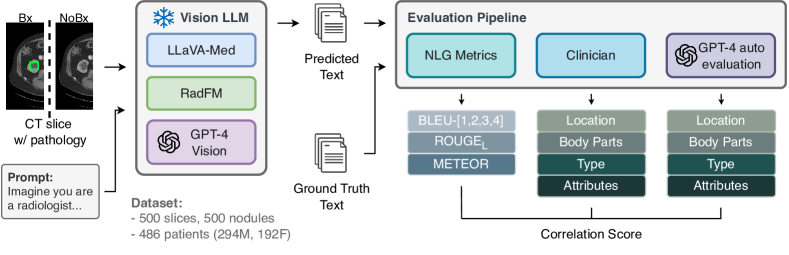
Decomposing Vision-based LLM Predictions for Auto-Evaluation: The proposed framework decomposes the predicted descriptions of CT-based findings (e.g., lesions) by a vision-based LLM, such that a language-centric LLM can auto-evaluate the predictions by comparing them against the ground-truth annotations. First, several prominent vision-based LLMs from recent literature were tasked with analyzing a CT slice with a known abnormality (lesion) and generating a free-text description of its characteristics. Next, GPT-4 parsed this prediction, and provided a score for each aspect by comparing them against the ground truth annotations from DeepLesion. The objective was to move beyond conventional natural language generation (NLG) metrics, which despite their linguistic coherence, are insufficient to evaluate predictions for clinical accuracy. Figure 1 illustrates the experimental design, consisting of three integral steps.
(1) Visual Context Integration: The abnormal findings within the CT slices were delineated with a bounding box prior to being input into the vision-based LLM. The clear visual context provided to the vision-based LLMs was expected to enhance the accuracy of the generated summaries of the findings.
(2) Text-Based Chain-of-Thought: In the Text-Based Chain of Thought (COT) approach, the vision-based LLM takes the input CT slice with an abnormality in it, and generates a free-text description of the abnormality. The output description should contain the following aspects: Body Part, Location (specific), Type, and Attributes. ‘Body Part’ is the larger anatomical region or organ of the body where the lesion or abnormality is situated. ‘Location’ refers to the precise area or specific site within a body part where a lesion or abnormality is located. ‘Type’ includes classifications, such as nodule, mass, or enlarged lymph node. ‘Attributes’ describe characteristics like size, shape, density (hypo or hyper), or calcification. The summary should be concise and clinically relevant, such that the characteristics of the findings can be pre-filled in the findings section of a radiology report. The summary should also lend itself to being auto-evaluated by a LLM (language model only). The prompt used for this task was designed to allow the model to concentrate on each aspect individually, thereby optimizing the use of its natural language generation capabilities to produce clinically relevant and informative descriptions of the findings. This approach contrasts with the one-shot methods [21] found in the literature, which attempt to generate entire radiology reports in a single step without explicit intermediate reasoning.
(3) Auto-Evaluation using GPT-4: This is the core of our framework that involves employing GPT-4 to mimic the evaluation process of radiologists. Inspired by [16], GPT-4 compared the predicted summary from the vision-based LLM against ground-truth annotations from DeepLesion and assigned a score for each aspect of the abnormality. The scores provided were: {“Correct,” “Partially Correct,” “Incorrect,”, “Not Applicable”}. “Correct” indicated that the interpretation was entirely accurate. “Partially Correct” signified that the interpretation was somewhat accurate, but lacked full precision or completeness. “Incorrect” meant that the interpretation did not match the correct answer in any way. Lastly, “Not Applicable” denoted that the question was not relevant to the situation and thus could not be graded. These scores enabled the automated evaluation of the accuracy of the predicted descriptions. The prompt provided to GPT-4, detailed in supplementary material, instructed the model to assess the AI-generated predictions in the same way as a clinician.
3 Experiments and Results
Baseline Models: Three vision-based LLMs from recent literature were evaluated for their capability to generate a summary of the CT-based abnormality. These models included GPT-4 Vision (GPT-4V) [24], LLaVA-Med [9], and RadFM [10]. For both LLaVA-Med and RadFM, the default configurations set by the respective authors were used for inference. OpenAI API version “2023-03-15-preview”, utilizing the “gpt-4” engine, was employed for GPT-4V. The configuration was set as follows: {”temperature”: 0.7, ”top_p”: 0.95, ”max_tokens”: 4000, ”model_version”: ”2024-02-15-preview”}.
Experiments: To determine the utility of GPT-4 for auto-evaluation of findings generated by vision-based LLMs, a baseline performance with respect to a clinician was required. In particular, 100 random lesions were chosen from the entire set of 500 lesions. As their characteristics were already verified previously by two board-certified radiologists, it was cumbersome to evaluate the free-text generations for all 500 lesions. Once their performance was quantified based on different metrics, the utility of using GPT-4 alone for auto-evaluation of the findings generated by vision-based LLMs compared against the ground-truth was determined. Evaluation of the generated findings was conducted based on four configurations: both with and without bounding boxes delineating the lesion in the CT slice, and combined with and without text-based COT.
Metrics: The linguistic quality of the generated summaries were evaluated using traditional NLG metrics, such as BLEU, METEOR, and ROUGE. However, considering the variability in reporting across different institutions, these metrics have limitations in capturing the clinical relevance of the generated summary (see example in supplementary material). Inspired by [16], auto-evaluation by GPT-4 was done by comparing the predictions against the ground-truth annotations. This evaluation was also compared against the evaluation performed by a clinician. Correlations between the evaluations done by the clinician and GPT-4 was considered to be a measure of the reliability of GPT-4 for auto-evaluation.
Results - Evaluation of Clinician vs. GPT-4: The clinician compared the ground truth against the free-text predictions generated by the vision-based LLMs for the 100 random lesions. The clinician provided a score for the prediction based on: body part, location, type, and attributes. The score fell into one of these categories: “Incorrect”: -1, “Partially Correct”: 0.5, “Correct”: 1. The “Not Applicable” category was excluded from the analysis as these responses did not contribute meaningful information regarding the accuracy or quality of the generated findings. Next, GPT-4 compared the same predictions against the ground-truth and provided the corresponding scores. Finally, NLG metrics were computed using these scores. In Fig. 2, heatmaps show the interaction between the various metrics computed based on the clinician and GPT-4 evaluations.
From Fig. 2, NLG metrics, such as BLEU and METEOR, revealed strong correlations amongst themselves (purple box), particularly at lower levels of precision like BLEU-1 and BLEU-2. This indicated a consistency in evaluating the linguistic quality of generated texts at these levels. However, the correlation weakened considerably at increased precision levels of BLEU-3 and BLEU-4. Particularly for LLaVA-Med, the scores were predominantly at 0, and exhibited no correlation. This pattern aligned with expectations and highlighted the limitations of BLEU metrics in capturing the nuanced accuracy of more complex sentences within radiology reports.
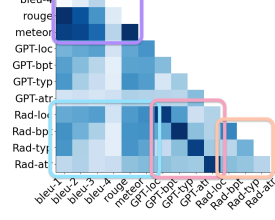
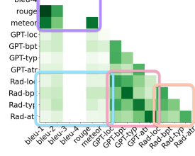
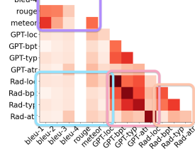
Furthermore, the decomposition of the evaluation into specific aspects (location, body part, lesion type, and attributes) revealed insightful patterns (peach box). These aspects showed a lack of strong correlation with one another, and other pairings also displayed no significant correlation. These observations affirmed the efficacy of the approach in dissecting the findings into their granular elements (except location and body part), such that the distinct parts of report quality can be isolated and assessed independently. Comparing NLG metrics with the clinician evaluations showed a weak correlation, suggesting that the NLG metrics may not serve as reliable indicators of clinical accuracy within radiology reports (blue box). This highlighted a potential gap in utilizing NLG metrics for assessing the clinical relevance of generated reports, pointing to the necessity for domain-specific evaluation methods.
Lastly, the comparison between evaluations conducted by GPT-4 and the clinician showed the strength of the automated evaluation framework (pink box), and summarised in Table 2. The results from auto-evaluation showed a strong correlation with those from a clinician based on Pearson’s correlation coefficient of 0.87 0.02 (p0.001). This suggests that GPT-4’s evaluation method closely aligned with the clinical assessment paradigms utilized by radiologists. This observation underscored the potential of language models like GPT-4 in accurately mirroring radiologists’ evaluations, offering promise for automating report assessment with a high degree of fidelity to clinical standards.
Results - Auto Evaluation using GPT-4: As there was a strong correlation between the evaluations conducted by GPT-4 and the clinician, GPT-4 was utilized to compare the predicted findings against the ground-truth for all the 500 lesions. Figure 3 shows grading scores from GPT-4 across four configurations: both with and without lesion bounding boxes in the CT slice, and combined with and without text-based Chain-of-Thought (COT). Notably, LLaVA-Med and RadFM lack the functionality to utilize text-based COT processes, which aligns with findings in literature [25]. This sets them apart from GPT-4V, which demonstrated enhanced performance when employing COT reasoning. Furthermore, the inclusion of bounding boxes generally lead to improved model performance as it provided guidance for the models, with GPT-4V showing the most significant enhancement. Amongst the evaluated models, GPT-4V outperformed its counterparts. However, despite these advancements, the overall quality of pre-filled findings for CT reporting remained suboptimal. This indicated a pressing need for further development of vision-based LLMs to meet clinical standards.
Consider the ground truth text ‘left mediastinal pleural enhancing mass’ and a predicted result by RadFM ‘Axial contrast enhanced images through the thoracic inlet demonstrating a homogeneous mass posterior to the trachea’. It is evident that the NLG scores are low owing to the minimal textual overlap. Furthermore, this sentiment extends to individuals without medical training, as an understanding of anatomy is necessary to recognize that both texts pertain to the chest and lung regions.
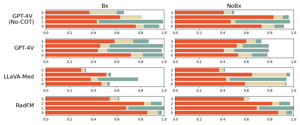
Color mapping = {orange: ‘Incorrect’, beige: ‘Partially Correct’, teal: ‘Correct’, white: ‘Not Applicable’}. Numeric labels = {0: ‘Location’, 1: ‘Body Part’, 2: ‘Type’, 3: ‘Attribute’}. X-axis denotes scores , = 500 samples.
![[Uncaptioned image]](/html/2403.05680/assets/images/001323_03_01_076.png)
GT: ‘right renal parapelvic cyst’
| GPT-4V: ‘Location’: ‘Right abdomen, near the kidney’, ‘Body Part’: ‘Abdomen, kidney’, ‘Type’: ‘Mass’, ‘Attributes’: ‘Well-circumscribed, homogenous’, ‘Impression’: ‘Well-circumscribed homogenous mass in the right abdomen, adjacent to the right kidney’ |
|---|
| LLaVA-Med: The image is an axial computed tomography (CT) scan that has been annotated with a bounding box by a radiologist. The bounding box is a rectangular box that encloses the lesion, which is indicated in green. This helps to highlight the area of interest and provides a clear view of the lesion’s location, size, and shape. |
| RadFM: { Lesion 1 } |
| Location | Body Part | Type | Attribute | Avg. | p-value | |
|---|---|---|---|---|---|---|
| GPT-4V | 0.86 | 0.90 | 0.84 | 0.86 | 0.87 0.02 | 0.001 |
| LLaVA-Med | 0.59 | 0.83 | 0.76 | 0.80 | 0.75 0.10 | 0.001 |
| RadFM | 0.99 | 0.92 | 0.82 | 0.89 | 0.90 0.07 | 0.001 |
4 Discussion and Conclusion
Through experiments, it was observed that LLaVA-Med and RadFM were unable to leverage text-based COT as shown in Table 1. This reflects the architectural or design constraints that prevented these models from effectively breaking down and processing information in a stepwise manner. GPT-4V’s improved performance with COT suggested its architecture was better suited to sequential reasoning, mimicking a radiologist’s thought process, thereby leading to more accurate generation of the characteristics of CT findings. We also noticed that the bounding box delineating the lesion improved the performance of most models, especially GPT-4V. We attribute this to the added visual cues that bounding boxes provided. These cues helped focus the model’s attention on a specific area of interest, thereby improving the accuracy of the abnormality summary. GPT-4V outperforming other models can be linked to its advanced language processing capabilities, likely benefiting from a more sophisticated understanding of context, superior text generation algorithms, and perhaps a more extensive training dataset that includes a diverse range of medical imaging scenarios.
With respect to the lesion characteristics (location, body part, type, attributes), a varied performance across the categories was seen. This reflected the differential impact of visual and textual guidance on model accuracy. For instance, the improvement in “Body Part” and “Type” with bounding boxes suggested these categories benefitted more from visual delineation. In contrast, the “Location” and “Attributes” categories, which may require more abstract reasoning or detailed textual information, showed mixed results. This indicated a complex interplay between model capabilities and the nature of the task.
Despite advancements, the overall performance of vision-based LLMs in describing the characteristics of findings in CT remains inadequate for clinical standards. This is largely due to the inherent complexity of medical images, the subtlety of pathologies, and the high standard of accuracy required for clinical diagnosis. Current vision-based LLMs are trained largely on natural images and lack sufficient training on diverse datasets. Thus, they still lack the ability to fully comprehend and articulate the complex interplay of visual features and clinical implications present in CT exams.
In summary, an framework was proposed for the auto-evaluation of AI-generated characteristics for findings in CT exams, which would be pre-filled into the findings section of radiology reports. Results from auto-evaluation showed a strong correlation with those from a clinician based on Pearson’s correlation coefficient of 0.87 0.02 (p0.001). GPT-4V outperformed other recent vision-language LLMs in predicting the characteristics of lesions in the dataset, and GPT-4 was sufficient for the auto-evaluation of the predictions from GPT-4V against ground-truth annotations. The evaluation approach pointed out particular weaknesses within various vision-based LLMs, and provided essential insights to enhance the precision and dependability of AI-generated interpretations from medical images. By addressing these identified gaps and focusing on comprehensive model development, there is potential to significantly improve the utility of vision- and language-based models in radiology.
5 Acknowledgements
This research was supported by the Intramural Research Program of the National Library of Medicine and Clinical Center at the NIH.
References
- [1] John J. Smith and Leonard Berlin. Signing a colleague’s radiology report. American Journal of Roentgenology, 176(1):27–30, 2001. PMID: 11133532.
- [2] Michael D Ringler, Brian C Goss, and Brian J Bartholmai. Syntactic and semantic errors in radiology reports associated with speech recognition software. Health Informatics Journal, 23(1):3–13, 2017. PMID: 26635322.
- [3] M. Mahesh, A. J. Ansari, and Jr Mettler, F. A. Patient exposure from radiologic and nuclear medicine procedures in the united states and worldwide: 2009-2018. Radiology, 301, 2023.
- [4] N. A. Fawzy, M. J. Tahir, A. Saeed, M. J. Ghosheh, T. Alsheikh, A. Ahmed, and Z. Lee, K. Y. Yousaf. Incidence and factors associated with burnout in radiologists: A systematic review. European journal of radiology open, 11:100530, 2023.
- [5] Zhihong Chen, Yan Song, Tsung-Hui Chang, and Xiang Wan. Generating radiology reports via memory-driven transformer. In Bonnie Webber, Trevor Cohn, Yulan He, and Yang Liu, editors, Proceedings of the 2020 Conference on Empirical Methods in Natural Language Processing (EMNLP), pages 1439–1449, Online, November 2020. Association for Computational Linguistics.
- [6] Zhihong Chen, Yaling Shen, Yan Song, and Xiang Wan. Cross-modal memory networks for radiology report generation. In Chengqing Zong, Fei Xia, Wenjie Li, and Roberto Navigli, editors, Proceedings of the 59th Annual Meeting of the Association for Computational Linguistics and the 11th International Joint Conference on Natural Language Processing (Volume 1: Long Papers), pages 5904–5914, Online, August 2021. Association for Computational Linguistics.
- [7] Akimichi Ichinose, Taro Hatsutani, Keigo Nakamura, Yoshiro Kitamura, Satoshi Iizuka, Edgar Simo-Serra, Shoji Kido, and Noriyuki Tomiyama. Visual grounding of whole radiology reports for 3d ct images. In Hayit Greenspan, Anant Madabhushi, Parvin Mousavi, Septimiu Salcudean, James Duncan, Tanveer Syeda-Mahmood, and Russell Taylor, editors, Medical Image Computing and Computer Assisted Intervention – MICCAI 2023, pages 611–621, Cham, 2023. Springer Nature Switzerland.
- [8] Josh Achiam, Steven Adler, Sandhini Agarwal, Lama Ahmad, Ilge Akkaya, Florencia Leoni Aleman, Diogo Almeida, Janko Altenschmidt, Sam Altman, Shyamal Anadkat, et al. Gpt-4 technical report. arXiv preprint arXiv:2303.08774, 2023.
- [9] Chunyuan Li, Cliff Wong, Sheng Zhang, Naoto Usuyama, Haotian Liu, Jianwei Yang, Tristan Naumann, Hoifung Poon, and Jianfeng Gao. LLaVA-Med: Training a large language-and-vision assistant for biomedicine in one day. Advances in Neural Information Processing Systems, 36, 2024.
- [10] Chaoyi Wu, Xiaoman Zhang, Ya Zhang, Yanfeng Wang, and Weidi Xie. Towards generalist foundation model for radiology. arXiv preprint arXiv:2308.02463, 2023.
- [11] Shubo Tian, Qiao Jin, Lana Yeganova, Po-Ting Lai, Qingqing Zhu, Xiuying Chen, Yifan Yang, Qingyu Chen, Won Kim, Donald C Comeau, et al. Opportunities and challenges for chatgpt and large language models in biomedicine and health. Briefings in Bioinformatics, 25(1):bbad493, 2024.
- [12] Harsha Nori, Nicholas King, Scott Mayer McKinney, Dean Carignan, and Eric Horvitz. Capabilities of gpt-4 on medical challenge problems. arXiv preprint arXiv:2303.13375, 2023.
- [13] Qiao Jin, Robert Leaman, and Zhiyong Lu. Retrieve, summarize, and verify: How will chatgpt impact information seeking from the medical literature? Journal of the American Society of Nephrology, pages 10–1681, 2023.
- [14] Qingqing Zhu, Tejas Sudharshan Mathai, Pritam Mukherjee, Yifan Peng, Ronald M. Summers, and Zhiyong Lu. Utilizing longitudinal chest x-rays and reports to pre-fill radiology reports. In MICCAI (5), volume 14224 of Lecture Notes in Computer Science, pages 189–198. Springer, 2023.
- [15] Jeremy Irvin, Pranav Rajpurkar, Michael Ko, Yifan Yu, Silviana Ciurea-Ilcus, Chris Chute, Henrik Marklund, Behzad Haghgoo, Robyn Ball, Katie Shpanskaya, et al. Chexpert: A large chest radiograph dataset with uncertainty labels and expert comparison. In Thirty-Third AAAI Conference on Artificial Intelligence, 2019.
- [16] Qingqing Zhu, Xiuying Chen, Qiao Jin, Benjamin Hou, Tejas Sudharshan Mathai, Pritam Mukherjee, Xin Gao, Ronald M Summers, and Zhiyong Lu. Leveraging professional radiologists’ expertise to enhance llms’ evaluation for radiology reports, 2024.
- [17] Qiao Jin, Fangyuan Chen, Yiliang Zhou, Ziyang Xu, Justin M Cheung, Robert Chen, Ronald M Summers, Justin F Rousseau, Peiyun Ni, Marc J Landsman, et al. Hidden flaws behind expert-level accuracy of gpt-4 vision in medicine. arXiv preprint arXiv:2401.08396, 2024.
- [18] Xiaosong Wang, Yifan Peng, Le Lu, Zhiyong Lu, Mohammadhadi Bagheri, and Ronald M Summers. Chestx-ray8: Hospital-scale chest x-ray database and benchmarks on weakly-supervised classification and localization of common thorax diseases. In Proceedings of the IEEE conference on computer vision and pattern recognition, pages 2097–2106, 2017.
- [19] Yifan Peng, Xiaosong Wang, Le Lu, Mohammadhadi Bagheri, Ronald Summers, and Zhiyong Lu. Negbio: a high-performance tool for negation and uncertainty detection in radiology reports. AMIA Summits on Translational Science Proceedings, 2018:188, 2018.
- [20] Yang Liu, Dan Iter, Yichong Xu, Shuohang Wang, Ruochen Xu, and Chenguang Zhu. G-eval: NLG evaluation using gpt-4 with better human alignment. In Houda Bouamor, Juan Pino, and Kalika Bali, editors, Proceedings of the 2023 Conference on Empirical Methods in Natural Language Processing, pages 2511–2522, Singapore, December 2023. Association for Computational Linguistics.
- [21] Jason Wei, Xuezhi Wang, Dale Schuurmans, Maarten Bosma, Fei Xia, Ed Chi, Quoc V Le, Denny Zhou, et al. Chain-of-thought prompting elicits reasoning in large language models. Advances in Neural Information Processing Systems, 35:24824–24837, 2022.
- [22] Ke Yan, Xiaosong Wang, Le Lu, and Ronald M. Summers. Deeplesion: Automated deep mining, categorization and detection of significant radiology image findings using large-scale clinical lesion annotations. CoRR, abs/1710.01766, 2017.
- [23] Ke Yan, Yifan Peng, Veit Sandfort, Mohammadhadi Bagheri, Zhiyong Lu, and Ronald M Summers. Holistic and comprehensive annotation of clinically significant findings on diverse ct images: learning from radiology reports and label ontology. In Proceedings of the IEEE/CVF Conference on Computer Vision and Pattern Recognition, pages 8523–8532, 2019.
-
[24]
OpenAI.
GPT-4V(ision) System Card.
https://cdn.openai.com/papers/GPTV\_System\_Card.pdf, 2023. - [25] Seungone Kim, Se June Joo, Doyoung Kim, Joel Jang, Seonghyeon Ye, Jamin Shin, and Minjoon Seo. The cot collection: Improving zero-shot and few-shot learning of language models via chain-of-thought fine-tuning. In The 2023 Conference on Empirical Methods in Natural Language Processing, 2023.