Multidimensional spectroscopy of nuclear spin clusters in diamond
Abstract
Optically active spin defects in solids offer promising platforms to investigate nuclear spin clusters with high sensitivity and atomic-site resolution. To leverage near-surface defects for molecular structure analysis in chemical and biological contexts using nuclear magnetic resonance (NMR), further advances in spectroscopic characterization of nuclear environments are essential. Here, we report Fourier spectroscopy techniques to improve localization and mapping of the testbed nuclear spin environment of individual, shallow nitrogen-vacancy centers at room temperature. We use multidimensional spectroscopy, well-known from classical NMR, in combination with weak measurements of single-nuclear-spin precession. We demonstrate two examples of multidimensional NMR: (i) improved nuclear spin localization by separate encoding of the two hyperfine components along spectral dimensions and (ii) spectral editing of nuclear-spin pairs, including measurement of internuclear coupling constants. Our work adds important tools for the spectroscopic analysis of molecular structures by single-spin probes.
Electronic spin defects in wide-bandgap dielectrics, including diamond, silicon carbide, or oxide crystals, provide exceptionally long spin coherence times, even at room temperature [1]. Combined with efficient optical spin preparation and readout, such defect spins can be used to analyze local nuclear spin environments [2] and conduct NMR at the single-spin level [3, 4, 5]. Particularly, nitrogen-vacancy (NV) defect spins in diamond have been used to detect and control individual spins in clusters comprising multiple nuclei, and offer potential routes to resolve the structure of proximal molecules with atomic resolution and elemental specificity [6, 7, 8, 9].
The naturally occurring 13C spin bath in diamond offers an ideal testbed for developing single-spin imaging techniques. Early work employed dynamical decoupling (DD) spectroscopy to detect 13C resonances corresponding to individual nuclei [10, 11, 12, 13]. Spectral assignment can be used to determine spatial positions or identify spin pairs [14, 15]. However, the spectral resolution of DD is limited by NV coherence time , and restricted to a few kHz unless under cryogenic conditions [16]. More recently, alternative methods such as correlation spectroscopy [17] and weak-measurement schemes [18, 5] have improved spectral resolution to the Hz range at room temperature.
These advances have enabled atomic-scale localization of 13C spins in the diamond lattice [19, 10, 20, 21, 22]. To date, clusters of roughly 20 nuclei at room temperature [23] and 50 nuclei at cryogenic temperatures [24] have been resolved. Despite this progress, mapping large nuclear spin networks remains challenging due to spectral overlap, positional ambiguities and internuclear interactions [23]. Multidimensional techniques have proven to reduce such ambiguities at cryogenic temperatures [2, 24], however, translating these approaches to ambient conditions is not straightforward because of reduced spin lifetimes.
In this work, we combine multidimensional Fourier spectroscopy with weak measurements of nuclear spin precession to establish a general route to improving the accuracy of spin localization. First, we present correlative mapping of the transverse and parallel hyperfine coupling parameters [25] to triangulate global nuclear spin positions. Second, we edit nuclear spin-spin couplings using a variant of -spectroscopy [26] to identify spin-pair configurations. In both cases, 2D spectra remove overlap present in 1D spectra and enable improved assignment of single nuclear peaks. We demonstrate our schemes on the testbed 13C environment of shallow NVs at room temperature, which is compatible with future experiments on surface-anchored molecules. Further work on stabilizing NV centers near the diamond surface [27, 28] and developing reliable chemical functionalization of the surface [29, 30, 31] will be needed to realize this exciting potential.
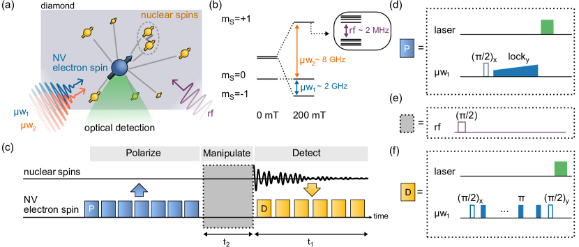
Our experimental scheme is shown in Fig. 1a. A central electronic NV spin is surrounded by a dilute bath ( natural abundance) of 13C nuclear spins. NVs are created by ion implantation in a single-crystalline diamond chip. Subsequent annealing results in shallow (-deep [31]) NVs. Following defect creation, nanopillar waveguides are fabricated to enhance optical collection efficiency [32]. Off-resonant (532 nm) laser pulses ( duration) are used to optically polarize and read out the spin state. A two-channel microwave generator connected to a broadband co-planar waveguide antenna serves to drive the two allowed electronic spin transitions at frequencies w1 and w2 (Fig. 1(b)). Typical -pulse durations are () for w1 (w2), respectively . Radio-frequency (rf) pulses for 13C manipulation are applied via a separate broadband microcoil antenna [33], with a typical -pulse duration of . Further details are given in the SM [34].
Fig. 1(c-f) revisit the protocol for 1D weak-measurement spectroscopy [5, 23]. The basic 1D protocol consists of three steps: First, the NV spin is polarized by a laser pump pulse followed by transfer of the NV polarization to the nuclei using the nuclear spin orientation via electron spin locking (NOVEL) protocol [35] (Fig. 1(d)). Since one transfer cycle can polarize a single 13C at most, this cycle is repeated times to polarize a nm-scale volume of 13C around the NV [23]. Second, a single rf pulse triggers nuclear precession (Fig. 1e). Third, precessing nuclear magnetization is detected via a series of weak measurements (Fig. 1f) [36, 18, 5]. The overall sequence is averaged over cycles to obtain sufficient signal-to-noise ratios. Resulting time-domain signals reflect the free-induction decay signal of nuclear precession [5]. Fourier transformation with respect to the free precession time then yields the 1D NMR spectrum of the NV’s 13C environment [23].
1D spectroscopy of the 13C nuclear spin bath has been used to determine spatial locations of nuclei surrounding NVs [19, 11, 21, 23]. Spatial mapping is achieved by analyzing the parallel and transverse components of the hyperfine interaction tensor, denoted by and , which uniquely define the radial distance and polar angle between the two spins (up to an inversion at the origin) [25]. Analysis of the nuclear precession phase permits extracting the azimuth [20, 22]. Together, the two measurements yield the full 3D location vector (,,) of a nuclear spin. While this approach has been successfully extended to several nuclear spins, mapping of larger clusters remains challenging because of spectral overlap and the presence of internuclear couplings. Further, unequal polarization of nuclear spins and varying local relaxation environments contribute to localization errors [23]. Therefore, 1D methods become imprecise for larger spin clusters (), necessitating more advanced methods for constraining positions.
In the following, we show that multidimensional spectroscopy can address some ambiguities and limitations associated with 1D methods. While multidimensional approaches have been considered in the context of NVs [25, 2], we take inspiration from 2D NMR [26] and show that weak-measurement spectroscopy can be extended to multiple spectral dimensions by introducing an indirect evolution time during the “manipulation” block (Fig. 1(c)). Using this approach, we demonstrate correlative measurement of hyperfine coupling constants and editing of dipolar-coupled spin pairs.
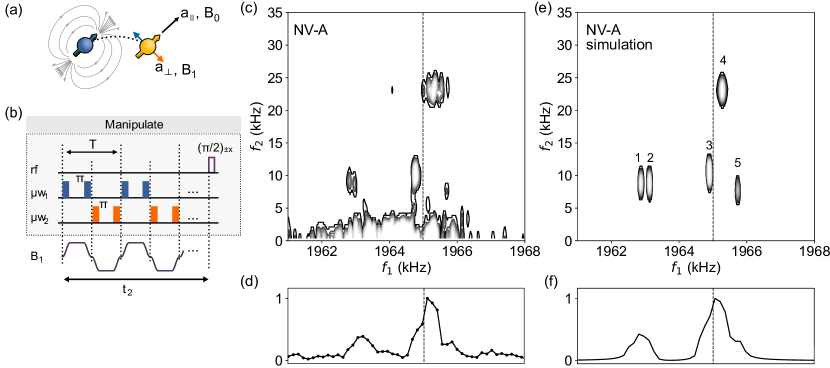
Fig. 2 presents a first example of multidimensional spectroscopy, where we correlate the two coupling parameters (,) of the hyperfine tensor. The goal of this sequence is to improve the accuracy of the measurement and resolve overlapping peaks with similar in 1D spectra. Specifically, we replace the rf pulse of the “manipulation” period (Fig. 1(e)) with a periodic inversion of the NV spin commensurate with the 13C Larmor precession (Fig. 2(b)). Periodic nuclear inversion results in modulation of the hyperfine coupling, which in turn causes Rabi driving of the nuclear spins. The amplitude of the driving field is given by
| (1) |
where is the duration of the electronic pulse, is the modulation period, and is the 13C gyromagnetic ratio. In contrast to previous Rabi driving experiments on single nuclei [37, 25], inversion of both electronic spin transitions is essential to drive nuclear ensembles. Because each nuclear spin experiences different static detunings by the term, inverting only one NV transition leads to large dispersion of Rabi frequencies due to off-resonant driving. Note that inverting both NV transitions doubles the amplitude (Fig. 2(b)).
To obtain 2D spectra, we acquire free-induction decays for a series of increments, where is the duration of the periodic inversion block. Applying 2D Fourier transformation with respect to and generates the desired 2D NMR spectrum.
Figs. 2(c,e) show a representative 2D spectrum for NV-A and corresponding numerical simulation. The horizontal axis represents the direct dimension (frequency ), while the vertical axis is the indirect dimension (frequency ) encoding the Rabi driving. In the horizontal direction, peaks are distributed between . Here, peak frequencies encode the parallel hyperfine coupling that shifts resonances away from the expected 13C frequency (no couplings present) at . The vertical direction corresponds to the Rabi driving amplitude , which is determined by the magnitude of the transverse hyperfine coupling . Isolated peaks with reflect proximal 13C with relative strong hyperfine interactions, while peaks with correspond to more distant 13C .
In the following, we focus our analysis on the five main peaks of Fig. 2(c). From the frequency coordinates of a peak we extract the hyperfine coupling parameters (,) according to [25],
| (2) | ||||
| (3) |
using the approximation . The (,) values may be converted to spatial position vectors by including the phase information (SM and Ref. [23]). To corroborate this procedure, we perform an ab initio density matrix simulation of a five-spin cluster, taking extracted spatial position vectors as input and accounting for dipolar interactions between nuclei (Fig. 2(e)). Excellent agreement to experimental peak positions are obtained with modest agreement to measured peak intensities. Variation in latter may be due to inconsistent polarization of nuclei. For comparison, Figs. 2(d,f) show corresponding 1D spectra, where the overlap between nearby 13C peaks with similar complicates spectral analysis.
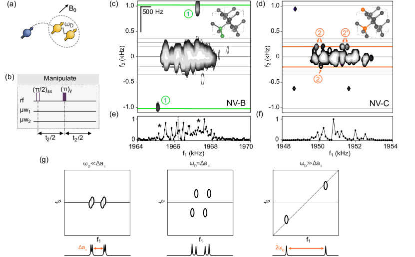
As a second application, we demonstrate identification of nuclear spin pairs. To detect spin pairs, we use a scheme derived from -spectroscopy [26] (Fig. 3(b)). The manipulation step of Fig. 1(c) is replaced by a nuclear spin echo, and we increment total echo duration . The spin echo eliminates static Zeeman and parallel hyperfine terms of the nuclear-spin Hamiltonian, such that evolution during is only due to nuclear spin-spin interactions. For two spins and with through-space distance forming an angle with the external bias field, coupling frequency is given by
| (4) |
Compared to conventional -spectroscopy, dipolar coupling dominates over scalar () coupling, while the spin Hamiltonian takes the same form except for different coupling constants. Acquiring a 2D dataset and applying a Fourier transform with respect to and , the coupling frequency of a spin pair can be extracted along the indirect dimension, and correlated with peak positions in the 1D spectrum.
Figs. 3(c-f) show 2D and projected 1D spectra of the 13C environment of NV-B and NV-C. Most peaks lie near the center line, signaling isolated 13C nuclei. A few satellite peaks with comparably large are visible in both spectra. Specifically, satellite peaks appear at for NV-B (Fig. 3(d)) and at for NV-C (Fig. 3(e)). Since the diamond lattice only permits discrete values of coupling frequencies corresponding to a specific 13C configurations, we use the measured to identify spin pairs. We assign the satellite peaks to three spin pairs: a nearest-neighbor pair along the crystal axis (NV-B), and two next-next-nearest neighbor pairs along (NV-C) (Figs. 3(c,d) insets).
To gain insight into the multiplet structure, we simulate the expected peak pattern for a spin pair with weak (), intermediate () and strong () coupling, where is the difference between the nuclear spin’s parameter. Qualitative behavior is shown in Fig. 3(g) and quantitative results are given in the SM [34]. In the weak-coupling case, each 13C resonance is split into a peak doublet separated by (left). As becomes comparable to (middle), the peaks are more evenly distributed and frequencies are not straightforward to interpret [34]. Once (right), only two peaks are observed, lying near the diagonal and separated by .
Revisiting the experimental spectra in Fig. 3(c,d), we qualitatively identify pair (1) near NV-B as a strong-coupling case, and pairs (2) and (2’) near NV-C as weak-to-intermediate coupling cases. However, only three out of four peaks (pair (2)) resp. two out for four peaks (pair (2’)) are visible in Fig. 3(d). This is because peak intensities are in general different [34] and since the signal-to-noise ratio is limited, some peaks fall below the detection threshold.
Both 2D spectra show structure near the center line, suggesting that more spin pairs are present. However, our spectral resolution () is insufficient to assign these peaks to specific pairs of lattice sites.
Unlike experiments conducted at cryogenic temperatures, where sub-Hz 13C resolution can be reached [2], our spectral resolution is limited by coherence times of the 13C spins (), bounded by the room-temperature time of the NV [38].
Future work will therefore need to focus on improving the spectral resolution. Unlike experiments conducted at cryogenic temperatures, where sub-Hz 13C resolution can be reached [2], extending the coherence at room temperature is not straightforward. In our experiments, the coherence time of the 13C spins () (and thus, the spectral resolution) is bounded by the room-temperature time of the NV and by bias field drifts [38]. One approach to prolonging is the use of dual-frequency driving similar to Fig. 2(b), where the weak measurement readout alternates between the two NV transitions combined with a periodic inversion of the nuclear spin. This approach requires higher w driving powers than currently available with our setup, but is the subject of future work. Alternatively, optical control of the NV spin and charge state [39, 40] could be explored to extend the 13C coherence.
In conclusion, we demonstrate a set of methods for extending single-spin NMR detection to multiple spectral dimensions. Our approach combines weak-measurement spectroscopy [5] with an additional, indirect evolution period well-known from multidimensional NMR spectroscopy [26]. We demonstrate precise measurement of the transverse component of the NV-13C hyperfine interaction, and selective editing of nuclear spin pairs including quantification of dipolar-coupling constants. The 2D spectra reduce spectral overlap and enable improved assignment of single 13C peaks.
Although the number of assigned peaks in this work (e.g., in Fig. 2(c)) is smaller compared to previous work where [23, 2, 24], we consider multi-dimensional techniques as an essential ingredient towards improved localization of intrinsic and extrinsic nuclear spins. To extend analysis to larger spin clusters, multidimensional cross-correlation methods derived from COSY or NOESY spectroscopy [41] could be adapted or higher spectral dimensions with additional indirect evolution periods included. Assuming challenges in stabilizing near-surface NV centers [27, 28] and molecular functionalization of diamond [29, 30, 31] can be overcome, multidimensional approaches as presented here will add an important tool for spectroscopic analysis of surface-anchored molecules.
The authors thank Jonathan Zopes for fruitful discussions. This work has been supported by Swiss National Science Foundation (SNFS) Project Grant No. 200020-175600, the National Center of Competence in Research in Quantum Science and Technology (NCCR QSIT), and the Advancing Science and TEchnology thRough dIamond Quantum Sensing (ASTERIQS) program, Grant No. 820394, of the European Commission. EJ acknowledges support from a Natural Sciences and Engineering Research Council of Canada (NSERC) postdoctoral fellowship (PDF-558200-2021).
References
- Atature et al. [2018] M. Atature, D. Englund, N. Vamivakas, S. Lee, and J. Wrachtrup, Material platforms for spin-based photonic quantum technologies, Nature Reviews Materials 3, 38 (2018).
- Abobeih et al. [2019] M. H. Abobeih, J. Randall, C. E. Bradley, H. P. Bartling, M. A. Bakker, M. J. Degen, M. Markham, D. J. Twitchen, and T. H. Taminiau, Atomic-scale imaging of a 27-nuclear-spin cluster using a quantum sensor, Nature 576, 411 (2019).
- Jelezko et al. [2004] F. Jelezko, T. Gaebel, I. Popa, M. Domhan, A. Gruber, and J. Wrachtrup, Observation of coherent oscillation of a single nuclear spin and realization of a two-qubit conditional quantum gate, Phys. Rev. Lett. 93, 130501 (2004).
- Childress et al. [2006] L. Childress, M. V. G. Dutt, J. M. Taylor, A. S. Zibrov, F. Jelezko, J. Wrachtrup, P. R. Hemmer, and M. D. Lukin, Coherent dynamics of coupled electron and nuclear spin qubits in diamond, Science 314, 281 (2006).
- Cujia et al. [2019] K. S. Cujia, J. M. Boss, K. Herb, J. Zopes, and C. L. Degen, Tracking the precession of single nuclear spins by weak measurements, Nature 571, 230 (2019).
- Degen [2008] C. L. Degen, Scanning magnetic field microscope with a diamond single-spin sensor, Appl. Phys. Lett. 92, 243111 (2008).
- Ajoy et al. [2015] A. Ajoy, U. Bissbort, M. D. Lukin, R. L. Walsworth, and P. Cappellaro, Atomic-scale nuclear spin imaging using quantum-assisted sensors in diamond, Phys. Rev. X 5, 011001 (2015).
- Kost et al. [2015] M. Kost, J. Cai, and M. B. Plenio, Resolving single molecule structures with nitrogen-vacancy centers in diamond, Scientific Reports 5, 11007 (2015).
- Perunicic et al. [2016] V. S. Perunicic, C. D. Hill, L. T. Hall, and L. Hollenberg, A quantum spin-probe molecular microscope, Nat. Commun. 7, 12667 (2016).
- Zhao et al. [2012] N. Zhao, J. Honert, B. Schmid, M. Klas, J. Isoya, M. Markham, D. Twitchen, F. Jelezko, R. Liu, H. Fedder, and J. Wrachtrup, Sensing single remote nuclear spins, Nature Nano. 7, 657 (2012).
- Taminiau et al. [2012] T. H. Taminiau, J. J. T. Wagenaar, T. V. der Sar, F. Jelezko, V. V. Dobrovitski, and R. Hanson, Detection and control of individual nuclear spins using a weakly coupled electron spin, Phys. Rev. Lett. 109, 137602 (2012).
- Kolkowitz et al. [2012] S. Kolkowitz, Q. P. Unterreithmeier, S. D. Bennett, and M. D. Lukin, Sensing distant nuclear spins with a single electron spin, Phys. Rev. Lett. 109, 137601 (2012).
- Bradley et al. [2019] C. Bradley, J. Randall, M. Abobeih, R. Berrevoets, M. Degen, M. Bakker, M. Markham, D. Twitchen, and T. Taminiau, A ten-qubit solid-state spin register with quantum memory up to one minute, Phys. Rev. X 9, 031045 (2019).
- Kalb et al. [2016] N. Kalb, J. Cramer, D. J. Twitchen, M. Markham, R. Hanson, and T. H. Taminiau, Experimental creation of quantum Zeno subspaces by repeated multi-spin projections in diamond, Nature Communications 7, 13111 (2016).
- Bartling et al. [2022] H. P. Bartling, M. H. Abobeih, B. Pingault, M. J. Degen, S. J. H. Loenen, C. E. Bradley, J. Randall, M. Markham, D. J. Twitchen, and T. H. Taminiau, Entanglement of spin-pair qubits with intrinsic dephasing times exceeding a minute, Physical Review X 12, 011048 (2022).
- Abobeih et al. [2018] M. H. Abobeih, J. Cramer, M. A. Bakker, N. Kalb, M. Markham, D. J. Twitchen, and T. H. Taminiau, One-second coherence for a single electron spin coupled to a multi-qubit nuclear-spin environment, Nature Communications 9, 2552 (2018).
- Laraoui et al. [2013] A. Laraoui, F. Dolde, C. Burk, F. Reinhard, J. Wrachtrup, and C. A. Meriles, High-resolution correlation spectroscopy of C-13 spins near a nitrogen-vacancy centre in diamond, Nature Commun. 4, 1651 (2013).
- Pfender et al. [2019] M. Pfender, P. Wang, H. Sumiya, S. Onoda, W. Yang, D. B. R. Dasari, P. Neumann, X. Pan, J. Isoya, R. Liu, and J. Wrachtrup, High-resolution spectroscopy of single nuclear spins via sequential weak measurements, Nature Communications 10, 594 (2019).
- Zhao et al. [2011] N. Zhao, J. L. Hu, S. W. Ho, J. T. K. Wan, and R. B. Liu, Atomic-scale magnetometry of distant nuclear spin clusters via nitrogen-vacancy spin in diamond, Nature Nanotechnology 6, 242 (2011).
- Sasaki et al. [2018] K. Sasaki, K. M. Itoh, and E. Abe, Determination of the position of a single nuclear spin from free nuclear precessions detected by a solid-state quantum sensor, Phys. Rev. B 98, 121405 (2018).
- Zopes et al. [2018a] J. Zopes, K. S. Cujia, K. Sasaki, J. M. Boss, K. M. Itoh, and C. L. Degen, Three-dimensional localization spectroscopy of individual nuclear spins with sub-angstrom resolution, Nat. Commun. 9, 4678 (2018a).
- Zopes et al. [2018b] J. Zopes, K. Herb, K. S. Cujia, and C. L. Degen, Three-dimensional nuclear spin positioning using coherent radio-frequency control, Phys. Rev. Lett. 121, 170801 (2018b).
- Cujia et al. [2022] K. S. Cujia, K. Herb, J. Zopes, J. M. Abendroth, and C. L. Degen, Parallel detection and spatial mapping of large nuclear spin clusters, Nature Communications 13, 1260 (2022).
- van de Stolpe et al. [2023] G. van de Stolpe, D. P. Kwiatkowski, C. Bradley, J. Randall, S. A. Breitweiser, L. C. Bassett, M. Markham, D. Twitchen, and T. Taminiau, Mapping a 50-spin-qubit network through correlated sensing, arXiv:2307.06939 (2023).
- Boss et al. [2016] J. M. Boss, K. Chang, J. Armijo, K. Cujia, T. Rosskopf, J. R. Maze, and C. L. Degen, One- and two-dimensional nuclear magnetic resonance spectroscopy with a diamond quantum sensor, Phys. Rev. Lett. 116, 197601 (2016).
- Aue et al. [1976] W. P. Aue, E. Bartholdi, and R. R. Ernst, Two-dimensional spectroscopy. application to nuclear magnetic resonance, J. Chem. Phys. 64, 2229 (1976).
- Sangtawesin et al. [2019] S. Sangtawesin, B. L. Dwyer, S. Srinivasan, J. J. Allred, L. V. Rodgers, K. D. Greve, A. Stacey, N. Dontschuk, K. M. ODonnell, D. Hu, D. A. Evans, C. Jaye, D. A. Fischer, M. L. Markham, D. J. Twitchen, H. Park, M. D. Lukin, and N. P. de Leon, Origins of diamond surface noise probed by correlating single-spin measurements with surface spectroscopy, Phys. Rev. X 9, 031052 (2019).
- Janitz et al. [2022] E. Janitz, K. Herb, L. A. Volker, W. S. Huxter, C. L. Degen, and J. M. Abendroth, Diamond surface engineering for molecular sensing with nitrogen-vacancy centers, Journal of Materials Chemistry C 10, 13533 (2022).
- Liu et al. [2022] K. S. Liu, A. Henning, M. W. Heindl, R. D. Allert, J. D. Bartl, I. D. Sharp, R. Rizzato, and D. B. Bucher, Surface NMR using quantum sensors in diamond, Proceedings of the National Academy of Sciences 119, e2111607119 (2022).
- Xie et al. [2022] M. Xie, X. Yu, L. V. H. Rodgers, D. Xu, I. Chi-Duran, A. Toros, N. Quack, N. P. de Leon, and P. C. Maurer, Biocompatible surface functionalization architecture for a diamond quantum sensor, Proceedings of the National Academy of Sciences 119, 10.1073/pnas.2114186119 (2022).
- Abendroth et al. [2022] J. M. Abendroth, K. Herb, E. Janitz, T. Zhu, L. A. Volker, and C. L. Degen, Single-nitrogen-vacancy NMR of amine-functionalized diamond surfaces, Nano Letters 22, 7294 (2022).
- Zhu et al. [2023] T. Zhu, J. Rhensius, K. Herb, V. Damle, G. Puebla-Hellmann, C. L. Degen, and E. Janitz, Multicone diamond waveguides for nanoscale quantum sensing, Nano Letters 23, 10110 (2023).
- Herb et al. [2020] K. Herb, J. Zopes, K. S. Cujia, and C. L. Degen, Broadband radio-frequency transmitter for fast nuclear spin control, Review of Scientific Instruments 91, 113106 (2020).
- [34] See Supplemental Material accompanying this manuscript .
- Henstra et al. [1988] A. Henstra, P. Dirksen, J. Schmidt, and W. Wenckebach, Nuclear spin orientation via electron spin locking (NOVEL), Journal of Magnetic Resonance 77, 389 (1988).
- Colangelo et al. [2017] G. Colangelo, F. M. Ciurana, L. C. Bianchet, R. J. Sewell, and M. W. Mitchell, Simultaneous tracking of spin angle and amplitude beyond classical limits, Nature 543, 525 (2017).
- Taminiau et al. [2014] T. H. Taminiau, J. Cramer, T. van der Sar, V. V. Dobrovitski, and R. Hanson, Universal control and error correction in multi-qubit spin registers in diamond, Nature Nano. 9, 171 (2014).
- Rosskopf et al. [2017] T. Rosskopf, J. Zopes, J. M. Boss, and C. L. Degen, A quantum spectrum analyzer enhanced by a nuclear spin memory, NPJ Quantum Information 3, 33 (2017).
- Maurer et al. [2012] P. C. Maurer, G. Kucsko, C. Latta, L. Jiang, N. Y. Yao, S. D. Bennett, F. Pastawski, D. Hunger, N. Chisholm, M. Markham, D. J. Twitchen, J. I. Cirac, and M. D. Lukin, Room-temperature quantum bit memory exceeding one second, Science 336, 1283 (2012).
- Pfender et al. [2017] M. Pfender, N. Aslam, P. Simon, D. Antonov, G. Thiering, S. Burk, F. F. de oliveira, A. Denisenko, H. Fedder, J. Meijer, J. A. Garrido, A. Gali, T. Teraji, J. Isoya, M. W. Doherty, A. Alkauskas, A. Gallo, A. Gruneis, P. Neumann, and J. Wrachtrup, Protecting a diamond quantum memory by charge state control, Nano Letters 17, 5931 (2017).
- Keeler [2011] J. Keeler, Understanding NMR Spectroscopy (Wiley, 2011).