3present address:]Eindhoven University of Technology, P.O. Box 513, 5600 MB Eindhoven, The Netherlands
Scanning tunnelling microscopy and X-ray photoemission studies of NdNiO2 infinite-layer nickelates films
Abstract
We report scanning tunnelling microscopy (STM) and x-ray photoemission spectroscopy (XPS) measurements on uncapped and SrTiO3 (STO) capped NdNiO2 realized by pulsed-laser deposition and topotactic reduction process. We find that untreated NdNiO2 surfaces are insulating and contain Ni mostly in a nominal Ni2+ oxidation state. Room temperature STM shows signatures of a striped-like topographic pattern, possibly compatible with recent reports of ordered oxygen vacancies in uncapped nickelates due to incomplete oxygen de-intercalation of the upper layers. A metallic surface and a full Ni1+ oxidation state is recovered by ultra high vacuum annealing at 250 ∘C, as shown by STM and XPS. STO-capped NdNiO2 films, on the other hand, show Ni mostly in Ni1+ configuration, but Nd 3d5/2 core level spectra have a relevant contribution from ligand 4f4L states, suggesting the formation of a NdTiNiOx layer at the interface with the STO. By in situ unit-cell by unit-cell Ar-ion sputtering removal of the STO capping and of the non stoichiometric interface layer, we were able to address the surface electronic properties of these samples, as shown by high resolution valence band photoemission spectroscopy. The results provide insights in the properties of infinite-layer NdNiO2 thin films prepared by the CaH2 topotactic reduction of perovskite NdNiO3 and suggest methods to improve their surface quality.
pacs:
Valid PACS appear hereI Introduction
Rare-earth nickelates RNiO3 (R= rare-earth) have been the leitmotiv of intense theoretical and experimental research efforts towards the realization of novel high-Tc superconductors mimicking the electronic structure of cuprates, as suggested by a seminal work Anisimov et al. (1999). The quest for Ni-based superconductivity has eventually led to a successful fabrication of superconducting Nd0.8Sr0.2NiO2 thin films deposited onto (001) SrTiO3 (STO) with a Tc = 15 K, obtained after topotactic reduction of perovskite NdNiO3 grown by pulsed laser deposition (PLD) Li et al. (2019). More recently, infinite-layers nickelates were also realized by molecular beam epitaxy (MBE), using an in situ thermal hydrogen cracker Parzyck et al. (2024) or a redox-process through a metal overlayer to reduce the perovskite phase Wei et al. (2023a, b), as in the case of MBE-grown superconducting Nd1-xEuxSrNiO2 samples capped by an Al film Wei et al. (2023b). While superconducting infinite-layer nickelates were also successfully synthesized without any capping layer Zeng et al. (2022), superconductivity was found particularly robust in films protected by STO, which might have an important role in the reduction mechanism during the topotactic process. In particular, recent studies demonstrated that the last few surface unit-cells of uncapped NdNiO2 (NNO) films might not be fully reduced, with excess oxygen ions arranged in a (short range) ordered pattern characterized by 1/3 reciprocal lattice units in plane periodicity Raji et al. (2023); Parzyck et al. (2024), possibly explaining some of the earlier evidence of charge order by resonant inelastic x-ray scattering measurements Krieger et al. (2022a); Tam et al. (2022); Rossi et al. (2022).
From these studies it emerges that the intrinsic electronic properties of nickelates cannot be easily accessed by surface-sensitive techniques, in particular, angle-resolved photoemission spectroscopy (ARPES) and scanning tunnelling spectroscopy (STS). It is then quite crucial to establish methods to restore the surface properties of these compounds in order to fully study their electronic properties. Among the important questions to be undisclosed, we cite the need to establish the role of rare-earth 5d-electronic statesHepting et al. (2020), the classification of the parent compounds as charge or Hubbard insulators Karp et al. (2020), and finally a clarification of the magnetic properties that might or might not be significantly different from cuprates Fowlie et al. (2022).
Here, we report a thorough study of the surface electronic properties of STO-capped and uncapped undoped NNO films realized by CaH2 topotactic reduction of the perovskite phase. We studied films realized at the CNRS-IPCMS in Strasbourg by the PLD technique followed by the topotactic reduction, and then introduced from the ambient atmosphere into surface analysis set-ups: lab-based x-ray photoemission spectroscopy (XPS) and scanning tunnelling microscopy (STM) at the CNR-SPIN Modular Oxide Deposition and analysis (MODA) Facility, and high-resolution valence band (VB) photoemission spectroscopy at the Cassiopee beamline of the SOLEIL synchrotron radiation facility.
We show that as received uncapped samples have insulating surfaces, which contain a substantial fraction of Ni in nominal Ni2+ electronic configuration, while in capped NdNiO2 Ni is mostly in Ni1+ valence state. However, the unit-cells at the interface with the STO overlayer of capped NdNiO2 are also not ideal, with some Ni in higher valence state and surprisingly Nd 3d5/2 core level spectra having a relevant contribution from ligand 4f4L states, thus differing from ideal Nd3+.
A metallic state with a robust Ni1+ valence in uncapped NNO is nevertheless achieved by ultra high vacuum (UHV) annealing at 250 ∘C, while the removal of the STO-capping-layer and of the interfacial NNO-layer by unit-cell by unit-cell Ar-ion sputtering in capped NNO gives rise to a metallic surface state and a substantial recover of Nd 3d5/2 spectra towards ideal Nd3+. The results provide insights into the properties of infinite-layer NdNiO2 thin films prepared by the CaH2 topotactic reduction of perovskite NdNiO3, and suggest methods to improve their surface quality.
II Experimental details
Perovskite NdNiO3 (NNO3) thin films of 10 nm in thickness were grown onto STO single crystal by PLD as described in Refs. Preziosi et al. (2017); Krieger et al. (2022b). In this work, the capping layer was composed of 3 STO unit cells in situ grown epitaxially on the NNO3 film. Both capped and uncapped NNO films were then realized by topotactic reduction processes using metal hydride (CaH2) as a reducing reagent Krieger et al. (2022b). The surface/interface valence states of uncapped and capped NNO were studied at room temperature by XPS (Scienta Omicron) using an Al Kα x-ray source (h=1486.6 eV), and a hemispherical energy analyzer, operated in large area mode with a pass energy of 50 eV. The angle between the source and the analyzer was 45∘. In most of the measurements, the sample surface-normal was at 45∘ to the source and the analyzer entrance slits, to maximize the intensity. Binding energies were determined by using Carbon-1s core-level as reference. The XPS spectra were corrected by a Shirley background and fitted using pseudo-Voigt functions by CASA-XPS software Fairley et al. (2021). Room temperature UHV-STM was carried out in situ using a Scienta Omicron VT-AFM, using electrochemically etched tungsten and iridium tips. The samples were also studied after in situ UHV ( pressure 5 10-10 Torr ) annealing at 250 ∘C for 12h. Finally, we studied the valence band of STO-capped NNO samples by photoelectron spectroscopy at 20 K at the SOLEIL Cassiopee beamline after removing the capping layer by Ar+ ion sputtering. Before the introduction into the UHV photoemission system, the sample surfaces were cleaned by oxygen plasma to remove the adventitious carbon layer. In order to minimize sample damage, Ar-implantation and off-stoichiometry, we employed low energy accelerated Ar+ ions of 800 eV, Ar-pressure of 10-6 mbar and ion current of 40 mA. Each cycle time was about 10 minutes. After 6 cycles, the 3 unit cells of the STO capping layer were completely removed and the underneath pristine infinite-layer nickelate was exposed at the surface. The VB spectra were acquired using horizontal polarized 600 eV photons at an incident angle of 20∘, with the sample in the vertical plane, and an energy resolution of 50 meV. The Fermi energy was calibrated by using a reference gold sample.
III Results and discussion


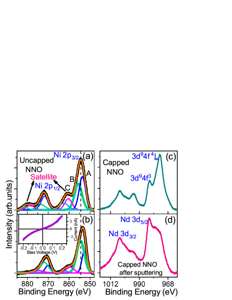
To study the surface structural properties of NNO samples, we performed UHV-STM measurements on uncapped samples. In Fig. 1, we show a large area STM image acquired on the uncapped NNO. Without any surface treatment, stable tunnelling conditions could be achieved only by gap voltages larger than 1 V and tunnelling currents below 1 nA. This result alone suggests that the surface layer is not metallic, as confirmed by local tunnelling spectroscopy showing semiconducting I-V spectra (Fig. 1b). The large area STM image in Fig. 1a) shows hints of step-terraces, in agreement with previous AFM data on similar samples Krieger et al. (2022b). On the other hand, the STM data, taken in constant current mode, show also the presence of elongated islands within each terrace, i.e. the surface is not atomically smooth. To better visualize any possible periodic pattern we show a high resolution topographic image in Fig. 1c, its Fast Fourier Transform (FFT) ( inset of Fig. 1c) and the averaged profiles along (100) and (010) directions of the square-root FFT magnitude (Fig. 1d) . High resolution topography shows locally on top of most of these islands, and in-between them, a periodic pattern along the (001) direction with a spacing between consecutive rows of nm, while along each row (010 direction), ordered atomic lines are unclear, and atomic-resolution 1x1 unit-cells are only locally observed. The FFT shows indeed that the surface lattice does not have a 1x1 long range order, while hints of a 1/3 pattern is also observed, suggesting a link with the HRTEM reports in refs Raji et al. (2023); Parzyck et al. (2024). This result was found in several areas of the sample, and on different uncapped NNO realized using the same method.
To get insight into the electronic properties of infinite-layers nickelates, and the effect of the topotactic process, in Figure 2 we compare core-level Ni 2p and Nd 3d XPS spectra of as-grown perovskite NNO3 (Fig. 2a,d), uncapped (Fig. 2b,e) and capped (Fig. 2c,f) NNO samples. The core-level Ni 2p spectra consist in 2p1/2 ( 871 eV) and 2p3/2 features ( 855 eV), composed by a lower-binding energy main peak and a higher energy satellite. The high energy satellite (labelled C) is due to final states related to fully unscreened 2p5 core-hole. The main peak, on the other hand, is related to final states where the 2p5 core-hole is screened by electrons belonging to neighbour ligand oxygen ions, or by electrons coming from neighbouring clusters van Veenendaal and Sawatzky (1993); van Veenendaal (2006). As a consequence, the 2p3/2 XPS spectra are decomposed in two peaks labelled A (non-local screened final states) and B (local screened final states). The fitting analysis shows that for the reference perovskite NNO3, local and non-local screening channels have similar spectral weights in good agreement with previous reports Preziosi et al. (2017); Fu et al. (2020). Since the perovskite NNO3 is regarded as a negative charge transfer system according to the Zaanen-Sawatzky-Allen scheme, at room temperature the ground state has mainly a 3d8L configuration (L denotes a ligand hole), with nominal Ni valence close to Ni2+ Bisogni et al. (2016). In infinite-layer nickelates, substantial changes in the energy and the spectral weights of A, B and C features were expected, in particular a shift of the main peak towards lower binding energy due to the lower overall oxidation state. However, in uncapped NNO, very surprisingly, the XPS Ni 2p core-level looks very similar to the perovskite (Fig. 2b), with only a few differences in the A/B ratio. This result suggests that, within the probing depth of 1.5 nm, uncapped NNO are barely reduced compared to the perovskite. On the other hand, capped NNO shows a clear shift towards lower binding energy of all features and substantial changes in the relative intensities (Fig. 2c). Additionally, the unscreened satellite peak (C) is characterized by a substantial decrease of spectral weight and shifts toward lower binding energies as-well, which indicates a lowering of Ni valence towards Ni1+ Higashi et al. (2021); Fu et al. (2020). The differences in the surface and interfacial Ni-valence between uncapped and capped nickelates confirm earlier X-ray absorption spectroscopy data Krieger et al. (2022a).
While a full understanding of the core-level Ni 2p spectra is beyond the scope of this work requiring sophisticated theoretical calculations, due to the formal Ni 3d9 ground state configuration of infinite-layer nickelates, it is reasonable to look for analogies between XPS Cu core-level data of undoped cuprates and those of the capped NNO sample. The latter exhibit a valence closer to Ni1+, thus a Ni 3d9 configuration similar to Cu2+ in the CuO2 planes. In cuprates, the features B and A in the 2p3/2 core level XPS have been interpreted as 2p53d10L local screened, and 2p53d10Z non-local screened final states, where Z stands as Zhang-Rice Singlet (ZRS) Taguchi et al. (2005). In the photo-excitation process, ZRS is an energetically favoured bound state between non-local electron screened hole and the pristine hole in neighbour clusters. Multiple cluster calculations are able to catch some general characteristics of the cuprate core-level spectra and in particular the relative intensities of features A and B van Veenendaal and Sawatzky (1994). It turns out that in cuprates the low-energy feature A dominates over feature B as a consequence of the low charge transfer energy, which favours the formation of ZRS bound states. On the other hand, since the charge transfer energy in infinite-layer nickelates is inherently larger, it is unclear if ZRS bound states are effectively stable or energetically favourable. As a matter of fact, the ratio between A and B spectral weights shown in Fig. 2c (bottom panel) in capped NNO is opposite to that of undoped cuprates, i.e. the low binding energy feature A has lower spectral weight compared to feature B. This result was also observed at the bare surface of NNO single crystals in refs. Fu et al. (2020); Higashi et al. (2021)and interpreted as a further indication that NNO is indeed a Hubbard and not a charge transfer insulator.
In order to get more insight into the electronic properties of as-grown capped and uncapped NNO, we performed core-level XPS Nd 3d measurements and compared the results to perovskite NNO3 samples, as shown in Figures 2d-f. The main core-level peaks at 982 and 1004.2 eV correspond to Nd 3d5/2 and 3d3/2 states, respectively, and reflect the Nd3+ oxidation state. Perovskite and uncapped NNO3 are characterized by similar Nd 3d core-level features, slightly shifted to higher binding energy by 0.9 eV in the case of uncapped NNO in good agreement with previous report on NNO single crystals Fu et al. (2020). On the other hand, capped NNO shows a very different Nd 3d5/2 feature, with 3d94f3 final state reduced in comparison to uncapped NNO, and a strongly enhanced 3d94f4L final state. This result shows that few NNO unit cells (1 or 2) at the interface with the STO capping layer are not ideal. There could be several reasons why the upper NiO2/TiO2 interface cannot be intrinsically sharp and ideal. For example, it is known that a NdTiNiO3 intermixed phase is created at the interface between NNO and the STO-single crystal substrate due to the atomic reconstruction that compensates the interfacial polar discontinuity, as demonstrated by combined high-resolution transmission electron microscopy (HR-TEM) and ab-initio calculations Goodge et al. (2023). As a consequence, it is not inconceivable to expect that a similar intermixed phase, maybe just including the interface, is created at the top NNO/STO interface. Furthermore, we do not exclude a possible accumulation of hydrogen at the interface after the topotactic process, which might help in explaining the anomalous Nd3+ core-level spectrum in as-received capped reduced nickelate films.
From the STM and XPS studies of untreated (uncapped and capped) NNO samples it clearly emerges that their surface and interface unit cells do not have an ideal NNO structure and stoichiometry, thus any surface/interface probe of their electronic properties would not be able to provide accurate insights about the differences between cuprates and nickelates. In the following, we suggest possible directions to address this problem.
First, we studied the non-ideal and barely reduced surface of uncapped NNO. To reduce excess oxygen and the amount of adsorbed species due to the ex-situ exposure to the atmosphere from the surface unit cells of uncapped NNO, the films were annealed in situ in UHV at 250 ∘C for 12h, as described above. Figure 3 shows XPS Ni 2p core-level spectra (Fig. 3a before and Fig. 3b after annealing). We do see pronounced changes in the core-level spectra: first, the main core-level features in the annealed sample shift by 0.9 eV towards lower binding energy; second, the spectral weight of the non-local screened feature A becomes (much) lower than the local screened final state B; finally, the intensity of the unscreened satellite peak (C) is strongly suppressed after annealing Fu et al. (2020); Higashi et al. (2021). The results suggest the reduction of the Ni2+ valence of as-grown uncapped NNO surface towards Ni1+ in UHV annealed NNO. At the same time, we find that the surface is characterized by a metallic surface state, as shown by in situ STM local I-V spectroscopy (inset in Fig. 3b). It is worth noting that local superconducting gap features in uncapped Sr-doped NNO were measured in uncapped samples after UHV annealing at a slightly reduced temperature in ref. Gu et al. (2020). However, annealing in UHV, while capable of recovering a metallic surface state and a Ni1+ oxidation state, is unable to change the overall morphology and to create a long-range ordered surface lattice. The irregular morphology on top of the terraces could not be modified substantially by the low temperature UHV annealing process.
We now go back to the capped NNO samples. To investigate the NNO electronic properties in capped NNO samples, and to possibly remove the non-ideal interfacial layer, we used gentle Ar+ ion-beam etching at the Cassiopee beamline, as described in the experimental details section. In Figures 3c,d we show the effect of Ar+ ion etching removal of the STO capping-layer on the Nd 3d spectra, comparing XPS data acquired before (3c) and after (3d) the capping removal. We can see that after etching, the NNO-surface state shows a Nd3+ spectra characterized by a substantial reduction of the prominent 3d94f4L final state feature observed before Ar+ ion etching. This result confirms that some kind of intermixed phase was formed at the NNO/STO interface. At the same time the Ni 2p spectrum (not shown) is essentially not affected by the Ar-ion etching process, i.e. the surface of the NNO sample after capping removal keeps the overall Ni1+ valence state.
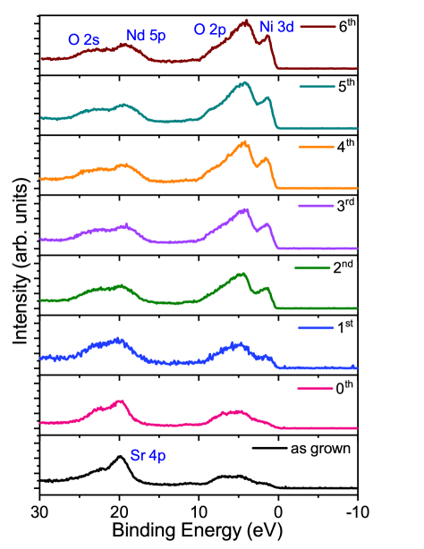
Figure 4 shows the valence band (VB) spectra of the capped NNO sample acquired with an incident photon energy of 600 eV and a resolution of 50 meV, including the VB (EF to -10 eV), core level Nd 5p and Sr 4p features after different cycles of Ar+ sputtering. In the pristine state, the spectrum shows mostly the O 2p VB of the topmost STO, with possible traces of in-gap Ti 3d and Ni 3d features below EF. Moreover, the Sr 4p peak is well visible around 20 eV of binding energy. After a few cycles of Ar+ ion etching, and at the end of the 6th cycle of sputtering, the Sr 4p (as well as all core levels related to Ti and Sr ions, not shown) is gradually suppressed, a Nd 5p feature around 18 eV binding energy appears, and the VB changes substantially. At the end of 6th cycle, the STO capping is removed together with the first intermixed NNO-STO unit cell. At the same time, the O 2p VB changes completely in shape, with a peak located between 6-8 eV, and Ni 3d states crossing the Fermi level show up. The final VB spectra after Ar+ sputtering have similarities with that one of uncapped PrNiO2 measured by soft X-ray photoemission spectroscopy, showing the appearance of Ni 3d states crossing the Fermi-level Chen et al. (2022). On the other hand, the VB peak is more intense than the Ni 3d feature. This can be possibly related to the contribution to the VB of Nd 5d and Nd 4f states together with O 2p states. From the data, we can tentatively estimate the differences in binding energy between O 2p and Ni 3d electronic states (E-E), which is found to be 2.72 eV, matching the values reported for PrNiO2 in ref.Chen et al. (2022). However, the contribution to the VB from states other than O 2p makes this estimation somewhat uncertain. Assuming the value of (E-E) found correct, following ref. Chen et al. (2022) the result suggests a Hubbard energy U lower than the charge transfer energy (U ).
IV Discussion and conclusions
In this work, we investigated the surface/interface electronic properties of uncapped and capped infinite-layer nickelates. The experimental results of our combined STM and photoemission spectroscopy studies show that the surface and interface unit cells of uncapped and capped NNO are not ideal. While uncapped NNO is characterized by an insulating surface state and a Ni-valence remarkably similar to reference NNO3 perovskite, capped NNO shows mostly Ni in Ni1+ configuration. On the other hand, at the interface with the STO capping an intermixed non-ideal layer, presumably of NdTiNiOx composition (and possibly excess hydrogen), is formed. A metallic surface with a Ni1+ valence state was restored via annealing in UHV at 250 ∘C in uncapped NNO, while an Ar+ ion sputtering process was necessary to access the electronic properties of capped NNO samples by removing the STO layers and the interfacial (non-ideal) unit cell. In this way, we could provide an experimental study of the VB of Ni1+ NdNiO2 nickelate and access the near Fermi level electronic properties, giving further support to the classification of these oxides as Hubbard or intermediate between Hubbard and charge transfer insulators.
Our work represents a step towards the goal of creating an ideal NdNiO2 surface state, which could allow deep investigations by surface spectroscopy methods like angle-resolved photoemission. While annealing in UHV and ion-sputtering are not yet sufficient to establish long-range atomically flat ideal NiO2 surfaces on samples prepared by ex-situ CaH2 topotactic reduction of perovskite NdNiO3 samples, their opportune combination and optimization are promising for future studies aimed at preserving not only the stoichiometry but also the long-range surface structural order. These methods combined with the novel in-situ approaches of realization of infinite layer nickelates, like hydrogen thermal cracker technique Parzyck et al. (2024) and metal overlayer deposition Wei et al. (2023a), or the investigation of other epitaxially-grown capping-layers seem promising in the direction to improve the surface quality of infinite-layer nickelates.
Acknowledgements.
This work was supported by the European Union’s Horizon 2022 research and innovation programme under the Marie Sklodowska-Curie project MODERN, grant agreement No. 101108695, and by the Ministry of University and Research project PRIN QTFLUO No.20207ZXT4Z. This work was funded by the French National Research Agency (ANR) through the ANR-JCJC FOXIES ANR-21-CE08-0021. This work was also done as part of the Interdisciplinary Thematic Institute QMat, ITI 2021 2028 program of the University of Strasbourg, CNRS and Inserm, and supported by IdEx Unistra (ANR 10 IDEX 0002), and by SFRI STRAT’US project (ANR 20 SFRI 0012) and EUR QMAT ANR-17-EURE-0024 under the framework of the French Investments for the Future Program. The synchrotron experiments were performed at SOLEIL under proposal number 20220304, and the staff of Cassiopee beamline is acknowledged for their technical support. We acknowledge Prof. G. Ghiringhelli for the discussions on the physics of nickelates.References
- Anisimov et al. (1999) V. I. Anisimov, D. Bukhvalov, and T. M. Rice, Phys. Rev. B 59, 7901 (1999).
- Li et al. (2019) D. Li, K. Lee, B. Y. Wang, M. Osada, S. Crossley, H. R. Lee, Y. Cui, Y. Hikita, and H. Y. Hwang, Nature 572, 624 (2019).
- Parzyck et al. (2024) C. T. Parzyck, N. K. Gupta, Y. Wu, V. Anil, L. Bhatt, M. Bouliane, R. Gong, B. Z. Gregory, A. Luo, R. Sutarto, F. He, Y.-D. Chuang, T. Zhou, G. Herranz, L. F. Kourkoutis, A. Singer, D. G. Schlom, D. G. Hawthorn, and K. M. Shen, Nat. Mater. (2024), 10.1038/s41563-024-01797-0.
- Wei et al. (2023a) W. Wei, K. Shin, H. Hong, Y. Shin, A. S. Thind, Y. Yang, R. F. Klie, F. J. Walker, and C. H. Ahn, Phys. Rev. Mater. 7, 013802 (2023a).
- Wei et al. (2023b) W. Wei, D. Vu, Z. Zhang, F. J. Walker, and C. H. Ahn, Sci. Adv. 9, eadh3327 (2023b).
- Zeng et al. (2022) S. W. Zeng, X. M. Yin, C. J. Li, L. E. Chow, C. S. Tang, K. Han, Z. Huang, Y. Cao, D. Y. Wan, Z. T. Zhang, Z. S. Lim, C. Z. Diao, P. Yang, A. T. S. Wee, S. J. Pennycook, and A. Ariando, Nat. Commun. 13, 743 (2022).
- Raji et al. (2023) A. Raji, G. Krieger, N. Viart, D. Preziosi, J.-P. Rueff, and A. Gloter, Small 19, 2304872 (2023).
- Krieger et al. (2022a) G. Krieger, L. Martinelli, S. Zeng, L. E. Chow, K. Kummer, R. Arpaia, M. Moretti Sala, N. B. Brookes, A. Ariando, N. Viart, M. Salluzzo, G. Ghiringhelli, and D. Preziosi, Phys. Rev. Lett. 129, 027002 (2022a).
- Tam et al. (2022) C. C. Tam, J. Choi, X. Ding, S. Agrestini, A. Nag, M. Wu, B. Huang, H. Luo, P. Gao, M. García-Fernández, L. Qiao, and K.-J. Zhou, Nat. Mater. 21, 1116 (2022).
- Rossi et al. (2022) M. Rossi, M. Osada, J. Choi, S. Agrestini, D. Jost, Y. Lee, H. Lu, B. Y. Wang, K. Lee, A. Nag, Y.-D. Chuang, C.-T. Kuo, S.-J. Lee, B. Moritz, T. P. Devereaux, Z.-X. Shen, J.-S. Lee, K.-J. Zhou, H. Y. Hwang, and W.-S. Lee, Nat. Phys. 18, 869 (2022).
- Hepting et al. (2020) M. Hepting, D. Li, C. J. Jia, H. Lu, E. Paris, Y. Tseng, X. Feng, M. Osada, E. Been, Y. Hikita, Y.-D. Chuang, Z. Hussain, K. J. Zhou, A. Nag, M. Garcia-Fernandez, M. Rossi, H. Y. Huang, D. J. Huang, Z. X. Shen, T. Schmitt, H. Y. Hwang, B. Moritz, J. Zaanen, T. P. Devereaux, and W. S. Lee, Nat. Mater. 19, 381 (2020).
- Karp et al. (2020) J. Karp, A. S. Botana, M. R. Norman, H. Park, M. Zingl, and A. Millis, Phys. Rev. X 10, 021061 (2020).
- Fowlie et al. (2022) J. Fowlie, M. Hadjimichael, M. M. Martins, D. Li, M. Osada, B. Y. Wang, K. Lee, Y. Lee, Z. Salman, T. Prokscha, J.-M. Triscone, H. Y. Hwang, and A. Suter, Nat. Phys. 18, 1043 (2022).
- Preziosi et al. (2017) D. Preziosi, A. Sander, A. Barthélémy, and M. Bibes, AIP Advances 7, 015210 (2017).
- Krieger et al. (2022b) G. Krieger, A. Raji, L. Schlur, G. Versini, C. Bouillet, M. Lenertz, J. Robert, A. Gloter, N. Viart, and D. Preziosi, J. Phys. D: Appl. Phys. 56, 024003 (2022b).
- Fairley et al. (2021) N. Fairley, V. Fernandez, M. Richard‐Plouet, C. Guillot-Deudon, J. Walton, E. Smith, D. Flahaut, M. Greiner, M. Biesinger, S. Tougaard, D. Morgan, and J. Baltrusaitis, Appl. Surf. Sci. Adv. 5, 100112 (2021).
- van Veenendaal and Sawatzky (1993) M. A. van Veenendaal and G. A. Sawatzky, Phys. Rev. Lett. 70, 2459 (1993).
- van Veenendaal (2006) M. van Veenendaal, Phys. Rev. B 74, 085118 (2006).
- Fu et al. (2020) Y. Fu, L. Wang, H. Cheng, S. Pei, X. Zhou, J. Chen, S. Wang, R. Zhao, W. Jiang, C. Liu, M. Huang, X. Wang, Y. Zhao, D. Yu, F. Ye, S. Wang, and J.-W. Mei, (2020), arXiv:1911.03177 [cond-mat.supr-con] .
- Bisogni et al. (2016) V. Bisogni, S. Catalano, R. J. Green, M. Gibert, R. Scherwitzl, Y. Huang, V. N. Strocov, P. Zubko, S. Balandeh, J.-M. Triscone, G. Sawatzky, and T. Schmitt, Nat. Commun. 7, 13017 (2016).
- Higashi et al. (2021) K. Higashi, M. Winder, J. Kuneš, and A. Hariki, Phys. Rev. X 11, 041009 (2021).
- Taguchi et al. (2005) M. Taguchi, A. Chainani, K. Horiba, Y. Takata, M. Yabashi, K. Tamasaku, Y. Nishino, D. Miwa, T. Ishikawa, T. Takeuchi, K. Yamamoto, M. Matsunami, S. Shin, T. Yokoya, E. Ikenaga, K. Kobayashi, T. Mochiku, K. Hirata, J. Hori, K. Ishii, F. Nakamura, and T. Suzuki, Phys. Rev. Lett. 95, 177002 (2005).
- van Veenendaal and Sawatzky (1994) M. A. van Veenendaal and G. A. Sawatzky, Phys. Rev. B 49, 3473 (1994).
- Goodge et al. (2023) B. H. Goodge, B. Geisler, K. Lee, M. Osada, B. Y. Wang, D. Li, H. Y. Hwang, R. Pentcheva, and L. F. Kourkoutis, Nat. Mater. 22, 466 (2023).
- Gu et al. (2020) Q. Gu, Y. Li, S. Wan, H. Li, W. Guo, H. Yang, Q. Li, X. Zhu, X. Pan, Y. Nie, and H.-H. Wen, Nat. Commun. 11, 6027 (2020).
- Chen et al. (2022) Z. Chen, M. Osada, D. Li, E. M. Been, S.-D. Chen, M. Hashimoto, D. Lu, S.-K. Mo, K. Lee, B. Y. Wang, F. Rodolakis, J. L. McChesney, C. Jia, B. Moritz, T. P. Devereaux, H. Y. Hwang, and Z.-X. Shen, Matter 5, 1806 (2022).