SpatialVisVR: An Immersive, Multiplexed Medical Image Viewer With Contextual Similar-Patient Search
Abstract.
In modern pathology, multiplexed immunofluorescence (mIF) and multiplex immunohistochemistry (mIHC) bring both vast opportunities and challenges. These techniques illuminate complex tumor microenvironment interactions, necessitating intuitive visualization tools. With the rise of electronic health records (EHR) and information overload for physicians, integrating advanced technologies becomes essential. Enter SpatialVisVR: a versatile VR platform for comparing medical images, adaptable for data privacy on embedded hardware. Clinicians can capture pathology slides in real-time via mobile, then SpatialVisVR employs a deep learning algorithm to match and display similar mIF images. This interface allows for adding or removing up to 100 multiplexed protein channels, aiding immuno-oncology decisions. Ultimately, SpatialVisVR aims to refine diagnostic processes, promoting a holistic, efficient approach to immuno-oncology research and treatment.
1. Introduction
Multiplexed immunofluorescence (mIF) and multiplex immunohistochemistry (mIHC) are revolutionizing the realm of pathology, especially in the field of immuno-oncology, by providing proteomic measurements spatial context in whole slide images (Harms et al., 2023). They offer unparalleled insights into the tumor microenvironment and various immune populations (Phillips et al., 2021). Notably, our SpatialVisVR platform focuses on visualizing CODEX images, a state of the art multiplexed tissue imaging technology.(Goltsev et al., 2018). Yet, these advanced mIF and mIHC images, showcasing about 60 markers, pose unique visualization hurdles (Black et al., 2021). With approximately 20,000 stainable proteins and each laboratory opting for different markers, standardized visualization becomes challenging. Moreover, most multiplex viewers available today are restricted to desktop or web platforms and typically involve subscription fees (Hoffer et al., 2020; Manz et al., 2022; Stefani et al., 2018).
Meanwhile, virtual reality (VR) is emerging as a transformative tool for data visualization. In the biomedical arena, applications such as visualizing nucleotide sequences and protein structure through BioVR are gaining traction (Zhang et al., 2019).
In the context of mIF’s increasing use, the dearth of publicly available mIF data becomes evident, especially when compared to the vast archive of traditional H&E stained images. Addressing this disparity, our app, SpatialVisVR, developed in Unity, implements multimodal search to visualize similar mIF images (compared to more standard, inputted H&E slides) that have useful proteomic context for tasks such as selecting cancer immunotherapy treatments. Through a mobile device, one can capture an H&E slide, subsequently initiating a segmentation to differentiate tissue from the backdrop. The tool then cross-references with its database, pinpointing analogous tissues, thus allowing juxtaposition with mIF-detailed tissues of similar cancer categories. As we advance our tool, we’re working towards ensuring its compatibility with lightweight platforms, especially the Jetson Nano. Our aspiration is to offer resource-efficient in-house imaging suitable even for clinics with restricted resources while emphasizing data confidentiality and minimizing transfer risks.
Driven by these trends, our research synergizes VR and mIF to redefine diagnostic paradigms and fortify immuno-oncology research, with a focus on comprehensive quantitative and spatial image examination. In alignment with our mission, we intend to incorporate enhanced quantitative diagnostic tools and delve deeper into cell segmentation or interactions within the cellular environment for more profound insights.
Key contributions of our endeavor include:
-
•
Pioneering VR and mIF Fusion with SpatialVisVR: To our knowledge, SpatialVisVR is the first VR app geared towards this specialized task, crafting a sophisticated interface for in-depth multiplexed image exploration.
-
•
On-the-Go Mobile Imaging with SpatialVisVR: Addressing the need for a bridge between traditional H&E images and mIF data, our software empowers users to effortlessly capture, segment, and cross-reference H&E slides. Weighing only 53 MB, it serves dual roles as both a multiplexed image viewer and an image search engine.
-
•
Striving Towards an Efficient Pipeline: In our journey towards fully embracing lightweight platforms, we’re taking strides with the Jetson Nano. Our vision is to optimize in-house imaging for all clinics, particularly those with restricted resources, without compromising patient data privacy.
Following this introduction, Section 2 discusses related works, Section 3 details our methodologies, Section 4 concludes the paper, and Section 5 discusses the future direction of this work.
2. Current State of the Field
In the burgeoning field of medical research, emerging technologies like Virtual Reality (VR) and Augmented Reality (AR) are gaining prominence due to their potential in revolutionizing clinical practice and medical education. Our ACM TEI WIP sits at the intersection of multiplexed imaging, machine learning in pathology, and the immersive capabilities of VR and AR, highlighting the advancements and challenges in these interconnected domains.
Histopathology has traditionally relied on H&E stained imaging for cellular and tissue structure visualization (Bejnordi et al., 2015). However, modern techniques like CODEX have driven a paradigm shift, enabling visualization of over 60 protein markers within a single tissue site, thus enhancing cellular-level understanding and facilitating sophisticated data amalgamation in complex fields like oncology (Black et al., 2021; Boehm et al., 2022; Cheerla and Gevaert, 2019).
The complexity of navigating through advanced imaging data, particularly Whole-Slide Imaging (WSIs), which are the ”low tech” workhorse of pathology and are still up to 50k by 50k pixels in size, poses a unique challenge (Yagi et al., 2012). Emerging solutions like Mistic, Viv, and Minerva are addressing this by offering intuitive navigation and efficient data access for multiplexed imaging, leveraging client-side GPU rendering and cloud-supported guided analyses (Manz et al., 2022; Hoffer et al., 2020).
VR and AR are at the forefront of modern medical imaging, with applications ranging from virtual surgeries to diagnostic advancements (Javaid and Haleem, 2020; Farahani et al., 2016). Innovations like ConfocalVR and ExMicroVR exemplify VR’s potential in rendering cellular structures, while AR continues to evolve in therapeutic and educational domains despite challenges in achieving optimal visual fidelity (Stefani et al., 2018; Cheng et al., 2023; Innocente et al., 2022).
The integration of traditional histopathology, advanced multiplexed imaging, and immersive tools like VR and AR heralds a new era in pathology imaging. Platforms like Minerva and Avivator are leading the way in rendering multiplexed visuals with unparalleled detail, envisioning a future where VR and AR significantly enhance user experience in exploring both traditional and multiplexed images (Hoffer et al., 2020; Keller et al., 2021).
3. Methods
This section outlines the utilized methodologies, including object detection, hardware setups, and Multimodal Pathology Image Retrieval (MPIR) integration for enhanced pathology data analysis and visualization, linking computational techniques with tangible user interactions.
3.1. SpatialVisVR App
3.1.1. VR Application:
VR was utilized for visualizing CODEX images as intricate 3D channel stacks. Flask, implemented server-side, cached user-selected CODEX images in RAM for faster data retrieval. APIs were created to return channel names and nth channel of specified CODEX images, enabling dynamic rendering in VR. Interactive coloring selection was facilitated via APIs, where user-selected channels were processed server-side based on color and threshold parameters before being sent to VR for 3D arrangement and visualization, enhanced by Unity shaders for better clarity and color vibrancy.
3.1.2. GUI Overview:
SpatialVisVR, designed for pathologists, focuses on a seamless user experience utilizing spatially multiplexed data to improve diagnostic workflow, as showcased in Figure 4. The interface comprises three tailored windows serving distinct purposes in the diagnostic workflow.
The left window visualizes H&E slides from edge detection of real-time captures, with an ’Update’ button to initiate search in the third window and a primary viewing area for real-time image processing.
The central window, the primary viewer, facilitates exploration of CODEX ome.tiff images, allowing up to seven channel views at once for understanding cellular interactions. Essential UI elements include a sliding carousel for channel selection, a dropdown for accessing stored CODEX images, and a navigation bar for channel management, each channel color-coded for differentiation.
The right window visualizes search results for similar CODEX images to an H&E slide, resembling the second window but displaying the five most similar CODEX images, aiding in further exploration.
Overall, the multi-window setup in SpatialVisVR enhances pathologists’ workflow and diagnostic decision-making by providing diverse data examination, critical insights, and a more efficient diagnostic process. An overview of of the interface depicted in the LABEL:frames.
3.1.3. API Implementation
We deployed a server-client model using RESTful APIs (Neumann et al., 2021) for communication between server-side application and VR. A Flask application was set up on the server end, and Unity was used for processing API call data, with shaders enhancing data visualization. To address VR headset limitations, resource-intensive tasks were shifted to the server, improving user experience. The server, equipped with a 4TB SSD, hosted CODEX datasets, with data transfer rate limited to 200 MB for better VR responsiveness.
3.2. ML Backbones
3.2.1. Object Detection Implementation
The object detection employed a ResNet-50 backbone using PyTorch, with the classification layer replaced by a segmentation head comprising a Conv2D layer, ReLU layer, and ConvTranspose layer. This setup processed the output of ResNet-50 through a 3x3 convolution, and scaled the image to a 2x640x640 output via ConvTranspose2D, distinguishing between pathology slide and non-pathology slide pixels. The architecture is presented in the LABEL:seg.
For training, image preprocessing entailed Resize, RandomHorizontalFlip, RandomInvert, RandomRotation, GaussianBlur, and ColorJitter transforms to enhance model robustness. Prediction preprocessing included image resizing to 640x640, padding, and subsequent resizing to address bounding area inconsistencies.
Performance varied with photo setup aspects like lighting, angle, and focus. Consistent lighting yielded better results, while shadows or inconsistent lighting led to suboptimal outputs. Optimal results were observed at 90-degree angles, and acceptable outcomes at shallow angles. However, steep angles or out-of-focus photos resulted in inconsistent detection and bounding, affecting overall output quality.
3.2.2. MPIR Implementation
We employed Multimodal Pathology Image Retrieval (MPIR) for retrieving similar codex images to a given H&E image slide(Hajighasemi et al., 2023). Utilizing a Variational Autoencoder (VAE), images were encoded into lower-dimension latent vectors. The VAE, comprising an encoder and decoder, maps input data to a probabilistic latent space and generates data samples from these latent variables, aiding in dimensionality reduction and data reconstruction.
Initially, VAE was trained on H&E images, followed by separate training on seven specific channels of codex images, chosen due to their relevance in indicating tumor and immune cell interactions. Preprocessing involved selecting useful patches from high-resolution images for training. During testing, latent vectors were obtained from codex and H&E image patches, with dynamic time warping algorithm(Sakoe and Chiba, 1978) used for batch effect correction. Subsequently, cosine similarity and search algorithm identified top 5 most similar patches of codex slides for each H&E image patch. Results across different channels were consolidated using rank choice voting algorithms to retrieve the most similar codex slides for the H&E input. This algorithm was also applied to camera-captured H&E images post edge detection processing.
To test the MPIR model, two H&E image inputs were used: the actual image and the edge detection algorithm result. For the actual image, the top five similar codex slides were obtained, and a different set was identified for the edge-detected image, with each slide having a voting number representing its similarity rank.
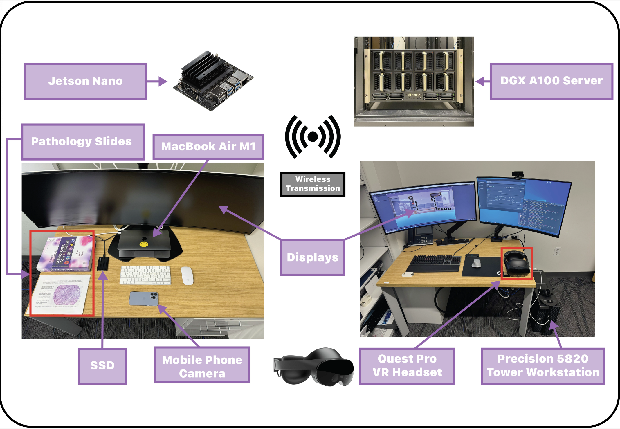
3.3. Hardware Setup
Various hardware components were utilized for this project. iOS and Android smartphones captured H&E slide images in real-time, functioning as IP Webcams over a Local Area Network (LAN) with the OpenCV Python library facilitating video stream interfacing due to its GStreamer integration and object detection capabilities.
For object detection model testing, a Jetson Nano 2GB Developer Kit was used for our representative segmentation model, ensuring real-time performance, while an Apple 13-inch MacBook Air with an M1 chip and 16GB RAM was employed for deployment. Post object detection, a DGX Server with eight NVIDIA A100 GPUs was used for MPIR algorithm testing and training, with the execution phase shifted to the MacBook Air.
Each time inference is run with our model, the top 5 candidates identified by the MPIR’s ranked choice voting algorithm are transmitted to a Meta Quest Pro headset via a Unity Development Environment on a Dell Precision 5820 Tower Workstation for final visualization by the physicians on our team, who gave feedback about user interface design. For the non work-in-progress future of this work, we aim to have the pipeline fully deployed only to Jetson Nano, ARM Cortex, and any VR headset. The LABEL:hardware represents an overview of different hardware that has been used in this research.
3.4. Data Used
VAE model was trained using H&E and mIF images from CODEX technology, comprising colorectal cancer samples from 35 patients with over 50 protein markers, imaged using a Keyence BZ-X710 microscope (Schürch et al., 2020). 109 stitched CODEX images from NIH HuBMAP, representing various organs, were visualized in VR (Snyder et al., [n. d.]). For object detection, a dataset of around 500 H&E slides from three universities and 195 from The Cancer Genome Atlas for breast cancer was used, digitized with Aperio or Ventana scanners (Cruz-Roa et al., 2018).
4. Conclusion
Traditional pathology primarily relies on immunohistochemistry staining for diagnostic or prognostic evaluations. The advent of multiplex immunohistochemistry staining has enriched insights on spatial cellular interactions, especially in oncology by elucidating tumor microenvironments (TME) (Black et al., 2021)(Allam et al., 2022)(Schürch et al., 2020). However, the 2D nature of multiplexed images constrains the spatial exploration to x and y axes. Recent attempts towards 3D reconstructions of multiplexed images will further the spatial understanding of disease processes, yet the 2D interfaces for visualization remain a challenge (Lin et al., 2023).
Our VR platform addresses this by enabling interactive visualization of multiplexed immunohistochemistry stains that is contextual to the environment of existing H&E slides, facilitating an intuitive exploration for researchers and clinicians. This capability enhances the analysis of spatial features in medical imaging, and provides a more interactive environment for navigating whole slide images in the context of clinically richer multiplexed immunofluorescent ones, aiding in better diagnostic and prognostic evaluations. Furthermore, our platform facilitates slide comparison and retrieval, directing pathologists towards potential diagnoses or treatment responses based on what other cliniciancs have observed in similar patients.
5. Future Directions
We aim to enhance the resolution of multiplexed slides in VR to display distinct cellular interactions. This advancement could enable manual manipulation of cellular interactions and prediction of downstream effects within VR.
Integration of features for swift extraction of diagnostic or prognostic information is also envisioned. For instance, facilitating quick calculations of HER2 expression in 3D to ascertain candidacy for HER2-directed therapies. We plan to broaden slide comparison and retrieval capabilities by expanding our spatial atlas of multiplex slides with contributions from end-users.
Transitioning towards exploring entire tumors in 3D, beyond individual multiplexed slides, will allow easy visualization of protein expression heterogeneity like HER2 or PDL1 in VR. This could uncover missed treatment opportunities from single biopsy slides, potentially impacting treatment avenues for millions globally.
6. Supplemental Video
A demonstrative video by the authors is available in the supplemental material section summarizing the method’s potential benefits for physicians.
7. Code and Data Availability
An anonymous version of our code sufficient to recreate our entire pipeline is available at:
https://anonymous.4open.science/r/SpatialVisVR-2023/readme.md. All imaging used is available from the NIH HubMAP consortium.
References
- (1)
- Allam et al. (2022) Mayar Allam, Thomas Hu, Jeongjin Lee, Jeffrey Aldrich, Sunil S Badve, Yesim Gökmen-Polar, Manali Bhave, Suresh S Ramalingam, Frank Schneider, and Ahmet F Coskun. 2022. Spatially variant immune infiltration scoring in human cancer tissues. NPJ Precision Oncology 6, 1 (2022), 60.
- Bejnordi et al. (2015) Babak Ehteshami Bejnordi, Geert Litjens, Nadya Timofeeva, Irene Otte-Höller, André Homeyer, Nico Karssemeijer, and Jeroen AWM Van Der Laak. 2015. Stain specific standardization of whole-slide histopathological images. IEEE transactions on medical imaging 35, 2 (2015), 404–415.
- Black et al. (2021) Sarah Black, Darci Phillips, John W Hickey, Julia Kennedy-Darling, Vishal G Venkataraaman, Nikolay Samusik, Yury Goltsev, Christian M Schürch, and Garry P Nolan. 2021. CODEX multiplexed tissue imaging with DNA-conjugated antibodies. Nature protocols 16, 8 (2021), 3802–3835.
- Boehm et al. (2022) Kevin M Boehm, Pegah Khosravi, Rami Vanguri, Jianjiong Gao, and Sohrab P Shah. 2022. Harnessing multimodal data integration to advance precision oncology. Nature Reviews Cancer 22, 2 (2022), 114–126.
- Cheerla and Gevaert (2019) Anika Cheerla and Olivier Gevaert. 2019. Deep learning with multimodal representation for pancancer prognosis prediction. Bioinformatics 35, 14 (2019), i446–i454.
- Cheng et al. (2023) Zhangyu Cheng, Caroline Stefani, Thomas Skillman, Aleksandra Klimas, Aramchan Lee, Emma F DiBernardo, Karina Mueller Brown, Tatyana Milman, Yuhong Wang, Brendan R Gallagher, et al. 2023. MicroMagnify: A Multiplexed Expansion Microscopy Method for Pathogens and Infected Tissues. Advanced Science (2023), 2302249.
- Cruz-Roa et al. (2018) Angel Cruz-Roa, Hannah Gilmore, Ajay Basavanhally, Michael Feldman, Shridar Ganesan, Natalie Shih, John Tomaszewski, Anant Madabhushi, and Fabio González. 2018. High-throughput adaptive sampling for whole-slide histopathology image analysis (HASHI) via convolutional neural networks: Application to invasive breast cancer detection. PloS one 13, 5 (2018), e0196828.
- Farahani et al. (2016) Navid Farahani, Robert Post, Jon Duboy, Ishtiaque Ahmed, Brian J Kolowitz, Teppituk Krinchai, Sara E Monaco, Jeffrey L Fine, Douglas J Hartman, and Liron Pantanowitz. 2016. Exploring virtual reality technology and the Oculus Rift for the examination of digital pathology slides. Journal of pathology informatics 7, 1 (2016), 22.
- Goltsev et al. (2018) Yury Goltsev, Nikolay Samusik, Julia Kennedy-Darling, Salil Bhate, Matthew Hale, Gustavo Vazquez, Sarah Black, and Garry P Nolan. 2018. Deep profiling of mouse splenic architecture with CODEX multiplexed imaging. Cell 174, 4 (2018), 968–981.
- Hajighasemi et al. (2023) Amir Hajighasemi, MD Saurav, Mohammad S Nasr, Jai Prakash Veerla, Aarti Darji, Parisa Boodaghi Malidarreh, Michael Robben, Helen H Shang, and Jacob M Luber. 2023. Multimodal Pathology Image Search Between H&E Slides and Multiplexed Immunofluorescent Images. arXiv preprint arXiv:2306.06780 (2023).
- Harms et al. (2023) Paul W Harms, Timothy L Frankel, Myrto Moutafi, Arvind Rao, David L Rimm, Janis M Taube, Dafydd Thomas, May P Chan, and Liron Pantanowitz. 2023. Multiplex immunohistochemistry and immunofluorescence: a practical update for pathologists. Modern Pathology 36, 7 (2023), 100197.
- Hoffer et al. (2020) John Hoffer, Rumana Rashid, Jeremy L Muhlich, Yu-An Chen, Douglas Peter William Russell, Juha Ruokonen, Robert Krueger, Hanspeter Pfister, Sandro Santagata, and Peter K Sorger. 2020. Minerva: a light-weight, narrative image browser for multiplexed tissue images. Journal of open source software 5, 54 (2020).
- Innocente et al. (2022) Chiara Innocente, Luca Ulrich, Sandro Moos, and Enrico Vezzetti. 2022. Augmented Reality: Mapping Methods and Tools for Enhancing the Human Role in Healthcare HMI. Applied Sciences 12 (04 2022), 4295. https://doi.org/10.3390/app12094295
- Javaid and Haleem (2020) Mohd Javaid and Abid Haleem. 2020. Virtual reality applications toward medical field. Clinical Epidemiology and Global Health 8, 2 (2020), 600–605.
- Keller et al. (2021) Mark S Keller, Ilan Gold, Chuck McCallum, Trevor Manz, Peter V Kharchenko, and Nils Gehlenborg. 2021. Vitessce: a framework for integrative visualization of multi-modal and spatially-resolved single-cell data. (2021).
- Lin et al. (2023) Jia-Ren Lin, Shu Wang, Shannon Coy, Yu-An Chen, Clarence Yapp, Madison Tyler, Maulik K Nariya, Cody N Heiser, Ken S Lau, Sandro Santagata, et al. 2023. Multiplexed 3D atlas of state transitions and immune interaction in colorectal cancer. Cell 186, 2 (2023), 363–381.
- Manz et al. (2022) Trevor Manz, Ilan Gold, Nathan Heath Patterson, Chuck McCallum, Mark S Keller, Bruce W Herr, Katy Börner, Jeffrey M Spraggins, and Nils Gehlenborg. 2022. Viv: multiscale visualization of high-resolution multiplexed bioimaging data on the web. Nature Methods 19, 5 (2022), 515–516.
- Neumann et al. (2021) Andy Neumann, Nuno Laranjeiro, and Jorge Bernardino. 2021. An Analysis of Public REST Web Service APIs. IEEE Transactions on Services Computing 14, 4 (2021), 957–970. https://doi.org/10.1109/TSC.2018.2847344
- Phillips et al. (2021) Darci Phillips, Christian M Schürch, Michael S Khodadoust, Youn H Kim, Garry P Nolan, and Sizun Jiang. 2021. Highly multiplexed phenotyping of immunoregulatory proteins in the tumor microenvironment by CODEX tissue imaging. Frontiers in Immunology 12 (2021), 687673.
- Sakoe and Chiba (1978) H. Sakoe and S. Chiba. 1978. Dynamic programming algorithm optimization for spoken word recognition. IEEE Transactions on Acoustics, Speech, and Signal Processing 26, 1 (1978), 43–49. https://doi.org/10.1109/TASSP.1978.1163055
- Schürch et al. (2020) Christian M Schürch, Salil S Bhate, Graham L Barlow, Darci J Phillips, Luca Noti, Inti Zlobec, Pauline Chu, Sarah Black, Janos Demeter, David R McIlwain, et al. 2020. Coordinated cellular neighborhoods orchestrate antitumoral immunity at the colorectal cancer invasive front. Cell 182, 5 (2020), 1341–1359.
- Snyder et al. ([n. d.]) Michael P. Snyder, Shin Lin, Amanda Posgai, Mark Atkinson, Aviv Regev, Jennifer Rood, Orit Rozenblatt-Rosen, Leslie Gaffney, Anna Hupalowska, Rahul Satija, Nils Gehlenborg, Jay Shendure, Julia Laskin, Pehr Harbury, Nicholas A. Nystrom, Jonathan C. Silverstein, Ziv Bar-Joseph, Kun Zhang, Katy Börner, Yiing Lin, Richard Conroy, Dena Procaccini, Ananda L. Roy, Ajay Pillai, Marishka Brown, Zorina S. Galis, Long Cai, Jay Shendure, Cole Trapnell, Shin Lin, Dana Jackson, Michael P. Snyder, Garry Nolan, William James Greenleaf, Yiing Lin, Sylvia Plevritis, Sara Ahadi, Stephanie A. Nevins, Hayan Lee, Christian Martijn Schuerch, Sarah Black, Vishal Gautham Venkataraaman, Ed Esplin, Aaron Horning, Amir Bahmani, Kun Zhang, Xin Sun, Sanjay Jain, James Hagood, Gloria Pryhuber, Peter Kharchenko, Mark Atkinson, Bernd Bodenmiller, Todd Brusko, Michael Clare-Salzler, Harry Nick, Kevin Otto, Amanda Posgai, Clive Wasserfall, Marda Jorgensen, Maigan Brusko, Sergio Maffioletti, Richard M. Caprioli, Jeffrey M. Spraggins, Danielle Gutierrez, Nathan Heath Patterson, Elizabeth K. Neumann, Raymond Harris, Mark deCaestecker, Agnes B. Fogo, Raf van de Plas, Ken Lau, Long Cai, Guo-Cheng Yuan, Qian Zhu, Ruben Dries, Peng Yin, Sinem K. Saka, Jocelyn Y. Kishi, Yu Wang, Isabel Goldaracena, Julia Laskin, DongHye Ye, Kristin E. Burnum-Johnson, Paul D. Piehowski, Charles Ansong, Ying Zhu, Pehr Harbury, Tushar Desai, Jay Mulye, Peter Chou, Monica Nagendran, Ziv Bar-Joseph, Sarah A. Teichmann, Benedict Paten, Robert F. Murphy, Jian Ma, Vladimir Yu. Kiselev, Carl Kingsford, Allyson Ricarte, Maria Keays, Sushma A. Akoju, Matthew Ruffalo, Nils Gehlenborg, Peter Kharchenko, Margaret Vella, Chuck McCallum, Katy Börner, Leonard E. Cross, Samuel H. Friedman, Randy Heiland, Bruce Herr, Paul Macklin, Ellen M. Quardokus, Lisel Record, James P. Sluka, Griffin M. Weber, Nicholas A. Nystrom, Jonathan C. Silverstein, Philip D. Blood, Alexander J. Ropelewski, William E. Shirey, Robin M. Scibek, Paula Mabee, W. Christopher Lenhardt, Kimberly Robasky, Stavros Michailidis, Rahul Satija, John Marioni, Aviv Regev, Andrew Butler, Tim Stuart, Eyal Fisher, Shila Ghazanfar, Jennifer Rood, Leslie Gaffney, Gokcen Eraslan, Tommaso Biancalani, Eeshit D. Vaishnav, Richard Conroy, Dena Procaccini, Ananda Roy, Ajay Pillai, Marishka Brown, Zorina Galis, Pothur Srinivas, Aaron Pawlyk, Salvatore Sechi, Elizabeth Wilder, and James Anderson. [n. d.]. The human body at cellular resolution: the NIH Human Biomolecular Atlas Program. 574, 7777 ([n. d.]), 187–192. https://doi.org/10.1038/s41586-019-1629-x Number: 7777 Publisher: Nature Publishing Group.
- Stefani et al. (2018) Caroline Stefani, Adam Lacy-Hulbert, and Thomas Skillman. 2018. ConfocalVR: immersive visualization for confocal microscopy. Journal of molecular biology 430, 21 (2018), 4028–4035.
- Yagi et al. (2012) Yukako Yagi, Shigeatsu Yoshioka, Hiroshi Kyusojin, Maristela Onozato, Yoichi Mizutani, Kiyoshi Osato, Hiroaki Yada, Eugene J Mark, Matthew P Frosch, and David N Louis. 2012. An ultra-high speed whole slide image viewing system. Analytical Cellular Pathology 35, 1 (2012), 65–73.
- Zhang et al. (2019) Jimmy F Zhang, Alex R Paciorkowski, Paul A Craig, and Feng Cui. 2019. BioVR: a platform for virtual reality assisted biological data integration and visualization. BMC bioinformatics 20 (2019), 1–10. https://doi.org/10.1186/s12859-019-2666-z