PneumoLLM: Harnessing the Power of Large Language Model for Pneumoconiosis Diagnosis
Abstract
The conventional pretraining-and-finetuning paradigm, while effective for common diseases with ample data, faces challenges in diagnosing data-scarce occupational diseases like pneumoconiosis. Recently, large language models (LLMs) have exhibits unprecedented ability when conducting multiple tasks in dialogue, bringing opportunities to diagnosis. A common strategy might involve using adapter layers for vision-language alignment and diagnosis in a dialogic manner. Yet, this approach often requires optimization of extensive learnable parameters in the text branch and the dialogue head, potentially diminishing the LLMs’ efficacy, especially with limited training data. In our work, we innovate by eliminating the text branch and substituting the dialogue head with a classification head. This approach presents a more effective method for harnessing LLMs in diagnosis with fewer learnable parameters. Furthermore, to balance the retention of detailed image information with progression towards accurate diagnosis, we introduce the contextual multi-token engine. This engine is specialized in adaptively generating diagnostic tokens. Additionally, we propose the information emitter module, which unidirectionally emits information from image tokens to diagnosis tokens. Comprehensive experiments validate the superiority of our methods and the effectiveness of proposed modules. Our codes can be found at https://github.com/CodeMonsterPHD/PneumoLLM/tree/main.
keywords:
\KWDLarge language model, Medical image diagnosis, Foundational model1 Introduction
In the computer-aided diagnosis community, the processing and analysis prowess applied to medical data is pivotal. It facilitates the diagnosis of potential diseases and the prediction of future clinical outcomes. With the rapid evolution of deep learning theories, researchers have designed sophisticated network architectures [15, 11] and have curated extensive, high-quality datasets [7, 56] to pretrain these powerful networks. Pretraining strategies endow networks with valuable knowledge by optimizing weight distribution, which, in turn, equips researchers to further refine the model with labeled data targeted at diagnosing specific diseases. When the data is abundant and accurately labeled, this classical paradigm typically achieves commendable results, particularly with common ailments. An example is EchoNet-Dynamic [43], which has surpassed medical specialists in cardiac function assessment.
However, the landscape shifts when we delve into occupational diseases such as pneumoconiosis [31, 10]. Individuals subjected to prolonged exposure in dust-laden environments—be it at construction sites or within the coal mines—without personal protective equipments are susceptible to pulmonary fibrosis, a precursor to pneumoconiosis [45, 8]. Areas with increased prevalence of pneumoconiosis often lack economical development, medical resources and infrastructure, and prefessional medical practitioners. Furthermore, there is a noticeable reticence towards disease screening and diagnosis, leading to an acute shortfall in clinical data for these diseases [50, 19]. This paucity of data renders the conventional pretraining-and-finetuning strategy ineffective.
Addressing the diagnostic challenges posed by data-scarce occupational diseases requires an inventive approach. It involves tapping into the rich knowledge wells [20] of foundational models and controlling the amount of learnable parameters to streamline the learning process. The advent of large language models (LLMs) [25, 2] has gained a bounty of knowledge from voluminous pretraining corpora, showcasing an impressive generalization capability for new tasks. The medical image diagnosis community has witnessed the emergence of its foundational models [68], with significant strides made in pathology image analysis [69, 22] and medical image segmentation [5].
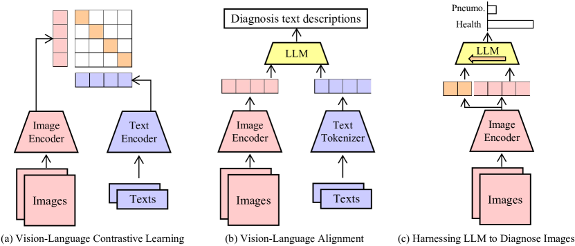
In the wake of such LLMs breakthroughs, concerted efforts continue to leverage knowledge from large-scale models to enhance image processing tasks, as depicted in Fig. 1. For instance, CLIP [47] embarks on vision-language contrastive learning to carefully align visual and language representations, as showcased in Fig. 1(a). The multimodal community, in turn, benefits from the integration of LLMs by interpreting visual tokens as a specialized form of language and devising adapters [29, 67] to convert visual inputs into comprehensible representations, as shown in Fig. 1(b). These works often employ advanced techniques, such as instruction tuning [49], to yield fluent and varied narrative outputs. The medical image diagnosis sector has also made notable advances by developing vision-language models [59, 65] or by constructing medical foundational models from scratch [39].
Nonetheless, the application of these existing vision-language alignment strategies to diagnose data-scarce occupational diseases poses several challenges. Firstly, the dependency on ample paired image-text data intensifies the complexity of data gathering, particularly when factoring in the constraints of patient privacy. Besides, processing text inputs through separate branches escalates computational demands substantially. Although the textual outputs are versatile, they may be unnecessarily complex for tasks that require simple binary outcomes, such as affirming the presence or absence of a specific disease.
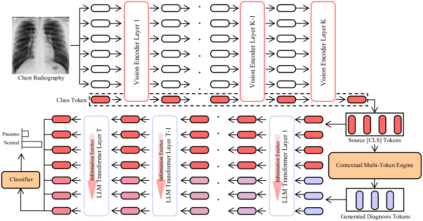
Our approach diverges from existing methodologies by eliminating the textual processing branch and directly harnessing LLMs to process images for the diagnosis of pneumoconiosis, as shown in Fig. 1(c). We hypothesize that LLMs, after extensive corpus learning, are adept at selecting salient visual tokens and filtering out irrelevant ones, thereby benefiting the medical image diagnostic process. Fig. 2 presents the proposed PneumoLLM framework. We revise the language prediction head into a classification head, transitioning from dialogue-based outputs to succinct disease classification. After freezing parameters of both the visual encoder and the LLM, we integrate the adapter module and manage the learnable parameters effectively. We ascertain that eliciting diagnostic knowledge from LLMs hinges on balancing the preservation of comprehensive image details with the progression towards specific diagnostic task. To navigate this balance, we introduce the contextual multi-token engine that generates diagnostic tokens conditioned on image tokens. This ensures that the source image tokens retain all the pertinent image details. Subsequently, the information emitter module is engineered to unidirectionally emit information from source tokens to diagnosis tokens, thus steering the learning trajectory towards accurate diagnosis.
In brief, this work contributes to the field by:
-
1.
Charting new paradigm in applying LLMs to medical image analysis, especially for data-scarce occupational diseases, thereby simplifying the diagnostic process while yielding promising results.
-
2.
Designing the novel contextual multi-token engine and information emitter module to meticulously draw out knowledge from LLMs, achieving a harmonious balance between preserving image representations and harnessing LLMs diagnostic intelligence.
-
3.
Demonstrating the superiority of our method in diagnosing pneumoconiosis through extensive experiments and validating the effectiveness of each designed module.
2 Related Work
2.1 Disease diagnosis based on X-ray
Recent advancements in deep learning have shown significant promise in the field of medical image diagnosis. [1] introduced a novel additive angular metric for few-shot classification of diverse endoscopy samples within a prototypical network framework. [24] proposed a method for intra- and inter-task consistent learning, enhancing model predictions across various related tasks and addressing inconsistencies inherent in such tasks. [36] developed a 3D sphere representation-based center-points matching detection network specifically for detecting pulmonary nodules in CT images. - In the realm of anomaly detection, [30] pioneered an unsupervised framework, SSL-AnoVAE, which leverages self-supervised learning to provide fine-grained semantic analysis for anomalies in retinal images. [21] introduced a transformer-based approach for classifying pneumoconiosis in 3D CT images, effectively combining intra-slice and inter-slice interaction information. [3] proposed a dynamic feature splicing strategy for few-shot diagnosis of rare diseases (e.g., hernia), employing similarity channel replacement at both low and high feature levels. [62] presented a two-stage diagnostic framework involving multi-modal learning and cross-modal distillation, addressing challenges of limited dataset size and structural variations. [64] combined deep learning and machine learning methods for segmentation and feature extraction, mimicking the workflow of experienced radiologists. [4] introduced OLFG, an orthogonal latent space learning approach with feature weighting and graph learning for multimodal Alzheimer’s Disease diagnosis. [37] developed MGCA-RAFFNet, a multi-graph cross-attention-based network for brain disorder diagnosis, utilizing multi-template analysis. [12] extended conventional siamese networks for low-shot learning, introducing a semi-supervised strategy that utilizes unlabeled data to enhance accuracy.
As for disease diagnosis based on X-ray images, [55] proposed COVID-Net, a ResNext50 network pre-trained on ImageNet and employing a lightweight PEPX design pattern. [71] restructured GoogLeNet using convolutional kernel decomposition. [57] utilized GoogleNet (Inception-v3) to detect pneumoconiosis. [9] employed two Convolutional Neural Network (CNN) models for feature extraction in pneumoconiosis CR images. [14] developed a vision transformer based on attention models and DenseNet for COVID-19 classification from 2D slices of 3D CT images. [16] proposed a CAE-Transformer framework for efficient classification of lung adenocarcinoma tumours using whole 3D CT images. However, the reliance on data-driven deep learning methods necessitates ample training data, presenting challenges for occupational diseases like pneumoconiosis.
2.2 Foundational models and applications to diagnosis
The emergence of foundational models in the natural language domain, exemplified by the pre-training of Transformer [53] and BERT [25], has demonstrated remarkable generalization abilities. Researchers have developed various parameter-efficient fine-tuning strategies, such as prefix tuning [27] and adapter methods [17], to leverage the potential of these models, often achieving competitive or superior performance in downstream tasks. The advent of CLIP, through its contrastive pre-training approach, has established a robust vision-language foundational model [47]. This development has significantly advanced zero-shot and few-shot learning tasks, facilitated by innovative prompt tuning strategies [73] and adapter techniques [18].
Building upon the aforementioned progress in vision-language models, BLIP-2 [29] integrates pre-existing vision and language models, freezing the original parameters while learning an additional transformation network, thereby generating strong vision-language representations. In the vision domain, recent advancements in foundational models have focused on providing general representations for a variety of downstream tasks [42] and enhancing performance in open-world environments, including segmenting any object [26] and recognizing diverse entities [70]. In the language domain, development efforts have led to the creation of large-scale foundational models, such as PaLM [6] and ChatGPT [40], which, being non-open-sourced, are accessible only through APIs. Conversely, other efforts have produced open-sourced models like LLaMA [51], opening new avenues for research.
The medical image diagnosis domain has also benefitted considerably from foundational models. For example, Med-PaLM [48] and DoctorGLM [63] infuse extensive medical knowledge into general foundational models. Similarly, MedCLIP [58] and CXR-CLIP [66] utilize X-ray images to pretrain foundational models specialized for disease diagnosis. Subsequent research has focused on exploring and harnessing the rich knowledge embedded in these models, developing disease-specific adaptations through methods like prompt-tuning [69], adaptation [54], and continual learning [65]. Additionally, efforts have been made to develop multimodal foundational models, such as PLIP [22] and RadFM [60], targeting a wide array of diagnostic tasks in a unified manner.
Despite the promising potential of foundational models, they typically require a substantial volume of paired image-text training data and often generate predictions in a dialogue format. In the context of pneumoconiosis diagnosis, the available data is limited to hundreds of images, and the annotations are classification labels rather than dialogue sentences. Therefore, this work represents an early exploration into harnessing the rich knowledge within foundational language models for direct application to image diagnosis tasks.
3 Method
3.1 Overview
The efficacy of computer-aided diagnosis systems is crucial in processing and analyzing medical data. However, these systems often face a significant shortfall in clinical data availability. Leveraging the rich knowledge reservoirs of foundational models is a promising strategy to address this data scarcity. Yet, the conventional pretraining-and-finetuning approach may compromise the representation capabilities of LLMs, due to substantial changes in their parameter spaces, leading to increased training time and memory overhead [51, 52, 41].
To mitigate these challenges, we introduce PneumoLLM, an innovative LLM-based framework tailored for diagnosing pneumoconiosis using chest radiographs. PneumoLLM begins by processing chest radiographs (Fig. 2) through a vision encoder to extract informative source tokens, which are subsequently input into the LLMs to derive the final classification results. During this process, to effectively meld the visual encoder with the LLM and harness the profound knowledge of foundational models, we propose the integration of additional tokens. These tokens are intended to provide contextual information, enhancing the adaptability of the LLM to specific diagnosis task while maintaining the original LLM’s structure. To achieve this, we design the contextual multi-token engine and information emitter modules. The former is responsible for generating the additional contextual diagnosis tokens, while the latter emits information from class source tokens to additional contextual diagnosis tokens, preserving complete radiography source details and consolidating valuable diagnostic information. Additionally, to avoid disrupting the LLM’s robust representation, we introduce adapter in both the vision encoder and the LLM model.
In detail, a chest radiography image, denoted as , is processed through a vision encoder . We utilize the image encoder from the pretrained CLIP-ViT [47] to capitalize on its intrinsic alignment with language data, thereby facilitating the comprehension by subsequent LLMs. The visual features, represented as , are extracted using [CLS] tokens from every fourth layer of the ViT, where is the number of extracted [CLS] tokens and is the feature dimension of each token. Subsequently, is processed by our contextual multi-token engine module, generating additional contextual diagnosis tokens . The original [CLS] tokens and the new contextual diagnosis tokens are then concatenated and passed through a simple adapter MLP layer. This layer, comprising a simple two-layer bottleneck structure (Linear-SiLU-Linear) with the hidden layer dimension reduced to 128, transforms the amalgamated visual tokens into dimensions compatible with the LLM, resulting in , where is the total number of tokens resulting from the concatenation of and , and is the feature dimension of each token in LLM.
The processed tokens are input into the pretrained LLM, sans its final classifier layer, to generate the final features . These features are then fed into our disease classification network , designed for pneumoconiosis diagnosis, yielding the final classification logit scores , where denotes the number of classification categories. In line with the previous approaches [35, 67], we integrate the adapter layer to each multi-head attention module in both the vision encoder and LLM layers. During training, the learnable parameters in the adapter, the contextual multi-token engine module, and the disease classification network undergo training via a cross-entropy loss function, while the rest of the PneumoLLM parameters remain fixed.
3.2 Contextual multi-token engine
In the field of vision-language alignment, some data-efficient approaches like CoOp [73] and CoCoOp [72] advocate prompt engineering to achieve better performance than fixed hand-crafted prompts, which serves as an inspiration for our design. The promote engineering may also be useful in directly guiding the LLMs to further complement the information of visual tokens beyond mere vision-language alignment. However, CoOp’s limitation lies in its uniform prompts for all samples, restricting instance flexibility during inference. CoCoOp extends this by learning a vision-conditional prompt token, moderately enhancing inference adaptability. Yet, this direct addition of fixed and flexible prompts might confuse LLMs and underutilize its potential. Our proposed contextual multi-token engine module aims generate new diagnosis tokens to seamlessly integrate and maximize the utility of information across diverse vision tokens. They will guide LLMs to generate more complementary features, leading to accurate diagnosis.
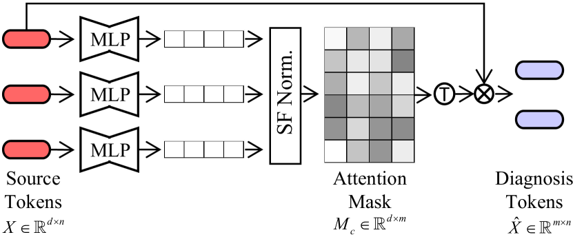
Specifically, as shown in Fig. 3, we employ a two-layer contextual multi-token MLP network (Linear-ReLU-Linear) denoted as , with the hidden layer reducing the input dimension by 12. This network, along with Softmax normalization, is utilized to generate the output contextual attention map conditioned on the original [CLS] tokens , where is a hyper-parameter representing the number of generated diagnosis tokens. Subsequently, we employ matrix multiplication to compute the output diagnosis tokens . The entire process can be described in Eq. (1).
| (1) |
3.3 Information emitter module
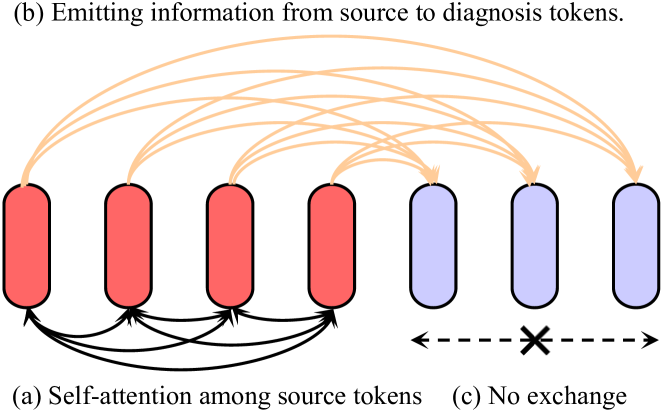
After obtaining the original [CLS] tokens and the new contextual diagnosis tokens , we concatenate and process them through a simple adapter MLP layer , aligning the combined visual tokens with LLM-compatible dimensions, resulting in . Subsequently, we develop the information emitter module (Fig.4) to preserve the original LLM’s information interactions for the source [CLS] tokens, while allowing the newly generated context-diagnostic tokens to extract and repurpose this information, thereby fostering novel insights for the diagnosis task.
Specifically, we improve the attention mechanism in each ViT layer of the LLM, preventing [CLS] token features from being altered by token features. We define a self-attention mask and configure its values as follows:
| (2) |
Subsequently, this mask guides the multihead self-attention process in each ViT layer, as formulated below:
| (3) |
According to the mask definition in Eq. (2), the attention calculation can be further formulated as:
| (4) |
where represents the self-attention fusion resulting from the information in the original [CLS] tokens. , , and represent the query, key, and value information extracted from the original [CLS] tokens. denotes the cross-attention fusion results, indicating the newly generated context diagnostic tokens learning from the original [CLS] tokens. represents the diagnostic query tokens, receiving the emitted information from source tokens.
By this design, we can ensure that the newly generated context diagnostic tokens will not affect the self-attention process of the original [CLS] tokens, but absorb emitted information from them and supplement their own information through the setting of mask. Besides, it should be noted that there is no information interaction between the newly generated context diagnostic tokens to ensure the uniqueness and diversity of promotes information.
3.4 Network training
| Method | Sens. (%) | Spec. (%) | Acc. (%) | AUC (%) | AVG (%) |
| ResNet [15] | 76.55 | 54.49 | 68.57 | 71.11 | 67.68 |
| ViT [11] | 73.56 | 51.96 | 65.72 | 64.23 | 63.87 |
| Swin Transformer [32] | 74.08 | 52.77 | 66.34 | 67.17 | 65.09 |
| Conformer [44] | 70.81 | 59.88 | 66.82 | 70.10 | 66.90 |
| ConvNeXt [33] | 69.35 | 62.84 | 66.99 | 67.64 | 66.70 |
| DINOv2 [42] | 75.56 | 57.23 | 68.89 | 70.40 | 68.02 |
| VAPFormer [23] | 63.04 | 59.26 | 61.15 | 63.39 | 61.71 |
| PneumoLLM | 80.54 | 67.66 | 75.87 | 78.98 | 75.76 |
During the training phase, we focus on training the adapter layers , the contextual multi-token engine network , the simple adapter MLP layer , and the disease classification network , employing the standard categorical cross-entropy loss. The optimization objective is formulated as:
| (5) |
where denotes the cross-entropy loss function, represents the ground-truth pneumoconiosis diagnose labels, represents the predicted results by our complete PneumoLLM framework, and are the learnable parameters to be optimized.
4 Experiments
4.1 Experimental setups
Dataset acquisition and splits
In this study, we utilized the posterior-anterior chest radiograph database from Jinneng Holding Coal Industry Group Co.Ltd Occupational Disease Precaution Clinic, comprising 630 chest radiographs in DICOM format, including 401 pneumoconiosis cases.
To ensure a balanced ratio of pneumoconiosis and normal samples in training and testing, we conduct five-fold cross-validation for experiments. Subsequently, we merge them into five distinct randomized datasets by patients (Datasets 1-5), and report the average performance of five experiments. Each dataset contains approximately 504 training and 126 testing radiographs, with pneumoconiosis cases constituting about 63% of each dataset.
Dataset pre-processing
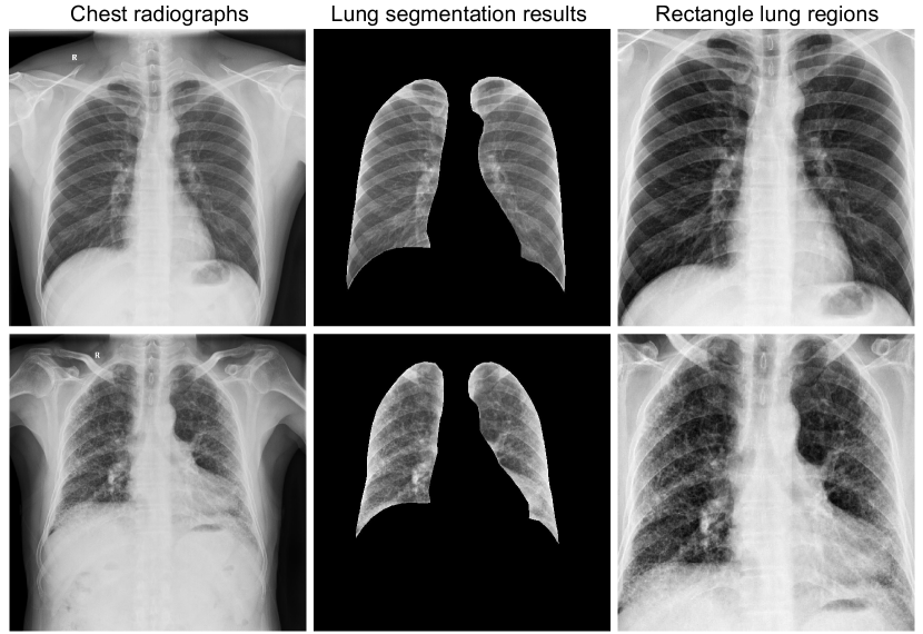
In the preprocessing phase, all chest radiographs are initially processed using the pyplot.imsave function to automatically adjust the contrast and brightness of the DICOM format (UInt16) pixel array, converting it to UInt8 PNG format. Next, we employ the CheXmask pipeline, introduced by Gaggion et al. [13], for lung segmentation. Based on the lung segmentation results, we use the maximum external rectangle extraction technique to isolate the rectangular lung regions in original chest radiographs. Finally, we resize them into a uniform size of 224224 pixels for analyses. Fig. 5 displays representative chest radiographs, showcasing both categories, their corresponding lung segmentation results, and the extracted rectangular lung regions.
Evaluation metrics
Following [3] and [46], we adapt Accuracy (Acc.), Sensitivity (Sens.), Specificity (Spec.), and Area Under the Curve (AUC) for quantitative analysis to comprehensively evaluate the performance of pneumoconiosis diagnosis. To facilitate analysis and comparison, we calculate the average of these metrics to assess the overall performance.
Implementation details
Consistent with [35], we employ the ViT-L/14 [11] of the pre-trained CLIP [47] as the vision encoder. We extract visual features as six [CLS] tokens from every fourth layer of ViT-L/14. The LLM utilize the LLaMA-7B [51] model. We set the visual neck dimension to 128 and the adapter dimension to 8. We employ AdamW [34] as the optimizer, training the model for 100 epochs with a cosine decay learning rate schedule. The batch size, learning rate, warmup epochs, and weight decay are set to 16, 310-4, 2 and 0.02, respectively.
4.2 Comparison experiments
To evaluate our proposed PneumoLLM, we compare it against established natural image classification methods, including ResNet [61], ViT [11], Swin Transformer [32], Conformer [44], ConvNeXt [33], and DINOv2 [42], as well as the advanced medical image diagnosis method VAPFormer [23]. All comparison models are implemented based on open-source configurations.
Pneumoconiosis diagnosis performance
Tab. 1 details the quantitative performance metrics of our PneumoLLM against the prestigious image classification methods which are pre-trained on ImageNet-1k [7] or LVD-142M [42]. Notably, among these prestigious image classification methods, models with inherent inductive biases like ResNet, Conformer, and ConvNeXt outperform transformer models like ViT and Swin Transformer in our limited Pneumoconiosis diagnosis dataset, while DINOv2 excels due to extensive pretraining data and prior information. Conversely, the medical image diagnosis method VAPFormer with prompts exhibits the least efficacy, which illustrates the big diagnostic challenges posed by data-scarce occupational disease. Our PneumoLLM, in constract, shows promising results by harnessing the power of LLM. In addition, we can observe that all the methods in Tab. 1 have significantly higher sensitivity and relatively lower specificity. The occurrence of this phenomenon is attributed to the unbalanced data distribution resulting from 401 out of the total 630 images in the dataset.
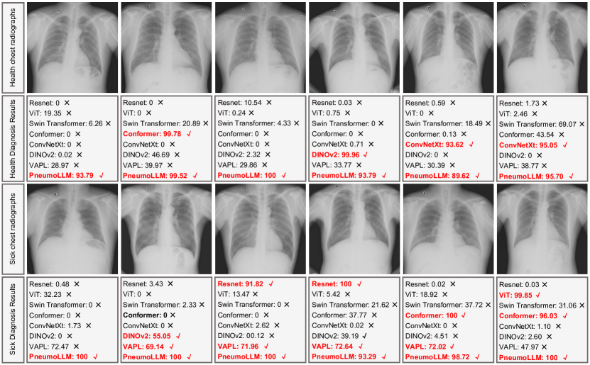
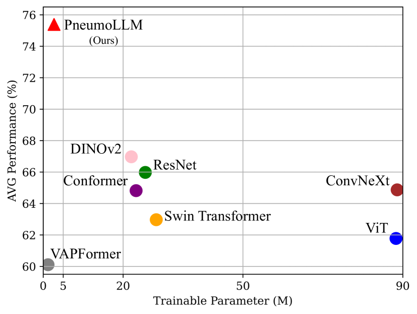
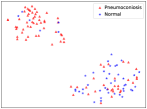
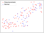
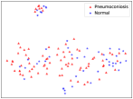
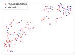
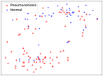
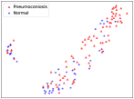
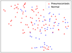
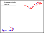
Besides, we perform some qualitative comparisons to provide insights into PneumoLLM’s effectiveness. When compared with different algorithms to diagnose pneumoconiosis, PneumoLLM showed higher confidence in accurate predictions (Fig. 6). PneumoLLM shows superior performance (Fig. 7) despite requiring fewer trainable parameters than all but VAPFormer, which had lowest diagnosis performance. Further, to evaluate feature representation quality, we employ t-SNE [38] method to project high-level feature representations onto a 2D plane (Fig. 8). Self-attention-based models like ViT and Swin Transformer underperformed with smaller datasets, whereas models like ResNet, Conformer, and ConvNeXt showed better class separation but larger within-class variations. PneumoLLM, however, demonstrates more compact intra-class representations and pronounced inter-class separations, indicative of enhanced feature representation. These observations align with our quantitative findings, further validating PneumoLLM’s effectiveness over existing methods.
| Models | L-para.(M) | Mem. (G) | Sens. (%) | Spec. (%) | Acc. (%) | AUC (%) | AVG (%) |
| PneumoLLM w/o LLaMA | 0.52 | 4.15 | 77.77 | 62.00 | 72.06 | 75.81 | 71.91 |
| PneumoLLM | 2.70 | 15.48 | 80.54 | 67.66 | 75.87 | 78.98 | 75.76 (+3.85) |
4.3 Ablation experiments
In our research, we perform a comprehensive ablation analysis of PneumoLLM, modifying one component at a time. Our PneumoLLM mainly comprises the utilization of LLM, contextual multi-token engine, and information emitter design. Initially, in Sec. 4.3.1, we analyze the model capacity, focusing on the essential role of the foundational LLaMA model, and the design of eliminating the textual processing branch by directly harnessing LLaMA to process visual features from the vision encoder. Subsequently, we evaluate the impact of various PneumoLLM components in Sec. 4.3.2, including the adapter, the contextual multi-token engine, and the information emitter module.
| Settings | L-para.(M) | Mem.(G) | Sens. (%) | Spec. (%) | Acc. (%) | AUC (%) | AVG (%) |
| LaVIN | 3.77 | 20.25 | 87.80 | 47.52 | 73.33 | 71.82 | 70.12 |
| Simplyfied PneumoLLM | 2.69 | 15.32 | 71.56 | 71.67 | 71.58 | 75.21 | 72.50 (+2.38) |
4.3.1 Model capacity
Ablation study on LLaMA utilization
In contrast to existing classification vision models, our PneumoLLM incorporates the LLaMA-7B model, harnessing its extensive knowledge and prior information to improve pneumoconiosis diagnosis. To validate this approach, we perform an ablation study examining the effects of LLaMA integration, detailed in Tab. 2. Our default PneumoLLM configuration integrates the ViT-L/14+LLaMA architecture with additional learnable adapter layers, contextual multi-token engine network, and classification network. Without LLaMA, PneumoLLM solely utilizes the ViT-L/14 architecture with adapter layers and classification network. As indicated in Table 2, the utilization of LLaMA demonstrates superior learning efficiency on small pneumoconiosis datasets, achieving improvements across all diagnosis metrics. However, this improvement comes at the cost of increased memory due to the LLM’s inherent model size.
Ablation on eliminating the textual processing branch in LLM
Contrasting with LaVIN [35], which approaches image classification as a question-answering task, requiring both question text and image as input to LLM, we assume that the question text inputs are redundant and useless for the vision classification task. Employing the ViT-L/14 from pre-trained CLIP as the vision encoder, we input the [CLS] tokens directly into the LLaMA model. This design aims to enhance pneumoconiosis diagnosis by capitalizing on CLIP’s ability to align image features with textual data and LLaMA’s proficient information processing capabilities. Our ablation studies aimed to validate this design.
We compare our PneumoLLM with image-only input (Fig. 1(c)) against LaVIN’s dual image-question input setup (Fig. 1(b)), simplifying our model by removing the contextual multi-token engine and information emitter module for fairness. In the LaVIN setup, pneumoconiosis diagnosis is treated as a question-answering task with a coincident question input: “Does the patient have pneumoconiosis?”. Meanwhile, we create ‘Yes’ or ‘No’ text answer labels to ensure consistency with the original LaVIN setting. Our PneumoLLM uses only the chest radiographs input and ‘Pneumoconiosis’ or ‘Normal’ labels. Tab. 3 demonstrates that our simplified PneumoLLM outperformed LaVIN in both efficiency and efficacy, supporting our hypothesis that question input is superfluous for diagnosing pneumoconiosis.
| Baseline | Adapter | CoOp | CoCoOp | Contextual Multi-Token Engine | Information Emitter | Sens. (%) | Spec. (%) | Acc. (%) | AUC (%) | AVG (%) |
| ✓ | 78.07 | 62.77 | 72.54 | 74.52 | 71.98 | |||||
| ✓ | ✓ | 71.56 | 71.67 | 71.58 | 75.21 | 72.50 | ||||
| ✓ | ✓ | ✓ | 73.54 | 68.56 | 71.74 | 75.56 | 72.35 | |||
| ✓ | ✓ | ✓ | 72.57 | 70.31 | 71.75 | 75.80 | 72.61 | |||
| ✓ | ✓ | ✓ | 78.29 | 66.82 | 74.13 | 76.42 | 73.92 | |||
| ✓ | ✓ | ✓ | ✓ | 80.54 | 67.66 | 75.87 | 78.98 | 75.76 |
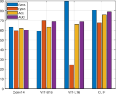
To further assess the impact of various vision encoders and the necessity of the usage of pre-trained CLIP network, we compare various vision encoders, including a standard convolutional layer, ViT-B/16 and ViT-L/16 pre-trained on ImageNet-21k, and ViT-L/14 from pre-trained CLIP [47]. Results in Fig. 9(a) indicate the superior performance of the ViT-L/14 from pre-trained CLIP. Notably, ViT-L/16 exhibited high sensitivity but low specificity, suggesting a tendency to over-diagnose. This ablation study highlights the significance of choosing appropriate pre-trained vision encoder for optimal integration with LLM.
4.3.2 Ablation on various PneumoLLM components
Due to the limited availability of pneumoconiosis patient data and patient privacy concerns, acquiring extensive pneumoconiosis datasets is challenging. Fine-tuning our entire PneumoLLM on the small pneumoconiosis dataset can make drastic changes in the LLM’s parameter spaces, leading to increased training time and huge memory requirements. In this scenario, the usage of lightweight adapters, our proposed contextual multi-token engine and information emitter module prove crucial in adapting LLM efficiently to pneumoconiosis diagnosis. To validate this, we conduct comprehensive ablation studies in Tab. 4. In this table, we use the simplified PneumoLLM in Tab. 3 without adapters as our baseline for comparing various component configurations.
Ablation on adapter usage
We conduct an ablation study to measure the effect of the usage of adapters. The results, as presented in the first two rows of Tab. 4, indicate that the incorporation of adapters significantly enhances diagnostic performance in our small pneumoconiosis dataset.
Ablation on the contextual multi-token engine
We compared our contextual multi-token engine with two recent prompt engineering methods, CoOp [73] and CoCoOp [72], as discussed in Sec. 3.2. As shown in Tab. 4, while CoOp and CoCoOp settings show limited improvement, our multi-token engine yields a significant performance increase (+1.98%). CoOp’s uniform prompts across samples hinder its adaptability, leading to a slight decrease in average performance. CoCoOp, though offering improved flexibility with vision-conditional prompts, faces limitations due to the mixed use of fixed and flexible prompts, yielding minimal gains. In contrast, our multi-token engine, designed to optimize the synergy between vision tokens and LLM, significantly enhances pneumoconiosis diagnosis.
Ablation on the information emitter module
Another ablation study focuses on our proposed information emitter module. Results in Tab. 4 demonstrate that combining the multi-token engine with the information emitter module achieves the best performance in pneumoconiosis diagnosis. This module successfully maintains the integrity of the original LLM’s information processing while emitting valuable information to context-diagnostic tokens, fostering enhanced diagnostic insights.
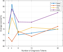
Additionally, the optimal number of generated diagnosis tokens is determined based on performance analysis (Fig. 9(b)), leading to the selection of four as the ideal number.
5 Conclusion
In this paper, we introduce PneumoLLM, a pioneering approach utilizing large language models for streamlined diagnostic processes in medical imaging. By discarding the text branch and transforming the dialogue head into a classification head, PneumoLLM simplifies the workflow for eliciting knowledge from LLMs. This innovation proves particular effectiveness when only classification labels are available for training, rather than extensive descriptive sentences. The streamlined process also significantly reduces the optimization space, facilitating learning with limited training data. Ablation studies further underscore the necessity and effectiveness of the proposed modules, especially in maintaining the integrity of source image details while advancing towards accurate diagnostic outcomes.
Looking ahead, we plan to expand PneumoLLM’s application to more imaging modalities beyond X-ray radiography, e.g., CT and MRI scans, aiming to broaden its diagnostic capabilities across a spectrum of diseases. The potential of a robust diagnostic system like PneumoLLM extends beyond disease diagnosis, offering promising avenues in other areas of medical image processing such as whole slide image classification [28] and pathology image analysis [22]. These future endeavors could enhance the capabilities of automated diagnostic systems, paving the way for more practical medical imaging analyses.
Acknowledgments
This work was supported in part by grants from the Chinese Academy of Medical Sciences Innovation Fund for Medical Sciences, China CIFMS2021-I2M-1-044, 2021-I2M-1-049, and the Non-profit Central Research Institute Fund of Chinese Academy of Medical Sciences, 2022-RC310-06, and the NSFC 62071382.
References
- Ali et al. [2020] Ali, S., Bhattarai, B., Kim, T.K., Rittscher, J., 2020. Additive angular margin for few shot learning to classify clinical endoscopy images, in: Proceedings of the International Workshop on Machine Learning in Medical Imaging, Springer. pp. 494–503.
- Brown et al. [2020] Brown, T., Mann, B., Ryder, N., Subbiah, M., Kaplan, J.D., Dhariwal, P., Neelakantan, A., Shyam, P., Sastry, G., Askell, A., et al., 2020. Language models are few-shot learners. Advances in Neural Information Processing Systems 33, 1877–1901.
- Chen et al. [2023a] Chen, Y., Guo, X., Pan, Y., Xia, Y., Yuan, Y., 2023a. Dynamic feature splicing for few-shot rare disease diagnosis. Medical Image Analysis 90, 102959.
- Chen et al. [2023b] Chen, Z., Liu, Y., Zhang, Y., Li, Q., Initiative, A.D.N., et al., 2023b. Orthogonal latent space learning with feature weighting and graph learning for multimodal alzheimer’s disease diagnosis. Medical Image Analysis 84, 102698.
- Cheng et al. [2023] Cheng, D., Qin, Z., Jiang, Z., Zhang, S., Lao, Q., Li, K., 2023. Sam on medical images: A comprehensive study on three prompt modes. arXiv preprint arXiv:2305.00035 .
- Chowdhery et al. [2022] Chowdhery, A., Narang, S., Devlin, J., Bosma, M., Mishra, G., Roberts, A., Barham, P., Chung, H.W., Sutton, C., Gehrmann, S., et al., 2022. Palm: Scaling language modeling with pathways. arXiv preprint arXiv:2204.02311 .
- Deng et al. [2009] Deng, J., Dong, W., Socher, R., Li, L.J., Li, K., Fei-Fei, L., 2009. Imagenet: A large-scale hierarchical image database, in: Proceedings of the IEEE Conference on Computer Vision and Pattern Recognition, IEEE. pp. 248–255.
- Devnath et al. [2022] Devnath, L., Fan, Z., Luo, S., Summons, P., Wang, D., 2022. Detection and visualisation of pneumoconiosis using an ensemble of multi-dimensional deep features learned from chest x-rays. International Journal of Environmental Research and Public Health 19, 11193.
- Devnath et al. [2021] Devnath, L., Luo, S., Summons, P., Wang, D., 2021. Automated detection of pneumoconiosis with multilevel deep features learned from chest x-ray radiographs. Computers in Biology and Medicine 129, 104125.
- Dong et al. [2022] Dong, H., Zhu, B., Zhang, X., Kong, X., 2022. Use data augmentation for a deep learning classification model with chest x-ray clinical imaging featuring coal workers’ pneumoconiosis. BMC Pulmonary Medicine 22, 1–14.
- Dosovitskiy et al. [2020] Dosovitskiy, A., Beyer, L., Kolesnikov, A., Weissenborn, D., Zhai, X., Unterthiner, T., Dehghani, M., Minderer, M., Heigold, G., Gelly, S., et al., 2020. An image is worth 16x16 words: Transformers for image recognition at scale, in: International Conference on Learning Representations, pp. 1–12.
- Fan et al. [2023] Fan, R., Bowd, C., Brye, N., Christopher, M., Weinreb, R.N., Kriegman, D.J., Zangwill, L.M., 2023. One-vote veto: Semi-supervised learning for low-shot glaucoma diagnosis. IEEE Transactions on Medical Imaging .
- Gaggion et al. [2023] Gaggion, N., Mosquera, C., Mansilla, L., Aineseder, M., Milone, D.H., Ferrante, E., 2023. Chexmask: a large-scale dataset of anatomical segmentation masks for multi-center chest x-ray images. arXiv preprint arXiv:2307.03293 .
- Gao et al. [2021] Gao, X., Qian, Y., Gao, A., 2021. Covid-vit: Classification of covid-19 from ct chest images based on vision transformer models. arXiv preprint arXiv:2107.01682 .
- He et al. [2016] He, K., Zhang, X., Ren, S., Sun, J., 2016. Deep residual learning for image recognition, in: Proceedings of the IEEE Conference on Computer Vision and Pattern Recognition, pp. 770–778.
- Heidarian et al. [2021] Heidarian, S., Afshar, P., Oikonomou, A., Plataniotis, K.N., Mohammadi, A., 2021. Cae-transformer: Transformer-based model to predict invasiveness of lung adenocarcinoma subsolid nodules from non-thin section 3d ct scans. arXiv preprint arXiv:2110.08721 .
- Houlsby et al. [2019] Houlsby, N., Giurgiu, A., Jastrzebski, S., Morrone, B., De Laroussilhe, Q., Gesmundo, A., Attariyan, M., Gelly, S., 2019. Parameter-efficient transfer learning for nlp, in: International Conference on Machine Learning, PMLR. pp. 2790–2799.
- Hu et al. [2021] Hu, E.J., Wallis, P., Allen-Zhu, Z., Li, Y., Wang, S., Wang, L., Chen, W., et al., 2021. Lora: Low-rank adaptation of large language models, in: International Conference on Learning Representations, pp. 1–16.
- Huang et al. [2023a] Huang, B., Liao, G., Lee, P.M.Y., Chan, C.K., Tai, L.b., Tsang, C.Y.J., Leung, C.C., Tse, L.A., 2023a. Association of circadian rhythm with mild cognitive impairment among male pneumoconiosis workers in hong kong: a cross-sectional study. Scientific Reports 13, 1650.
- Huang et al. [2023b] Huang, Q., Wang, D., Lu, Z., Zhou, S., Li, J., Liu, L., Chang, C., 2023b. A novel image-to-knowledge inference approach for automatically diagnosing tumors. Expert Systems with Applications 229, 120450.
- Huang et al. [2022] Huang, Y., Si, Y., Hu, B., Zhang, Y., Wu, S., Wu, D., Wang, Q., 2022. Transformer-based factorized encoder for classification of pneumoconiosis on 3d ct images. Computers in Biology and Medicine 150, 106137.
- Huang et al. [2023c] Huang, Z., Bianchi, F., Yuksekgonul, M., Montine, T.J., Zou, J., 2023c. A visual–language foundation model for pathology image analysis using medical twitter. Nature Medicine , 1–10.
- Kang et al. [2023] Kang, L., Gong, H., Wan, X., Li, H., 2023. Visual-attribute prompt learning for progressive mild cognitive impairment prediction, in: International Conference on Medical Image Computing and Computer-Assisted Intervention, Springer. pp. 547–557.
- Kang et al. [2022] Kang, Q., Lao, Q., Li, Y., Jiang, Z., Qiu, Y., Zhang, S., Li, K., 2022. Thyroid nodule segmentation and classification in ultrasound images through intra-and inter-task consistent learning. Medical Image Analysis 79, 102443.
- Kenton and Toutanova [2019] Kenton, J.D.M.W.C., Toutanova, L.K., 2019. Bert: Pre-training of deep bidirectional transformers for language understanding, in: Proceedings of the 2019 Conference of the North American Chapter of the Association for Computational Linguistics: Human Language Technologies, pp. 4171–4186.
- Kirillov et al. [2023] Kirillov, A., Mintun, E., Ravi, N., Mao, H., Rolland, C., Gustafson, L., Xiao, T., Whitehead, S., Berg, A.C., Lo, W.Y., et al., 2023. Segment anything. arXiv preprint arXiv:2304.02643 .
- Lester et al. [2021] Lester, B., Al-Rfou, R., Constant, N., 2021. The power of scale for parameter-efficient prompt tuning, in: Proceedings of the 2021 Conference on Empirical Methods in Natural Language Processing, pp. 3045–3059.
- Li et al. [2021] Li, J., Chen, W., Huang, X., Yang, S., Hu, Z., Duan, Q., Metaxas, D.N., Li, H., Zhang, S., 2021. Hybrid supervision learning for pathology whole slide image classification, in: International Conference on Medical Image Computing and Computer-Assisted Intervention, Springer. pp. 309–318.
- Li et al. [2023a] Li, J., Li, D., Savarese, S., Hoi, S., 2023a. BLIP-2: bootstrapping language-image pre-training with frozen image encoders and large language models, in: International Conference on Machine Learning, PMLR. pp. 1–13.
- Li et al. [2023b] Li, Y., Lao, Q., Kang, Q., Jiang, Z., Du, S., Zhang, S., Li, K., 2023b. Self-supervised anomaly detection, staging and segmentation for retinal images. Medical Image Analysis 87, 102805.
- Li et al. [2023c] Li, Z.G., Li, B.C., Li, Z.W., Hu, H.Y., Ma, X., Cao, H., Yu, Z.H., Dai, H.P., Wang, J., Wang, C., et al., 2023c. The potential diagnostic biomarkers for the igg subclass in coal workers’ pneumoconiosis. Journal of Immunology Research 2023.
- Liu et al. [2021] Liu, Z., Lin, Y., Cao, Y., Hu, H., Wei, Y., Zhang, Z., Lin, S., Guo, B., 2021. Swin transformer: Hierarchical vision transformer using shifted windows, in: Proceedings of the IEEE/CVF International Conference on Computer Vision, pp. 10012–10022.
- Liu et al. [2022] Liu, Z., Mao, H., Wu, C.Y., Feichtenhofer, C., Darrell, T., Xie, S., 2022. A convnet for the 2020s, in: Proceedings of the IEEE Conference on Computer Vision and Pattern Recognition, pp. 11976–11986.
- Loshchilov and Hutter [2017] Loshchilov, I., Hutter, F., 2017. Decoupled weight decay regularization. arXiv preprint arXiv:1711.05101 .
- Luo et al. [2023] Luo, G., Zhou, Y., Ren, T., Chen, S., Sun, X., Ji, R., 2023. Cheap and quick: Efficient vision-language instruction tuning for large language models. arXiv preprint arXiv:2305.15023 .
- Luo et al. [2022] Luo, X., Song, T., Wang, G., Chen, J., Chen, Y., Li, K., Metaxas, D.N., Zhang, S., 2022. Scpm-net: An anchor-free 3d lung nodule detection network using sphere representation and center points matching, in: Medical Image Analysis, Elsevier. p. 102287.
- Ma et al. [2023] Ma, Y., Cui, W., Liu, J., Guo, Y., Chen, H., Li, Y., 2023. A multi-graph cross-attention based region-aware feature fusion network using multi-template for brain disorder diagnosis. IEEE Transactions on Medical Imaging .
- Van der Maaten and Hinton [2008] Van der Maaten, L., Hinton, G., 2008. Visualizing data using t-sne. Journal of Machine Learning Research 9.
- Moor et al. [2023] Moor, M., Banerjee, O., Abad, Z.S.H., Krumholz, H.M., Leskovec, J., Topol, E.J., Rajpurkar, P., 2023. Foundation models for generalist medical artificial intelligence. Nature 616, 259–265.
- OpenAI [2023a] OpenAI, 2023a. Chatgpt. https://chat.openai.com. Accessed: [November 7th, 2023].
- OpenAI [2023b] OpenAI, 2023b. Gpt-4 technical report. arXiv preprint arXiv:2303.08774 .
- Oquab et al. [2023] Oquab, M., Darcet, T., Moutakanni, T., Vo, H., Szafraniec, M., Khalidov, V., Fernandez, P., Haziza, D., Massa, F., El-Nouby, A., et al., 2023. Dinov2: Learning robust visual features without supervision. arXiv preprint arXiv:2304.07193 .
- Ouyang et al. [2020] Ouyang, D., He, B., Ghorbani, A., Yuan, N., Ebinger, J., Langlotz, C.P., Heidenreich, P.A., Harrington, R.A., Liang, D.H., Ashley, E.A., et al., 2020. Video-based ai for beat-to-beat assessment of cardiac function. Nature 580, 252–256.
- Peng et al. [2021] Peng, Z., Huang, W., Gu, S., Xie, L., Wang, Y., Jiao, J., Ye, Q., 2021. Conformer: Local features coupling global representations for visual recognition, in: Proceedings of the IEEE/CVF international conference on computer vision, pp. 367–376.
- Qi et al. [2021] Qi, X.M., Luo, Y., Song, M.Y., Liu, Y., Shu, T., Liu, Y., Pang, J.L., Wang, J., Wang, C., 2021. Pneumoconiosis: current status and future prospects. Chinese Medical Journal 134, 898–907.
- Qu et al. [2023] Qu, J., Wei, X., Qian, X., 2023. Generalized pancreatic cancer diagnosis via multiple instance learning and anatomically-guided shape normalization. Medical Image Analysis 86, 102774.
- Radford et al. [2021] Radford, A., Kim, J.W., Hallacy, C., Ramesh, A., Goh, G., Agarwal, S., Sastry, G., Askell, A., Mishkin, P., Clark, J., et al., 2021. Learning transferable visual models from natural language supervision, in: International Conference on Machine Learning, PMLR. pp. 8748–8763.
- Singhal et al. [2023] Singhal, K., Azizi, S., Tu, T., Mahdavi, S.S., Wei, J., Chung, H.W., Scales, N., Tanwani, A., Cole-Lewis, H., Pfohl, S., et al., 2023. Large language models encode clinical knowledge. Nature 620, 172–180.
- Stiennon et al. [2020] Stiennon, N., Ouyang, L., Wu, J., Ziegler, D., Lowe, R., Voss, C., Radford, A., Amodei, D., Christiano, P.F., 2020. Learning to summarize with human feedback. Advances in Neural Information Processing Systems 33, 3008–3021.
- Sun et al. [2023] Sun, W., Wu, D., Luo, Y., Liu, L., Zhang, H., Wu, S., Zhang, Y., Wang, C., Zheng, H., Shen, J., et al., 2023. Expertnet: Defeat noisy labels by deep expert consultation paradigm for pneumoconiosis staging on chest radiographs. Expert Systems with Applications , 120710.
- Touvron et al. [2023a] Touvron, H., Lavril, T., Izacard, G., Martinet, X., Lachaux, M.A., Lacroix, T., Rozière, B., Goyal, N., Hambro, E., Azhar, F., et al., 2023a. Llama: Open and efficient foundation language models. arXiv preprint arXiv:2302.13971 .
- Touvron et al. [2023b] Touvron, H., Martin, L., Stone, K., Albert, P., Almahairi, A., Babaei, Y., Bashlykov, N., Batra, S., Bhargava, P., Bhosale, S., et al., 2023b. Llama 2: Open foundation and fine-tuned chat models. arXiv preprint arXiv:2307.09288 .
- Vaswani et al. [2017] Vaswani, A., Shazeer, N., Parmar, N., Uszkoreit, J., Jones, L., Gomez, A.N., Kaiser, Ł., Polosukhin, I., 2017. Attention is all you need. Advances in Neural Information Processing Systems 30.
- Wang et al. [2023] Wang, D., Wang, X., Wang, L., Li, M., Da, Q., Liu, X., Gao, X., Shen, J., He, J., Shen, T., et al., 2023. A real-world dataset and benchmark for foundation model adaptation in medical image classification. Scientific Data , 1–9.
- Wang et al. [2020a] Wang, L., Lin, Z.Q., Wong, A., 2020a. Covid-net: A tailored deep convolutional neural network design for detection of covid-19 cases from chest x-ray images. Scientific Reports 10, 19549.
- Wang et al. [2017] Wang, X., Peng, Y., Lu, L., Lu, Z., Bagheri, M., Summers, R.M., 2017. Chestx-ray8: Hospital-scale chest x-ray database and benchmarks on weakly-supervised classification and localization of common thorax diseases, in: Proceedings of the IEEE Conference on Computer Vision and Pattern Recognition, pp. 2097–2106.
- Wang et al. [2020b] Wang, X., Yu, J., Zhu, Q., Li, S., Zhao, Z., Yang, B., Pu, J., 2020b. Potential of deep learning in assessing pneumoconiosis depicted on digital chest radiography. Occupational and Environmental Medicine 77, 597–602.
- Wang et al. [2022] Wang, Z., Wu, Z., Agarwal, D., Sun, J., 2022. Medclip: Contrastive learning from unpaired medical images and text, in: Proceedings of the 2022 Conference on Empirical Methods in Natural Language Processing, pp. 3876–3887.
- Wen et al. [2023] Wen, S., Fang, G., Zhang, R., Gao, P., Dong, H., Metaxas, D., 2023. Improving compositional text-to-image generation with large vision-language models. arXiv preprint arXiv:2310.06311 .
- Wu et al. [2023] Wu, C., Zhang, X., Zhang, Y., Wang, Y., Xie, W., 2023. Towards generalist foundation model for radiology. arXiv preprint arXiv:2308.02463 .
- Xie et al. [2017] Xie, S., Girshick, R., Dollár, P., Tu, Z., He, K., 2017. Aggregated residual transformations for deep neural networks, in: Proceedings of the IEEE Conference on Computer Vision and Pattern Recognition, pp. 1492–1500.
- Xing et al. [2023] Xing, X., Chen, Z., Hou, Y., Yuan, Y., 2023. Gradient modulated contrastive distillation of low-rank multi-modal knowledge for disease diagnosis. Medical Image Analysis , 102874.
- Xiong et al. [2023] Xiong, H., Wang, S., Zhu, Y., Zhao, Z., Liu, Y., Wang, Q., Shen, D., 2023. Doctorglm: Fine-tuning your chinese doctor is not a herculean task. arXiv preprint arXiv:2304.01097 .
- Xu et al. [2023] Xu, X., Jia, Q., Yuan, H., Qiu, H., Dong, Y., Xie, W., Yao, Z., Zhang, J., Nie, Z., Li, X., et al., 2023. A clinically applicable ai system for diagnosis of congenital heart diseases based on computed tomography images. Medical Image Analysis 90, 102953.
- Yi et al. [2023] Yi, H., Qin, Z., Lao, Q., Xu, W., Jiang, Z., Wang, D., Zhang, S., Li, K., 2023. Towards general purpose medical ai: Continual learning medical foundation model. arXiv preprint arXiv:2303.06580 .
- You et al. [2023] You, K., Gu, J., Ham, J., Park, B., Kim, J., Hong, E.K., Baek, W., Roh, B., 2023. Cxr-clip: Toward large scale chest x-ray language-image pre-training, in: International Conference on Medical Image Computing and Computer-Assisted Intervention, Springer. pp. 101–111.
- Zhang et al. [2023a] Zhang, R., Han, J., Zhou, A., Hu, X., Yan, S., Lu, P., Li, H., Gao, P., Qiao, Y., 2023a. Llama-adapter: Efficient fine-tuning of language models with zero-init attention. arXiv preprint arXiv:2303.16199 .
- Zhang and Metaxas [2023] Zhang, S., Metaxas, D., 2023. On the challenges and perspectives of foundation models for medical image analysis. arXiv preprint arXiv:2306.05705 .
- Zhang et al. [2023b] Zhang, Y., Gao, J., Zhou, M., Wang, X., Qiao, Y., Zhang, S., Wang, D., 2023b. Text-guided foundation model adaptation for pathological image classification, in: International Conference on Medical Image Computing and Computer-Assisted Intervention, Springer. pp. 272–282.
- Zhang et al. [2023c] Zhang, Y., Huang, X., Ma, J., Li, Z., Luo, Z., Xie, Y., Qin, Y., Luo, T., Li, Y., Liu, S., et al., 2023c. Recognize anything: A strong image tagging model. arXiv preprint arXiv:2306.03514 .
- Zheng et al. [2019] Zheng, R., Deng, K., Jin, H., Liu, H., Zhang, L., 2019. An improved cnn-based pneumoconiosis diagnosis method on x-ray chest film, in: International Conference on Human Centered Computing, Springer. pp. 647–658.
- Zhou et al. [2022a] Zhou, K., Yang, J., Loy, C.C., Liu, Z., 2022a. Conditional prompt learning for vision-language models, in: Proceedings of the IEEE Conference on Computer Vision and Pattern Recognition, pp. 16816–16825.
- Zhou et al. [2022b] Zhou, K., Yang, J., Loy, C.C., Liu, Z., 2022b. Learning to prompt for vision-language models. International Journal of Computer Vision 130, 2337–2348.