- AP
- Average Precision
- ADC
- Apparent diffusion coefficient
- ASSD
- Average Symmetric Surface Distance
- AUC
- Area Under the Curve
- BraTS-M
- ASNR-MICCAI BraTS Brain Metastasis Challenge
- BraTS-G
- RSNA-ASNR-MICCAI BraTS Continuous Evaluation Challenge
- CNN
- Convolutional Neural Network
- CCA
- Connected Component Analysis
- CT
- Computed Tomography
- clDice
- Centerline Dice Similarity Coefficient
- DL
- Deep Learning
- DQ
- Detection Quality
- TP
- True Positive
- FP
- False Positive
- FN
- False Negatives
- DSC
- Dice Similarity Coefficient
- DWI
- Diffusion Weighted Imaging
- ET
- Enhancing Tumor
- GAN
- Generative Adversarial Network
- gvDSC
- global volumetric Dice Similarity Coefficient
- HD
- Hausdorff Distance
- ISLES
- Ischemic Stroke Lesion Segmentation 2022 challenge
- IOU
- Intersection over Union
- InS
- Instance Segmentation
- ML
- Machine Learning
- MM
- Matching Metric
- MONAI
- Medical Open Network for Artificial Intelligence
- MRI
- Magnetic Resonance Imaging
- MS
- Matching Score
- NETC
- Nonenhancing Tumor Core
- NN
- Neural Network
- NSD
- Normalized Surface Dice
- PGT
- Peak Ground Truth
- PQ
- Panoptic Quality
- RQ
- Recognition Quality
- RWMP
- Real World Model Performance
- SQ
- Segmentation Quality
- PanS
- Panoptic Segmentation
- SNFH
- Surrounding Non-enhancing FLAIR Hyperintensity
- SemS
- Semantic Segmentation
- SQASSD
- Segmentation Quality Average Symmetric Surface Distance
- t1c
- post-contrast T1-weighted
- t1w
- pre-contrast T1-weighted
- t2f
- T2-weighted Fluid Attenuated Inversion Recovery
- FLAIR
- T2-weighted Fluid Attenuated Inversion Recovery
- t2w
- T2-weighted
- VerSe
- Large Scale Vertebrae Segmentation Challenge
panoptica – instance-wise evaluation of
3D semantic and instance segmentation maps
Abstract
This paper introduces panoptica, a versatile and performance-optimized package designed for computing instance-wise segmentation quality metrics from 2D and 3D segmentation maps. panoptica addresses the limitations of existing metrics and provides a modular framework that complements the original IOU-based Panoptic Quality with other metrics, such as the distance metric ASSD. The package is open-source, implemented in Python, and accompanied by comprehensive documentation and tutorials. panoptica employs a three-step metrics computation process to cover diverse use cases. We demonstrate the efficacy of panoptica on various real-world biomedical datasets, where an instance-wise evaluation is instrumental for an accurate representation of the underlying clinical task. Overall, we envision panoptica as a valuable tool facilitating in-depth evaluation of segmentation methods.
1 Introduction
Segmentation is mostly formulated as a task related to either stuff, representing all pixels/voxels belonging to a single class with the same label ( Semantic Segmentation (SemS)) or things, labeling each pixel/voxel of a class (e.g., metastatic tumor lesions) with an individual label ( Instance Segmentation (InS)) [19].
For many biomedical segmentation problems, an instance-wise evaluation focusing on individual lesions is highly relevant and desirable for both scientific research and clinical applications. This significance is evident in the context of multiple sclerosis, an inflammatory disease characterized by a variable number of white matter lesions in the brain. In the prediction of disease activity, a graph neural network leveraging representations from individual lesions has been suggested [30]. From a clinical perspective, the occurrence of a single new inflammatory lesion is crucial in establishing dissemination in time, playing a pivotal role in the diagnosis and ongoing monitoring of multiple sclerosis patients [37]. Both the scientific modeling of disease activity as well as the clinical monitoring of patients are clearly dependent on accurate lesion-wise InS.
Despite the apparent need for reliable instance-wise segmentation, many biomedical segmentation problems are still addressed as SemS problems, partly due to the lack of appropriate instance labels and partly due to the advances in semantic segmentation methodology:
The biomedical image analysis community has successfully developed a plethora of (mostly U-Net-based [32, 14]) methods for solving the problem of SemS across a range of imaging modalities in diverse applications such as primary brain tumors [2, 21, 20], brain metastases [5, 6], vascular lesions [36], multiple sclerosis [22] or stroke [13] in brain MRI, liver tumors [4, 22] or the spine [33] in CT or light-sheet microscopy tasks [18, 3].
In view of the need for reliable instance-wise segmentation outlined above, there have been attempts to optimize instance awareness in SemS, e.g., [22]. Reuniting stuff and things by simultaneously evaluating SemS and InS is, therefore, a relevant but unmet need, especially in biomedical image analysis.
Contribution: We introduce panoptica, a modular, performance-optimized package to compute instance-wise segmentation quality metrics for 2D and 3D semantic- and instance segmentation maps. On an instance level, panoptica allows reporting detection metrics, such as Recognition Quality (RQ) (equivalent to F1-score), and segmentation quality metrics, such as Segmentation Quality (SQ) and Panoptic Quality (PQ). Furthermore, additional metrics such as Average Symmetric Surface Distance (ASSD) are available. panoptica is an open-source Python package, and comprehensive documentation and tutorials are available (cf. Section 3).
To overcome limitations in the PQ metric, which may obscure information related to either detection or segmentation quality [31, 26], panoptica focuses on presenting all aspects relevant to instance segmentation tasks by complementing PQ with other pertinent performance metrics.
The metric computation process in panoptica is divided into three steps, offering flexibility for various use cases, cf. Sections 3 and 4. In contrast to existing solutions, panoptica facilitates the evaluation of semantic segmentation methods at an instance level. We showcase the capabilities of panoptica across multiple biomedical data sets featuring real-world segmentation challenges. For these experiments, instance-wise evaluation proves crucial in capturing the nuances of the underlying biomedical tasks. The instance-wise analysis reveals aspects of segmentation quality that may remain concealed when solely evaluating the entire image domain,cf. Section 4.
2 Related Work
Kirillov et al. [19] introduce the task of Panoptic Segmentation (PanS) as combination of Semantic Segmentation (SemS) and Instance Segmentation (InS).
They define a PanS task as both assigning each pixel to a class-ID and an instance ID, for SemS and InS respectively.
Further, they propose the Panoptic Quality (PQ) metric, cf. Section 3.3.
Although the metric was originally proposed for PanS tasks, the Metrics Reloaded initiative [26] recommended employing the metric also for InS tasks.
In this context, PQ allows capturing both detection and segmentation quality in one score and is an alternative to the -score assessing per-class detection quality.
The PQ metric is implemented as part of the Medical Open Network for Artificial Intelligence (MONAI) framework [7] and torchmetrics [10]. However, these implementations only support 2D and not 3D input data, and the torchmetrics implementation provides no means for instance matching. Therefore, it is only applicable to already matched instance maps. Further, the Metrics Reloaded initiative [26] provides a 3D implementation in a side package of MONAI. While the program can compute PQ, SQ, and RQ, it is designed to always return a single metric. Hence, multiple calls are necessary if all three metrics are required. This is a conscious design choice as the program assumes that the user selects the most suitable metrics via the Metrics Reloaded framework [26]. Naturally, the computational overhead of multiple calls leads to a massive accumulation of processing times, prohibiting the evaluation of large-scale data sets. Further, even though computed internally, the package offers no means to return True Positive (TP), False Positive (FP), False Negatives (FN). Unlike panoptica, the implementation of the Metrics Reloaded initiative, lacking instance approximation capabilities, cannot calculate instance-wise metrics for semantic inputs.
3 Methods
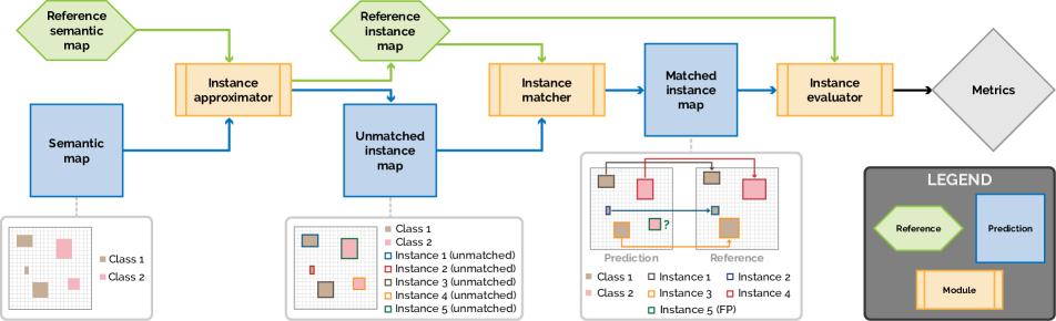
panoptica supports computing instance-wise segmentation, and object detection metrics from semantic-, as well as unmatched and matched segmentation maps, cf. Figure 1.
panoptica separates the metric computation process into three steps. This allows building modular metric computation pipelines, as illustrated in Figure 1. Due to its modular software architecture, resembling this three-step process, panoptica can easily be extended with additional algorithms for instance approximation, - matching, and metrics. To this end, a framework that automatically validates each input and output and defines all datatypes to be used is established. Therefore, a user extending panoptica with a custom algorithm receives immediate and understandable feedback regarding framework fit.
panoptica is an open-source package written in Python and included in the BrainLes project. It can be directly installed from PyPI , and the source code is available on GitHub . Further, panoptica is designed to process large-scale data-sets. To achieve fast computation times, it implements parallel computations by leveraging Python’s native multiprocessing library. Additionally, as many segmentation tasks only contain small foreground areas in an otherwise large image, inputs are cropped down to the combined bounding box of both prediction and reference to speed up computations. Detailed performance reports are available in the Supplemental Material, cf. Section 42.2.
The BrainLes project features several Jupyter notebook tutorials to illustrate the different use cases for panoptica with instance- and semantic segmentation maps. Further, Supplemental Material Section 42.3 illustrates how similarity metric curves can be analyzed in dependency of Intersection over Union (IOU) thresholds.
3.1 Instance approximation
While instance-wise metrics can directly be calculated from instance segmentation maps, semantic segmentation maps, by nature, do not contain instance information. Therefore, at the time of release, the instance approximator module provides means to approximate instances via Connected Component Analysis (CCA). For this CCA users can select between cc3d [35] and scipy backends. For most hardware configurations, cc3d is expected to perform better on 3D images, while scipy promises better 2D performance. Therefore, without user input, panoptica defaults to the respective backend for 2D and 3D inputs. For a detailed benchmark of CCA backends, cf. Supplemental Material Section 3.1.
3.2 Instance matching
The instance matcher module offers means to match instances based on a specified overlap metric. At the time of this writing, we support IOU and the Dice Similarity Coefficient (DSC). For simplicity of explanation, we will denote the selected metric as Matching Metric (MM), its value as Matching Score (MS). The task of the instance matcher is to assign each prediction instance to either a reference instance or as FP. Each FP is relabeled to an unused label so it persists in the map.
We only ever match instances of the prediction to the reference, leaving any given reference map untouched. Depending on the algorithm, we do support many-to-one matching, meaning multiple prediction instances can be matched to the same reference instance.
At the time of release, we offer two distinct instance-matching algorithms. However, the software architecture allows to easily extend panoptica with more sophisticated matching algorithms.
3.2.1 Naive Threshold Matcher
The naive threshold matcher assigns all prediction instances that exceed a certain user-given MS threshold with the reference instance.
If multiple predictions exceed the threshold for the same reference instance, only the one with the highest MS is matched to that reference. If the user specified that multiple predictions are allowed to be matched to the same reference instance, each of those matches are kept instead. Should a prediction instance exceed the threshold for multiple reference instances, it is only mapped to the reference instance with the highest MS.
3.2.2 Maximize Many-to-One Matcher
This many-to-one matching algorithm tries to maximize the MM by allowing multiple prediction instances to be assigned to the same reference instance if and only if the combined prediction instances have a greater MS than the individual instances. To this end, for each reference instance, it finds the best prediction instance based on the MM and then tests for all other instances that have an MS greater zero if including it would increase the score. Therefore, this algorithm maximizes the individual specified MM when matching but does not provide an overall optimal MS solution across all instances.
3.3 Instance evaluation
The instance evaluator module calculates instance-wise metrics by comparing reference to matched instance segmentation maps, cf. Figure 1.
Kirillov et al. [19] originally define the Panoptic Quality (PQ) metric as Equations 1 and 2:
| PQ | (1) |
| PQ | (2) |
where refers to the reference instances and to the predicted instances, and is the similarity metric.
In the original paper Kirillov et al. [19] propose to use IOU as similarity metric, while panoptica also implements PQ based on DSC and ASSD in addition to IOU. Note that the code can easily be modified to integrate further desired metrics, cf. Section 3.
Edge case handling: For some edge cases, segmentation metrics are not defined. For example, if both reference and prediction are empty DSC and ASSD become NaN, however, some might argue that DSC should be defined as for this case. A similar problem occurs for distance-based metrics such as ASSD, which become Inf if either prediction or reference is empty. NaN and Inf make it challenging to report aggregated metrics such as the mean. Therefore, practitioners developed task-specific mitigation strategies such as assigning a theoretically possible maximum distance for distance-based metrics [2]. In the absence of a general mitigation strategy producing satisfying results across all use cases, panoptica equips users with control over edge case handling.
4 Evaluation experiments
We conduct three biomedical evaluation experiments to showcase panoptica for various use cases. Across all experiments, we report the global volumetric Dice Similarity Coefficient (gvDSC) and the instance-wise Recognition Quality (RQ), Segmentation Quality (SQ), Panoptic Quality (PQ) and Segmentation Quality Average Symmetric Surface Distance (SQASSD). Note that SQASSD becomes NaN for cases with empty prediction and reference, and Inf for cases with either empty prediction or empty reference or only unmatched instances, cf. Section 3.3. Therefore, our mean and standard deviation cases are only based on cases that return a number.
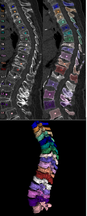
4.1 VerSe experiment
The Large Scale Vertebrae Segmentation Challenge (VerSe) challenge is about localization, labeling, and segmentation of vertebrae in CT scans of arbitrary fields of view [33]. For the segmentation part, the references were instance maps with labels that correspond to their anatomical label (i.e., 20 for L1). A simple volumetric DSC calculated on a binary version of this mask shows an overall semantic segmentation performance. However, instance-wise metrics are needed to measure how well an algorithm can actually distinguish the different vertebrae. Additionally, as in some cases, the actually different instance labels may be deemed relevant. If a model segments everything correctly but has all labels shifted by one, the resulting overall semantic similarity metric would be terrible, as this is not taken into account. panoptica allows the calculation of both metrics dynamically by stating whether the labels of the input maps are assumed to be already matched or not.
Segmentation Task: The task in the challenge is that of multi-class instance segmentation, though mainly tackled as a semantic segmentation task by the participants, cf. Figure 2.
Data: The dataset features Computed Tomography (CT) scans of various scanners, resolutions, and fields of view. We evaluate the segmentation algorithms on the hidden test set of the challenge consisting of subjects. The number of instances can vary between and (mean ) per scan.
Procedure: We use the top three submissions from the VerSe challenge and evaluate their predictions with the corresponding reference maps. We consider the input to panoptica to be unmatched instance maps, as we are not interested in evaluating the labeling capabilities but the segmentation metrics irrespective of their actual predicted vertebra labels. Table 1 illustrates the results of our analysis.
| Global Metrics | Instance-wise Metrics | ||||
|---|---|---|---|---|---|
| Submission | gvDSC | RQ | SQ | PQ | SQASSD |
| Payer et al. [29] | 0.932 ± 0.041 | 0.97 ± 0.046 | 0.912 ± 0.028 | 0.886 ± 0.048 | 0.37 ± 0.055 |
| deepreasoningai [8] | 0.937 ± 0.036 | 0.982 ± 0.038 | 0.898 ± 0.046 | 0.882 ± 0.063 | 0.414 ± 0.098 |
| jdlu [33] | 0.901 ± 0.037 | 0.947 ± 0.058 | 0.865 ± 0.039 | 0.818 ± 0.059 | 0.601 ± 0.41 |
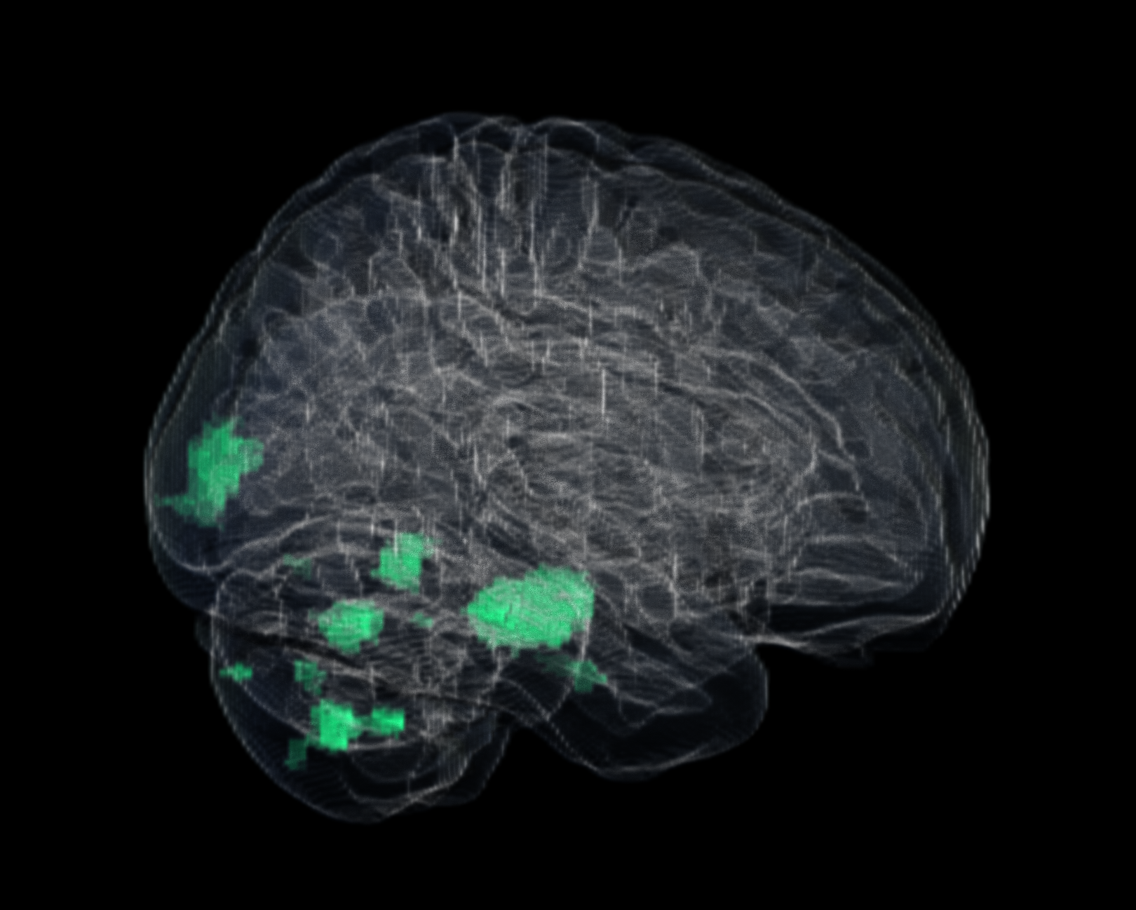
4.2 ISLES experiment
The Ischemic Stroke Lesion Segmentation 2022 challenge (ISLES) experiment serves as a practical example for the instance-wise evaluation of semantic segmentation maps. The segmentation of ischemia, addressed as a SemS task in Machine Learning (ML), is the focus of the ISLES’22 challenge. Despite this, the task and associated data encompass a wide variability of instances (lesions) requiring accurate delineation. Consequently, instance-wise analysis is valuable for tackling the problem. Specifically, the task involves scans featuring multiple non-overlapping embolic brain lesions that exhibit significant variations in terms of size, quantity, and anatomical location.
Segmentation Task: The ISLES challenge features a binary semantic segmentation task illustrated in Figure 3. The goal is to segment ischemic stroke brain lesions from multi-modal MRI data. Unlike this work, the original challenge evaluates the submissions’ performance using gvDSC, F1-score as instance-wise (here equals lesion-wise) detection, and further clinical metrics such as the differences in absolute lesion volume and absolute lesion count.
Data: We analyze the test set of the ISLES’22 MICCAI challenge [13]. The dataset consists of multi-center, multi-scanner MR scans. Each exam features three Magnetic Resonance Imaging (MRI) sequences, namely Diffusion Weighted Imaging (DWI), Apparent diffusion coefficient (ADC), and T2-weighted Fluid Attenuated Inversion Recovery (FLAIR), accompanied by a label mask encoding the stroke lesions. The data includes a wide variability of image acquisition and stroke patterns. The reference segmentations are obtained through an iterative hybrid algorithm-expert approach and are double-checked by experienced neuroradiologists. The number of non-overlapping instances (i.e., lesions) varies from none (i.e., healthy scans) to a hundred-twenty-six [13].
Procedure: In this work, we conduct a complementary analysis to the one conducted in the ISLES’22 challenge by assessing the top-three ranked methods with instance-wise metrics obtained through panoptica. We evaluate the submissions from team seals (rank 1) [12], nvauto (rank 2) and swan (rank 3). Team seals employs an ensemble of nnU-Nets [14]; team nvauto applies the Auto3dSeg pipeline [34] from MONAI [7], while the team swan utilizes a non-negative matrix factorization neural network [1]. Performance metrics obtained for each of these algorithms are summarized in Table Table 2.
| Global Metrics | Instance-wise Metrics | ||||
|---|---|---|---|---|---|
| Submission | gvDSC | RQ | SQ | PQ | SQASSD |
| seals [12] | 0.781 ± 0.181 | 0.52 ± 0.285 | 0.669 ± 0.217 | 0.380 ± 0.215 | 0.312 ± 0.175 |
| nvauto [34] | 0.783 ± 0.177 | 0.485 ± 0.278 | 0.669 ± 0.217 | 0.357 ± 0.211 | 0.307 ± 0.166 |
| swan [1] | 0.763 ± 0.194 | 0.462 ± 0.285 | 0.634 ± 0.253 | 0.334 ± 0.213 | 0.353 ± 0.292 |
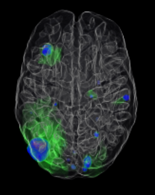
4.3 BraTS Mets experiment
The BraTS Mets experiment represents a use case for instance-wise evaluation of semantic segmentation maps. Although metastasis segmentation is an instance segmentation task from a clinical perspective, similar to stroke segmentation cf. Section 4.2, it is typically operationalized as a SemS task in ML, cf. Section 1.
Segmentation Task: The original segmentation task of the ASNR-MICCAI BraTS Brain Metastasis Challenge (BraTS-M) [28] is a multi-class hierarchical semantic segmentation task, cf. Figure 4.
Data: We evaluate on the publicly available training set of the BraTS-M [28]. The dataset consists of examinations, each containing four MRI sequences, namely a pre-contrast T1-weighted (t1w), a post-contrast T1-weighted (t1c), a T2-weighted (t2w), and a T2-weighted Fluid Attenuated Inversion Recovery (t2f) sequence combined with a multi-class semantic segmentation label, cf. Figure 4. Metastases counts in the dataset hugely vary between and (mean ) per exam.
Procedure: To explore how the glioma segmentation algorithms are able to generalize to cancer metastasis segmentation, we conduct the following off-label use experiment. We segment the whole dataset with five publicly available glioma SemS algorithms distributed through BraTS Toolkit [20], namely hnfnetv1-20 [16], isen-20 [15], sanet0-20 [39], scan_lite-20 [27], and yixinmpl-20 [38]. Additionally, we derive a sixth segmentation map named simple by fusing the five previously obtained algorithms using the implementation of the SIMPLE [25] fusion algorithm in BraTS Toolkit [20]. We then compute the gvDSC and the instance-wise metrics using panoptica, cf. Section 4. The results of this analysis are illustrated in Table 3.
| Global Metrics | Instance-wise Metrics | ||||
|---|---|---|---|---|---|
| Submission | gvDSC | RQ | SQ | PQ | SQASSD |
| hnfnetv1-20 [16] | 0.52 ± 0.39 | 0.33 ± 0.35 | 0.50 ± 0.39 | 0.27 ± 0.28 | 0.65 ± 0.37 |
| isen-20 [15] | 0.54 ± 0.39 | 0.37 ± 0.36 | 0.52 ± 0.38 | 0.29 ± 0.30 | 0.64 ± 0.32 |
| sanet0-20 [39] | 0.53 ± 0.38 | 0.35 ± 0.35 | 0.53 ± 0.38 | 0.28 ± 0.29 | 0.65 ± 0.35 |
| scan_lite-20 [27] | 0.49 ± 0.37 | 0.32 ± 0.35 | 0.46 ± 0.38 | 0.25 ± 0.28 | 0.70 ± 0.38 |
| yixinmpl-20 [38] | 0.52 ± 0.38 | 0.34 ± 0.35 | 0.50 ± 0.37 | 0.27 ± 0.28 | 0.70 ± 0.34 |
| simple [25] | 0.52 ± 0.39 | 0.35 ± 0.36 | 0.51 ± 0.38 | 0.28 ± 0.29 | 0.68 ± 0.36 |
5 Discussion
panoptica is an open-source, performance-optimized package written in Python. It allows the computation of instance-wise segmentation quality metrics, for instance- and semantic segmentation maps. Especially in the biomedical domain, for which we demonstrate three example use cases, instance-wise evaluation can be crucial to achieving a better representation of the underlying clinical task. Beyond our examples, there are countless segmentation problems, such as cell counting, that can profit from an instance-wise evaluation. As this also applies to the segmentation of natural images, panoptica is not limited to applications in the biomedical domain.
Limitations and future work: Transforming semantic segmentation problems into instance segmentation outputs via CCA comes with the drawback of typically discarding predicted class scores by relying on integer values as label maps. However, predicted class scores can be important and relevant for the validation. Luckily, instance-wise class scores can be retained from the SemS as shown in [31].
The metrics provided by panoptica can be easily extended to further metrics such as more selection for overlap- and boundary-based metrics, for example, by including Centerline Dice Similarity Coefficient (clDice), Normalized Surface Dice (NSD), or Hausdorff Distance (HD). In addition, the package could be extended to include multi-threshold metrics [26] such as Average Precision (AP) and [9] to assess detection quality in more detail. With a more comprehensive list of metrics, the package will be able to offer a more extensive performance assessment from different viewpoints and allow users to select the most suitable choices for their application.
The current package implements several overlap-based strategies. Another worthy addition would be to implement more matching strategies to cover a bigger variety of use cases. For example, if predicted class scores are available for each instance, the Greedy by Score matching [11] could be applied. This strategy orders predicted instances by their predicted class scores and identifies the matching reference instance by choosing the reference instance with the highest Matching Score (MS). Next, the assigned reference instance will be removed, such that this instance matcher remains a one-to-one matcher. If class scores are not available, the strategy can be simplified to the Greedy by Localization Criterion matching [26], which orders predicted instances by the MS. Furthermore, more advanced instance matchers like the Optimal Hungarian Matching [24] can be applied, which minimizes a cost function dependent on the Matching Metric (MM). Another interesting idea would be to implement matching based on topological features.
Similarly, we aim to extend the type of MM to include all localization criteria mentioned in the Metrics Reloaded framework [26], as it has been shown [26] that IOU is not always the best option for a localization criterion. For a comprehensive analysis of performance variability, we aim to expand the notion of mean and standard deviation to confidence intervals, for example, constructed via bootstrapping.
References
- Ashtari et al. [2023] Pooya Ashtari, Diana M Sima, Lieven De Lathauwer, Dominique Sappey-Marinier, Frederik Maes, and Sabine Van Huffel. Factorizer: A scalable interpretable approach to context modeling for medical image segmentation. Medical image analysis, 84:102706, 2023.
- Bakas et al. [2019] Spyridon Bakas, Mauricio Reyes, Andras Jakab, Stefan Bauer, Markus Rempfler, Alessandro Crimi, Russell Takeshi Shinohara, Christoph Berger, Sung Min Ha, Martin Rozycki, Marcel Prastawa, Esther Alberts, Jana Lipkova, John Freymann, Justin Kirby, Michel Bilello, Hassan Fathallah-Shaykh, Roland Wiest, Jan Kirschke, Benedikt Wiestler, Rivka Colen, Aikaterini Kotrotsou, Pamela Lamontagne, Daniel Marcus, Mikhail Milchenko, Arash Nazeri, Marc-Andre Weber, Abhishek Mahajan, Ujjwal Baid, Elizabeth Gerstner, Dongjin Kwon, Gagan Acharya, Manu Agarwal, Mahbubul Alam, Alberto Albiol, Antonio Albiol, Francisco J. Albiol, Varghese Alex, Nigel Allinson, Pedro H. A. Amorim, Abhijit Amrutkar, Ganesh Anand, Simon Andermatt, Tal Arbel, Pablo Arbelaez, Aaron Avery, Muneeza Azmat, Pranjal B., W Bai, Subhashis Banerjee, Bill Barth, Thomas Batchelder, Kayhan Batmanghelich, Enzo Battistella, Andrew Beers, Mikhail Belyaev, Martin Bendszus, Eze Benson, Jose Bernal, Halandur Nagaraja Bharath, George Biros, Sotirios Bisdas, James Brown, Mariano Cabezas, Shilei Cao, Jorge M. Cardoso, Eric N Carver, Adrià Casamitjana, Laura Silvana Castillo, Marcel Catà, Philippe Cattin, Albert Cerigues, Vinicius S. Chagas, Siddhartha Chandra, Yi-Ju Chang, Shiyu Chang, Ken Chang, Joseph Chazalon, Shengcong Chen, Wei Chen, Jefferson W Chen, Zhaolin Chen, Kun Cheng, Ahana Roy Choudhury, Roger Chylla, Albert Clérigues, Steven Colleman, Ramiro German Rodriguez Colmeiro, Marc Combalia, Anthony Costa, Xiaomeng Cui, Zhenzhen Dai, Lutao Dai, Laura Alexandra Daza, Eric Deutsch, Changxing Ding, Chao Dong, Shidu Dong, Wojciech Dudzik, Zach Eaton-Rosen, Gary Egan, Guilherme Escudero, Théo Estienne, Richard Everson, Jonathan Fabrizio, Yong Fan, Longwei Fang, Xue Feng, Enzo Ferrante, Lucas Fidon, Martin Fischer, Andrew P. French, Naomi Fridman, Huan Fu, David Fuentes, Yaozong Gao, Evan Gates, David Gering, Amir Gholami, Willi Gierke, Ben Glocker, Mingming Gong, Sandra González-Villá, T. Grosges, Yuanfang Guan, Sheng Guo, Sudeep Gupta, Woo-Sup Han, Il Song Han, Konstantin Harmuth, Huiguang He, Aura Hernández-Sabaté, Evelyn Herrmann, Naveen Himthani, Winston Hsu, Cheyu Hsu, Xiaojun Hu, Xiaobin Hu, Yan Hu, Yifan Hu, Rui Hua, Teng-Yi Huang, Weilin Huang, Sabine Van Huffel, Quan Huo, Vivek HV, Khan M. Iftekharuddin, Fabian Isensee, Mobarakol Islam, Aaron S. Jackson, Sachin R. Jambawalikar, Andrew Jesson, Weijian Jian, Peter Jin, V Jeya Maria Jose, Alain Jungo, B Kainz, Konstantinos Kamnitsas, Po-Yu Kao, Ayush Karnawat, Thomas Kellermeier, Adel Kermi, Kurt Keutzer, Mohamed Tarek Khadir, Mahendra Khened, Philipp Kickingereder, Geena Kim, Nik King, Haley Knapp, Urspeter Knecht, Lisa Kohli, Deren Kong, Xiangmao Kong, Simon Koppers, Avinash Kori, Ganapathy Krishnamurthi, Egor Krivov, Piyush Kumar, Kaisar Kushibar, Dmitrii Lachinov, Tryphon Lambrou, Joon Lee, Chengen Lee, Yuehchou Lee, M Lee, Szidonia Lefkovits, Laszlo Lefkovits, James Levitt, Tengfei Li, Hongwei Li, Wenqi Li, Hongyang Li, Xiaochuan Li, Yuexiang Li, Heng Li, Zhenye Li, Xiaoyu Li, Zeju Li, XiaoGang Li, Wenqi Li, Zheng-Shen Lin, Fengming Lin, Pietro Lio, Chang Liu, Boqiang Liu, Xiang Liu, Mingyuan Liu, Ju Liu, Luyan Liu, Xavier Llado, Marc Moreno Lopez, Pablo Ribalta Lorenzo, Zhentai Lu, Lin Luo, Zhigang Luo, Jun Ma, Kai Ma, Thomas Mackie, Anant Madabushi, Issam Mahmoudi, Klaus H. Maier-Hein, Pradipta Maji, CP Mammen, Andreas Mang, B. S. Manjunath, Michal Marcinkiewicz, S McDonagh, Stephen McKenna, Richard McKinley, Miriam Mehl, Sachin Mehta, Raghav Mehta, Raphael Meier, Christoph Meinel, Dorit Merhof, Craig Meyer, Robert Miller, Sushmita Mitra, Aliasgar Moiyadi, David Molina-Garcia, Miguel A. B. Monteiro, Grzegorz Mrukwa, Andriy Myronenko, Jakub Nalepa, Thuyen Ngo, Dong Nie, Holly Ning, Chen Niu, Nicholas K Nuechterlein, Eric Oermann, Arlindo Oliveira, Diego D. C. Oliveira, Arnau Oliver, Alexander F. I. Osman, Yu-Nian Ou, Sebastien Ourselin, Nikos Paragios, Moo Sung Park, Brad Paschke, J. Gregory Pauloski, Kamlesh Pawar, Nick Pawlowski, Linmin Pei, Suting Peng, Silvio M. Pereira, Julian Perez-Beteta, Victor M. Perez-Garcia, Simon Pezold, Bao Pham, Ashish Phophalia, Gemma Piella, G. N. Pillai, Marie Piraud, Maxim Pisov, Anmol Popli, Michael P. Pound, Reza Pourreza, Prateek Prasanna, Vesna Prkovska, Tony P. Pridmore, Santi Puch, Élodie Puybareau, Buyue Qian, Xu Qiao, Martin Rajchl, Swapnil Rane, Michael Rebsamen, Hongliang Ren, Xuhua Ren, Karthik Revanuru, Mina Rezaei, Oliver Rippel, Luis Carlos Rivera, Charlotte Robert, Bruce Rosen, Daniel Rueckert, Mohammed Safwan, Mostafa Salem, Joaquim Salvi, Irina Sanchez, Irina Sánchez, Heitor M. Santos, Emmett Sartor, Dawid Schellingerhout, Klaudius Scheufele, Matthew R. Scott, Artur A. Scussel, Sara Sedlar, Juan Pablo Serrano-Rubio, N. Jon Shah, Nameetha Shah, Mazhar Shaikh, B. Uma Shankar, Zeina Shboul, Haipeng Shen, Dinggang Shen, Linlin Shen, Haocheng Shen, Varun Shenoy, Feng Shi, Hyung Eun Shin, Hai Shu, Diana Sima, M Sinclair, Orjan Smedby, James M. Snyder, Mohammadreza Soltaninejad, Guidong Song, Mehul Soni, Jean Stawiaski, Shashank Subramanian, Li Sun, Roger Sun, Jiawei Sun, Kay Sun, Yu Sun, Guoxia Sun, Shuang Sun, Yannick R Suter, Laszlo Szilagyi, Sanjay Talbar, Dacheng Tao, Dacheng Tao, Zhongzhao Teng, Siddhesh Thakur, Meenakshi H Thakur, Sameer Tharakan, Pallavi Tiwari, Guillaume Tochon, Tuan Tran, Yuhsiang M. Tsai, Kuan-Lun Tseng, Tran Anh Tuan, Vadim Turlapov, Nicholas Tustison, Maria Vakalopoulou, Sergi Valverde, Rami Vanguri, Evgeny Vasiliev, Jonathan Ventura, Luis Vera, Tom Vercauteren, C. A. Verrastro, Lasitha Vidyaratne, Veronica Vilaplana, Ajeet Vivekanandan, Guotai Wang, Qian Wang, Chiatse J. Wang, Weichung Wang, Duo Wang, Ruixuan Wang, Yuanyuan Wang, Chunliang Wang, Guotai Wang, Ning Wen, Xin Wen, Leon Weninger, Wolfgang Wick, Shaocheng Wu, Qiang Wu, Yihong Wu, Yong Xia, Yanwu Xu, Xiaowen Xu, Peiyuan Xu, Tsai-Ling Yang, Xiaoping Yang, Hao-Yu Yang, Junlin Yang, Haojin Yang, Guang Yang, Hongdou Yao, Xujiong Ye, Changchang Yin, Brett Young-Moxon, Jinhua Yu, Xiangyu Yue, Songtao Zhang, Angela Zhang, Kun Zhang, Xuejie Zhang, Lichi Zhang, Xiaoyue Zhang, Yazhuo Zhang, Lei Zhang, Jianguo Zhang, Xiang Zhang, Tianhao Zhang, Sicheng Zhao, Yu Zhao, Xiaomei Zhao, Liang Zhao, Yefeng Zheng, Liming Zhong, Chenhong Zhou, Xiaobing Zhou, Fan Zhou, Hongtu Zhu, Jin Zhu, Ying Zhuge, Weiwei Zong, Jayashree Kalpathy-Cramer, Keyvan Farahani, Christos Davatzikos, Koen van Leemput, and Bjoern Menze. Identifying the best machine learning algorithms for brain tumor segmentation, progression assessment, and overall survival prediction in the brats challenge, 2019.
- Bhatia et al. [2022] Harsharan Singh Bhatia, Andreas-David Brunner, Furkan Öztürk, Saketh Kapoor, Zhouyi Rong, Hongcheng Mai, Marvin Thielert, Mayar Ali, Rami Al-Maskari, Johannes Christian Paetzold, et al. Spatial proteomics in three-dimensional intact specimens. Cell, 185(26):5040–5058, 2022.
- Bilic et al. [2023] Patrick Bilic, Patrick Christ, Hongwei Bran Li, Eugene Vorontsov, Avi Ben-Cohen, Georgios Kaissis, Adi Szeskin, Colin Jacobs, Gabriel Efrain Humpire Mamani, Gabriel Chartrand, et al. The liver tumor segmentation benchmark (lits). Medical Image Analysis, 84:102680, 2023.
- Buchner et al. [2023a] Josef A Buchner, Florian Kofler, Lucas Etzel, Michael Mayinger, Sebastian M Christ, Thomas B Brunner, Andrea Wittig, Björn Menze, Claus Zimmer, Bernhard Meyer, et al. Development and external validation of an mri-based neural network for brain metastasis segmentation in the aurora multicenter study. Radiotherapy and Oncology, 178:109425, 2023a.
- Buchner et al. [2023b] Josef A Buchner, Jan C Peeken, Lucas Etzel, Ivan Ezhov, Michael Mayinger, Sebastian M Christ, Thomas B Brunner, Andrea Wittig, Bjoern H Menze, Claus Zimmer, et al. Identifying core mri sequences for reliable automatic brain metastasis segmentation. Radiotherapy and Oncology, 188:109901, 2023b.
- Cardoso et al. [2022] M Jorge Cardoso, Wenqi Li, Richard Brown, Nic Ma, Eric Kerfoot, Yiheng Wang, Benjamin Murrey, Andriy Myronenko, Can Zhao, Dong Yang, et al. Monai: An open-source framework for deep learning in healthcare. arXiv preprint arXiv:2211.02701, 2022.
- Chen et al. [2015] Hao Chen, Chiyao Shen, Jing Qin, Dong Ni, Lin Shi, Jack C. Y. Cheng, and Pheng-Ann Heng. Automatic localization and identification of vertebrae in spine ct via a joint learning model with deep neural networks. In Medical Image Computing and Computer-Assisted Intervention – MICCAI 2015, pages 515–522, Cham, 2015. Springer International Publishing.
- Chen et al. [2023] Long Chen, Yuli Wu, Johannes Stegmaier, and Dorit Merhof. Sortedap: Rethinking evaluation metrics for instance segmentation. In Proceedings of the IEEE/CVF International Conference on Computer Vision, pages 3923–3929, 2023.
- Detlefsen et al. [2022] Nicki Skafte Detlefsen, Jiri Borovec, Justus Schock, Ananya Harsh Jha, Teddy Koker, Luca Di Liello, Daniel Stancl, Changsheng Quan, Maxim Grechkin, and William Falcon. Torchmetrics-measuring reproducibility in pytorch. Journal of Open Source Software, 7(70):4101, 2022.
- Everingham et al. [2015] Mark Everingham, SM Ali Eslami, Luc Van Gool, Christopher KI Williams, John Winn, and Andrew Zisserman. The pascal visual object classes challenge: A retrospective. International journal of computer vision, 111(1):98–136, 2015.
- Gao [2022] Shengbo Gao. SEALS- ISLES’22 participating method. https://github.com/Tabrisrei/ISLES22_SEALS, 2022. [Online; accessed 16-November-2023].
- Hernandez Petzsche et al. [2022] Moritz R Hernandez Petzsche, Ezequiel de la Rosa, Uta Hanning, Roland Wiest, Waldo Valenzuela, Mauricio Reyes, Maria Meyer, Sook-Lei Liew, Florian Kofler, Ivan Ezhov, et al. Isles 2022: A multi-center magnetic resonance imaging stroke lesion segmentation dataset. Scientific data, 9(1):762, 2022.
- Isensee et al. [2021a] Fabian Isensee, Paul F Jaeger, Simon AA Kohl, Jens Petersen, and Klaus H Maier-Hein. nnu-net: a self-configuring method for deep learning-based biomedical image segmentation. Nature methods, 18(2):203–211, 2021a.
- Isensee et al. [2021b] Fabian Isensee, Paul F Jäger, Peter M Full, Philipp Vollmuth, and Klaus H Maier-Hein. nnu-net for brain tumor segmentation. In Brainlesion: Glioma, Multiple Sclerosis, Stroke and Traumatic Brain Injuries: 6th International Workshop, BrainLes 2020, Held in Conjunction with MICCAI 2020, Lima, Peru, October 4, 2020, Revised Selected Papers, Part II 6, pages 118–132. Springer, 2021b.
- Jia et al. [2021] Haozhe Jia, Weidong Cai, Heng Huang, and Yong Xia. H^ 2 2 nf-net for brain tumor segmentation using multimodal mr imaging: 2nd place solution to brats challenge 2020 segmentation task. In Brainlesion: Glioma, Multiple Sclerosis, Stroke and Traumatic Brain Injuries: 6th International Workshop, BrainLes 2020, Held in Conjunction with MICCAI 2020, Lima, Peru, October 4, 2020, Revised Selected Papers, Part II 6, pages 58–68. Springer, 2021.
- Jungo et al. [2021] Alain Jungo, Olivier Scheidegger, Mauricio Reyes, and Fabian Balsiger. pymia: A python package for data handling and evaluation in deep learning-based medical image analysis. Computer methods and programs in biomedicine, 198:105796, 2021.
- Kaltenecker et al. [2023] Doris Kaltenecker, Rami Al-Maskari, Moritz Negwer, Luciano Hoeher, Florian Kofler, Shan Zhao, Mihail Todorov Todorov, Johannes Christian Paetzold, Benedikt Wiestler, Julia Geppert, et al. Virtual reality empowered deep learning analysis of brain activity. bioRxiv, pages 2023–05, 2023.
- Kirillov et al. [2019] Alexander Kirillov, Kaiming He, Ross Girshick, Carsten Rother, and Piotr Dollár. Panoptic segmentation. In Proceedings of the IEEE/CVF conference on computer vision and pattern recognition, pages 9404–9413, 2019.
- Kofler et al. [2020] Florian Kofler, Christoph Berger, Diana Waldmannstetter, Jana Lipkova, Ivan Ezhov, Giles Tetteh, Jan Kirschke, Claus Zimmer, Benedikt Wiestler, and Bjoern H Menze. Brats toolkit: translating brats brain tumor segmentation algorithms into clinical and scientific practice. Frontiers in neuroscience, page 125, 2020.
- Kofler et al. [2021] Florian Kofler, Ivan Ezhov, Fabian Isensee, Fabian Balsiger, Christoph Berger, Maximilian Koerner, Johannes Paetzold, Hongwei Li, Suprosanna Shit, Richard McKinley, et al. Are we using appropriate segmentation metrics? identifying correlates of human expert perception for cnn training beyond rolling the dice coefficient. arXiv preprint arXiv:2103.06205, 2021.
- Kofler et al. [2023a] Florian Kofler, Suprosanna Shit, Ivan Ezhov, Lucas Fidon, Izabela Horvath, Rami Al-Maskari, Hongwei Bran Li, Harsharan Bhatia, Timo Loehr, Marie Piraud, et al. blob loss: instance imbalance aware loss functions for semantic segmentation. In International Conference on Information Processing in Medical Imaging, pages 755–767. Springer Nature Switzerland Cham, 2023a.
- Kofler et al. [2023b] Florian Kofler, Johannes Wahle, Ivan Ezhov, Sophia J Wagner, Rami Al-Maskari, Emilia Gryska, Mihail Todorov, Christina Bukas, Felix Meissen, Tingying Peng, et al. Approaching peak ground truth. In 2023 IEEE 20th International Symposium on Biomedical Imaging (ISBI), pages 1–6. IEEE, 2023b.
- Kuhn [1955] Harold W Kuhn. The hungarian method for the assignment problem. Naval research logistics quarterly, 2(1-2):83–97, 1955.
- Langerak et al. [2010] Thomas Robin Langerak, Uulke A van der Heide, Alexis NTJ Kotte, Max A Viergever, Marco Van Vulpen, and Josien PW Pluim. Label fusion in atlas-based segmentation using a selective and iterative method for performance level estimation (simple). IEEE transactions on medical imaging, 29(12):2000–2008, 2010.
- Maier-Hein et al. [2023] Lena Maier-Hein, Annika Reinke, Patrick Godau, Minu D. Tizabi, Florian Buettner, Evangelia Christodoulou, Ben Glocker, Fabian Isensee, Jens Kleesiek, Michal Kozubek, Mauricio Reyes, Michael A. Riegler, Manuel Wiesenfarth, A. Emre Kavur, Carole H. Sudre, Michael Baumgartner, Matthias Eisenmann, Doreen Heckmann-Nötzel, A. Tim Rädsch, Laura Acion, Michela Antonelli, Tal Arbel, Spyridon Bakas, Arriel Benis, Matthew Blaschko, M. Jorge Cardoso, Veronika Cheplygina, Beth A. Cimini, Gary S. Collins, Keyvan Farahani, Luciana Ferrer, Adrian Galdran, Bram van Ginneken, Robert Haase, Daniel A. Hashimoto, Michael M. Hoffman, Merel Huisman, Pierre Jannin, Charles E. Kahn, Dagmar Kainmueller, Bernhard Kainz, Alexandros Karargyris, Alan Karthikesalingam, Hannes Kenngott, Florian Kofler, Annette Kopp-Schneider, Anna Kreshuk, Tahsin Kurc, Bennett A. Landman, Geert Litjens, Amin Madani, Klaus Maier-Hein, Anne L. Martel, Peter Mattson, Erik Meijering, Bjoern Menze, Karel G. M. Moons, Henning Müller, Brennan Nichyporuk, Felix Nickel, Jens Petersen, Nasir Rajpoot, Nicola Rieke, Julio Saez-Rodriguez, Clara I. Sánchez, Shravya Shetty, Maarten van Smeden, Ronald M. Summers, Abdel A. Taha, Aleksei Tiulpin, Sotirios A. Tsaftaris, Ben Van Calster, Gaël Varoquaux, and Paul F. Jäger. Metrics reloaded: Recommendations for image analysis validation, 2023.
- McKinley et al. [2021] Richard McKinley, Micheal Rebsamen, Katrin Dätwyler, Raphael Meier, Piotr Radojewski, and Roland Wiest. Uncertainty-driven refinement of tumor-core segmentation using 3d-to-2d networks with label uncertainty. In Brainlesion: Glioma, Multiple Sclerosis, Stroke and Traumatic Brain Injuries: 6th International Workshop, BrainLes 2020, Held in Conjunction with MICCAI 2020, Lima, Peru, October 4, 2020, Revised Selected Papers, Part I 6, pages 401–411. Springer, 2021.
- Moawad et al. [2023] Ahmed W Moawad, Anastasia Janas, Ujjwal Baid, Divya Ramakrishnan, Leon Jekel, Kiril Krantchev, Harrison Moy, Rachit Saluja, Klara Osenberg, Klara Wilms, et al. The brain tumor segmentation (brats-mets) challenge 2023: Brain metastasis segmentation on pre-treatment mri. ArXiv, 2023.
- Payer. et al. [2020] Christian Payer., Darko Štern., Horst Bischof., and Martin Urschler. Coarse to fine vertebrae localization and segmentation with spatialconfiguration-net and u-net. In Proceedings of the 15th International Joint Conference on Computer Vision, Imaging and Computer Graphics Theory and Applications (VISIGRAPP 2020) - Volume 5: VISAPP, pages 124–133. INSTICC, SciTePress, 2020.
- Prabhakar et al. [2023] Chinmay Prabhakar, Hongwei Bran Li, Johannes C Paetzold, Timo Loehr, Chen Niu, Mark Mühlau, Daniel Rueckert, Benedikt Wiestler, and Bjoern Menze. Self-pruning graph neural network for predicting inflammatory disease activity in multiple sclerosis from brain mr images. In International Conference on Medical Image Computing and Computer-Assisted Intervention, pages 226–236. Springer, 2023.
- Reinke et al. [2021] Annika Reinke, Minu D Tizabi, Carole H Sudre, Matthias Eisenmann, Tim Rädsch, Michael Baumgartner, Laura Acion, Michela Antonelli, Tal Arbel, Spyridon Bakas, et al. Common limitations of image processing metrics: A picture story. arXiv preprint arXiv:2104.05642, 2021.
- Ronneberger et al. [2015] Olaf Ronneberger, Philipp Fischer, and Thomas Brox. U-net: Convolutional networks for biomedical image segmentation. In Medical Image Computing and Computer-Assisted Intervention – MICCAI 2015, pages 234–241, Cham, 2015. Springer International Publishing.
- Sekuboyina et al. [2021] Anjany Sekuboyina, Malek E Husseini, Amirhossein Bayat, Maximilian Löffler, Hans Liebl, Hongwei Li, Giles Tetteh, Jan Kukačka, Christian Payer, Darko Štern, et al. Verse: a vertebrae labelling and segmentation benchmark for multi-detector ct images. Medical image analysis, 73:102166, 2021.
- Siddique et al. [2022] Md Mahfuzur Rahman Siddique, Dong Yang, Yufan He, Daguang Xu, and Andriy Myronenko. Automated ischemic stroke lesion segmentation from 3d mri. arXiv preprint arXiv:2209.09546, 2022.
- Silversmith [2021] William Silversmith. cc3d: Connected components on multilabel 3D & 2D images. Zenodo, 2021.
- Sudre et al. [2022] Carole H Sudre, Kimberlin Van Wijnen, Florian Dubost, Hieab Adams, David Atkinson, Frederik Barkhof, Mahlet A Birhanu, Esther E Bron, Robin Camarasa, Nish Chaturvedi, et al. Where is valdo? vascular lesions detection and segmentation challenge at miccai 2021. arXiv preprint arXiv:2208.07167, 2022.
- Thompson et al. [2018] Alan J Thompson, Brenda L Banwell, Frederik Barkhof, William M Carroll, Timothy Coetzee, Giancarlo Comi, Jorge Correale, Franz Fazekas, Massimo Filippi, Mark S Freedman, et al. Diagnosis of multiple sclerosis: 2017 revisions of the mcdonald criteria. The Lancet Neurology, 17(2):162–173, 2018.
- Wang et al. [2021] Yixin Wang, Yao Zhang, Feng Hou, Yang Liu, Jiang Tian, Cheng Zhong, Yang Zhang, and Zhiqiang He. Modality-pairing learning for brain tumor segmentation. In Brainlesion: Glioma, Multiple Sclerosis, Stroke and Traumatic Brain Injuries: 6th International Workshop, BrainLes 2020, Held in Conjunction with MICCAI 2020, Lima, Peru, October 4, 2020, Revised Selected Papers, Part I 6, pages 230–240. Springer, 2021.
- Yuan [2021] Yading Yuan. Automatic brain tumor segmentation with scale attention network. In Brainlesion: Glioma, Multiple Sclerosis, Stroke and Traumatic Brain Injuries: 6th International Workshop, BrainLes 2020, Held in Conjunction with MICCAI 2020, Lima, Peru, October 4, 2020, Revised Selected Papers, Part I 6, pages 285–294. Springer, 2021.
42 Supplemental Material
42.1 CCA Benchmark: cc3d vs. scipy
Table 4 summarizes the comparison of CCA backends. The code for the comparison is publicly available.
| Dimensionality | Volume Size | Scipy Time [s] | CC3D Time [s] |
|---|---|---|---|
| 2D | 0.0350 | 0.0552 | |
| 0.1344 | 0.2268 | ||
| 0.5121 | 0.8542 | ||
| 2.1542 | 3.4198 | ||
| 9.5657 | 16.4068 | ||
| 3D | 0.0204 | 0.0144 | |
| 0.1564 | 0.1160 | ||
| 1.2277 | 0.9043 | ||
| 20.7657 | 15.3032 | ||
| 195.1927 | 145.1652 |
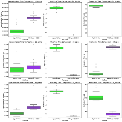
42.2 Computation times
We benchmark the three modules from panoptica on exemplary samples. Across all these performance tests, we use instance approximation based on CCA, instance matching with the naive one-to-one setting (cf. Section 3.2) using IOU as MM, and evaluate the instance-wise metrics RQ, SQ, PQ and ASSD. For all experiments, we use a IOU threshold of both for matching and evaluation. The following test setups are used:
-
•
Work station: Ubuntu 22.04, AMD Ryzen 9 5900x, 128GB RAM
-
•
Apple Macbook Pro with M1 Max 10-Core processor, 32GB RAM
The code to run the benchmark is publicly available. The exemplary samples include:
-
•
2d_simple: A simulated (50,50) 2D input consisting of two instances both in prediction and reference.
-
•
3d_spine: A real MRI spine segmentation sample of shape (170, 512, 17) consisting of 89 prediction instances and 87 reference instances. 73 instances are successfully matched.
-
•
3d_dense: A (100, 100, 100) shaped image densely filled with 40 simulated instances both in prediction and reference.
Figure 5 shows the result of our tests.
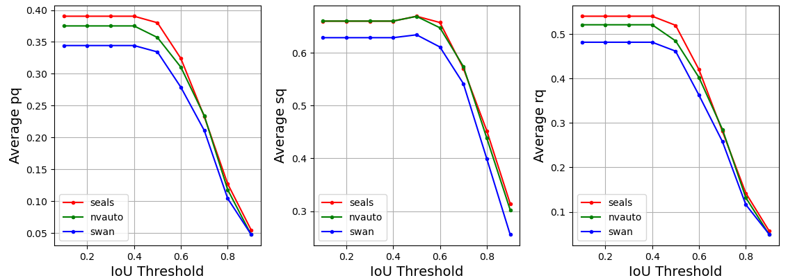
42.3 Segmentation Performance over varying IOU-thresholds
With panoptica performance measures can easily be computed for various IOU-thresholds, cf. Figure 6. Note that different thresholds could be selected for instance matching and instance evaluation.
Acknowledgement
Bjoern Menze, Benedikt Wiestler, and Florian Kofler are supported through the SFB 824, subproject B12. Supported by Deutsche Forschungsgemeinschaft (DFG) through TUM International Graduate School of Science and Engineering (IGSSE), GSC 81. Supported by Anna Valentina Lioba Eleonora Claire Javid Mamasani. Bjoern Menze acknowledges support by the Helmut Horten Foundation. Ivan Ezhov is supported by the Translational Brain Imaging Training Network (TRABIT) under the European Union’s ‘Horizon 2020’ research & innovation program (Grant agreement ID: 765148). Fabian Isensee is supported by Helmholtz Imaging (HI), a platform of the Helmholtz Incubator on Information and Data Science. Jan Kirschke has received Grants from the ERC, DFG, BMBF and is a Co-Founder of Bonescreen GmbH. Benedikt Wiestler is supported by the NIH (R01CA269948).