Wearable Technologies for Monitoring
Upper Extremity Functions During Daily Life
in Neurologically Impaired Individuals
Abstract
Neurological disorders, including stroke, spinal cord injuries, multiple sclerosis, and Parkinson’s disease, generally lead to diminished upper extremity (UE) function, impacting individuals’ independence and quality of life. Traditional assessments predominantly focus on standardized clinical tasks, offering limited insights into real-life UE performance. In this context, this review focuses on wearable technologies as a promising solution to monitor UE function in neurologically impaired individuals during daily life activities. Our primary objective is to categorize the different sensors, data collection and data processing approaches employed. What comes to light is that the majority of studies involved stroke survivors, and predominantly employed inertial measurement units and accelerometers to collect kinematics. Most analyses in these studies were performed offline, focusing on activity duration and frequency as key metrics. Although wearable technology shows potential in monitoring UE function in real-life scenarios, an ideal solution that combines non-intrusiveness, lightweight design, detailed hand and finger movement capture, contextual information, extended recording duration, ease of use, and privacy protection remains an elusive goal. Furthermore, it stands out a growing necessity for a multimodal approach in capturing comprehensive data on UE function during real-life activities to enhance the personalization of rehabilitation strategies and ultimately improve outcomes for these individuals.
Index Terms:
Upper Extremities, Monitoring, Wearable Technologies, Rehabilitation, AssistanceI Introduction
The functional use of the upper extremities (UEs) is a paramount aspect of daily life for every human being, as it directly correlates with the ability to independently conduct activities of daily living (ADLs) [1, 2]. Neurological disorders such as spinal cord injuries (SCI), stroke, multiple sclerosis (MS), and Parkinson’s disease (PD) have direct consequences on the ability to use the UEs, resulting in reduced independence, diminished quality of life, and limited social participation [3, 4, 5, 6]. Over the years, various rehabilitative approaches, including technologies like functional electrical stimulation (FES), transcutaneous spinal cord stimulation (tSCS), and exoskeletons, in conjunction with targeted physiotherapy and occupational therapy, have made notable strides in improving UE function following neurological disorders [7, 8, 9, 10, 11].
Despite significant progress in the field of rehabilitation therapies and assistive technologies, a substantial challenge persists. Traditional rehabilitation assessments and monitoring technologies predominantly concentrate on evaluating the capacity domain of UE use, as defined by the International Classification of Functioning, Disability, and Health (ICF) framework [12]. This domain focuses on an individual’s ability to perform standardized tasks in controlled environments. However, the heart of neurorehabilitation lies in the crucial aspect of translating the UE function improvements observed in clinical settings into real-life enhancements [13, 14, 15, 16]. This translation is essential for enabling individuals with neurological impairments to achieve increased independence. To achieve this, it is crucial to assess the performance domain of the ICF, which gauges how individuals carry out activities in their typical daily environment [17].
To gain a comprehensive understanding of the impact of rehabilitative interventions on individuals with neurological disorders affecting UE functions and to tailor rehabilitation strategies that enhance their independence and social participation, it is imperative to bridge the gap between capacity and performance evaluations [18, 19, 20]. This is where the potential of monitoring individuals during their daily activities becomes evident. Observing and quantifying how patients with neurological impairments use their UEs outside of clinical environments can provide invaluable insights for fine-tuning rehabilitation programs, with the ultimate goal of improving their independence and quality of life. This approach is pivotal for crafting personalized rehabilitation strategies that address the specific needs and challenges faced every day by these individuals.
To achieve this goal, we stand at the crossroads of rehabilitation science, wearable technology, and data science. The advent of wearable sensor technologies presents an encouraging opportunity to track UE usage in individuals with neurological conditions as they go about their everyday lives. These technologies hold the promise of revealing a wealth of data that was previously unattainable within the limits of clinical environments. Moreover, thanks to recent developments in machine learning, this data can be efficiently processed and conveyed to clinicians, offering valuable insights into patients’ real-life progress [21].
This literature review focuses on the latest advancements in wearable technologies designed to monitor UE function in real-life, unconstrained situations (i.e., not limited to standardized tasks), and across various neurological conditions, a perspective that has yet to be comprehensively covered in the existing literature. While similar reviews in the field of neurorehabilitation recognized the significance of wearable sensors for telemonitoring and telerehabilitation, they primarily focused on specific aspects. For instance, Toh et al. (2023) [22] investigated the effectiveness of wearable technologies in home-based physical rehabilitation for stroke, while Gopal et al. (2022) [23] examined the use of wearables, smartphone-, and tablet-based apps for standardized clinical assessments of hand function in chronic neurological disorders. Other reviews delved into the transition of inertial sensors from laboratory to community settings for monitoring upper and lower limb functions in individuals with PD [24], explored wearable sensor data’s role in stroke rehabilitation [25], and discussed wearable solutions in the context of MS [26].
By focusing on the pivotal aspect of monitoring UEs during the unstructured activities of daily life, we want to identify and categorize the most prevalent types of wearable sensors employed for monitoring UE use during daily life activities, describe the way these were used, and how collected data was processed. Beyond presenting a comprehensive state of the art, this review will offer valuable guidelines and insights to researchers and practitioners alike. We will explore the advantages and potential pitfalls associated with monitoring UE use in home environments, providing a roadmap for future research endeavors and acknowledging that true functional recovery extends beyond the clinic.
II Methods
The review was conducted by searching major scientific databases (Scopus, Google Scholar), using various combinations of keywords related to wearable technology, upper limb monitoring, and real-world applications. The search was limited to English manuscripts and journal articles only.
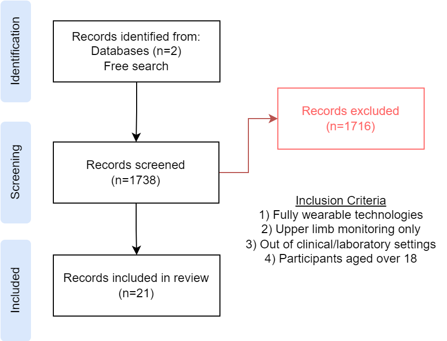
In the initial database search covering the period until April 2023, we retrieved a total of 1,738 titles. The first screening was performed based on the title and abstract, applying specific inclusion and exclusion criteria. Inclusion criteria comprised: 1) fully wearable technologies, 2) upper limb monitoring, 3) experiments conducted outside clinical or laboratory settings, and 4) the inclusion of adult participants aged over 18. On the contrary, we excluded studies involving only healthy individuals, non-neurological upper limb impairments (e.g., amputation, traumatic orthopedic conditions), papers focusing solely on lower limb monitoring, physiological signal monitoring (e.g., cardiovascular, brain, respiration), and studies conducted solely in clinical or laboratory environments.
In cases of uncertainty, we reviewed the full text before making inclusion/exclusion decisions. Additionally, we included other documents found through a manual search of references from existing papers. Finally, 21 papers – published between 2007 and April 2023 – met the criteria for this review.
III State of the Art
The included studies underwent a data extraction process to identify key domains for categorizing the literature in this research field. These domains were 1) the specific neurological impairments studied for monitoring the UE functions (application); 2) the hardware employed for UE function monitoring ; 3) the data processing techniques utilized to extract valuable measures from raw out-of-clinic data; and 4) the study protocols implemented for recording individuals with neurological impairments at home. In the subsequent sections, we will delve into each of these four domains. Table I details these studies and their main characteristics.
III-A Application
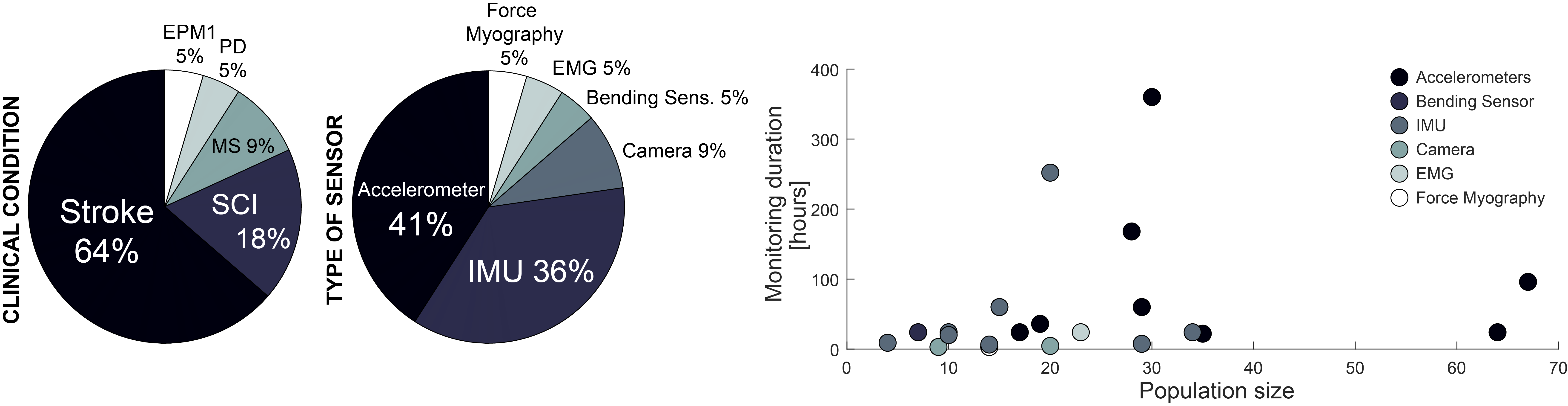
Among the studies included in our review, it is notable that the vast majority of investigations focused on individuals with stroke (15 out of 22, 68%). Stroke affects approximately 1 in 4 people worldwide [27] and it is the third-leading cause of death and disability worldwide [28]. One of the most common consequences of stroke is UE impairment [29], which makes it a significant area of research in the wearable technology domain. Following, studies examining SCI constituted 4 out of 22, MS accounted for 2 out of 22, PD and progressive myoclonic epilepsy type 1 (EPM1) both for 1 out of 22, all conditions affecting less than 1% of the global population, according to data sourced from the World Health Organization [30, 31, 32]. On average, the sample size was participants per study. Only two studies [14, 39] enrolled more than 50 participants, while 10 out of 22 studies enrolled less than 20 individuals. Among the studies enrolling post-stroke individuals, 31% of studies enrolled acute or sub-acute individuals (i.e., within 8 weeks since the event). This data is particularly interesting and in contrast with assistive technologies studies (e.g., robotic exoskeletons), where most of the available literature is on chronic patients [11].
III-B Hardware
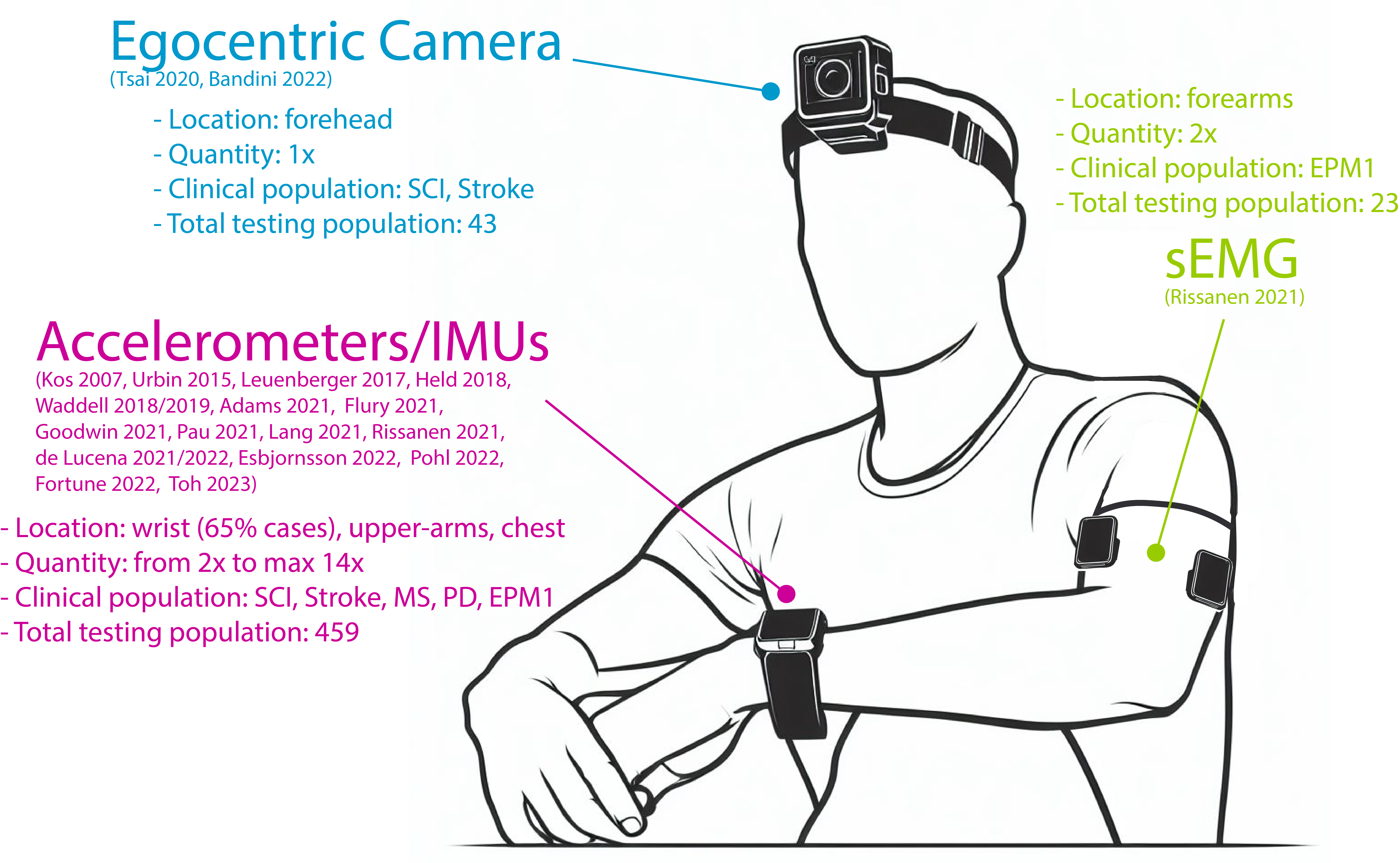
From a hardware standpoint, the most common strategies to monitor UE functions in individuals with neurological impairments are the use of accelerometers and inertial measurement units (IMUs), the latter being composed of an accelerometer, a goniometer, and – depending on the sensors – a magnetometer (see Figure 2). These sensors can track the UE kinematics and are widely spread thanks to the possibility to record continuously for hours, easiness of use, low cost, and compactness. They were placed mostly on the wrist (unilaterally [34, 48, 49] or bilaterally [37, 38, 14, 45, 50, 52, 40, 39, 35]), forearms [38, 42, 43, 46], upper-arms [38, 44, 51], and on the chest [38, 44, 42, 43, 51], mostly as a reference to compensate for trunk movements. Interestingly, De Lucena et al. [48, 49] embedded an IMU and four external magnetometers into a wrist-watch, and used this in combination with a magnetic ring on the index finger to track index and wrist movements.
Only 4 out of 22 studies did not use these sensors: Bandini et al. (2022) [18] and Tsai et al. (2020) [41] both used an egocentric camera, mounted on the forehead, activities that involved the use of the hands during daily life; Saleh et al. (2016) [36] used conductive ink-based bending sensors integrated on the index and middle fingers of a glove to monitor hand movements; Yang et al. (2021) [47] used force myography to detect the state of the hand by monitoring the surface (i.e., skin) stiffness on the wrist musculo-tendonous complex. A few research groups designed their devices from scratch [34, 36, 48, 49], while most of the investigations were carried out using off-the-shelf technology. Despite the number of studies, only in one of them [50] data collected was available remotely to the research team, and only in four of them [36, 50, 48, 49] data were available in real-time to the participants as a feedback of their performance.
III-C Data processing
As a consequence of the hardware selection, in the majority of research papers UE activity measures were derived from accelerometry data alone or in conjunction with other signals, including angles [37], and angular velocity [51]. If considering IMUs as a single-mode sensing technology (IMUs fuse data from 2-3 sources but the resulting outcome is purely kinematic), interestingly, only Rissanen et al. [46] implemented a true multimodal sensing strategy: they used data both from accelerometers and sEMG placed on both forearms of 23 individuals with EPM1. Noticeably, De Lucena et al. [48, 49], who used in their device an IMU, four magnetometers, and a magnetic ring on the index finger, while still collecting purely kinematics information, can also be considered multimodal.
The majority of studies opted for an offline data analysis, where data were only recorded and stored during the intervention, while processed only after the monitoring session has concluded [42, 48, 43, 44, 14, 45, 46, 47, 18, 51, 38, 34, 35, 39, 40]. Interestingly, however, a subset of studies employed real-time analysis, allowing for immediate processing and feedback during monitoring [36, 49, 50]. Saleh et al. [36] equipped their glove with an array of light-emitting diodes (LED) to inform the user about the finger flexion ROM. De Lucena et al. [49] had a screen in their wrist-watch showing the hand movement count and hand use intensity metrics (see below for more information), and an emoji representing their recent performance towards their daily goals. Esbjornsson et al. [50], instead, in case of an imbalance in arm movements, made their bracelets vibrate, requiring participants to take an app-based test on their smartphone to detect the onset of a stroke.
Measures extracted from the recordings
Regarding the measures used to quantify UE activity, the most prevalent metrics were activity count and activity duration. Activity count metrics encompass various measures that quantify the number of upper limb actions during daily life, typically expressed as the number of actions per unit of time. Activity duration metrics, on the other hand, quantify the duration of these activities in time units. Other less common measures included the type of activity performed, frequency metrics that described upper limb motion, and traditional biomechanical measurements such as range of motion (ROM) and muscle activity.
Study Clinical Sample Technology, Sensor Number Multimodal Monitoring Remote Real-Time Reference Condition Size and Sensor Location Sensing Duration Access Feedback Kos et al. 2007 [34] MS 19 Accelerometer (2x) on wrist and ankle 3 days Urbin et al. 2015 [35] Stroke 35 Accelerometers (2x) on wrists 22 hours Saleh et al. 2016 [36] Stroke 6 Bending sensors (2x) on index and 2 days ✓ 1 middle fingers 2 weeks Leuenberger et al. 2017 [37] Stroke 10 IMU (5x) on wrists, shanks and waist 2 days Held et al. 2018 [38] Stroke 4 IMU (14x) full-body suit (1x foot, 9 hours 2x leg, 3x arm, 2x torso) Waddell et al. 2018 [39] Stroke 64 Accelerometers (2x) on wrists 2 days Waddell et al. 2019 [40] 29 5 days Tsai et al. 2020 [41] Stroke 9 Egocentric camera (1x) on the head 3 hours SCI 14 Adams et al. 2021 [42] PD 17 Accelerometers (5x) on thighs, 2 days forearms and trunk Flury et al. 2021 [43] Stroke 15 IMU (6x) on chest, forearms, legs (3x) 5 hours Goodwin et al. 2021 [44] SCI 34 IMU (3x) on chest and upper arms 2 days Lang et al. 2021 [14] Stroke 67 Accelerometers (2x) on wrists 8 days Pau et al. 2021 [45] MS 28 Accelerometers (2x) on wrists 2 weeks Rissanen et al. 2021 [46] EPM1 23 EMG (2x) and accelerometers (2x) on ✓ 2 days forearms Yang et al. 2021 [47] Stroke 14 Force Myography (1x) on wrist 3 days Bandini et al. 2022 [18] SCI 20 Egocentric camera (1x) on the head 4.5 hours de Lucena et al. 2021 [48] Stroke 29 IMU (1x) and magnetometers (4x) on wrist ✓ 6-9 hours ✓ ✓ de Lucena et al. 2022 [49] 20 and magnetic ring (1x) on the index finger 3 weeks Esbjornsson et al. 2022 [50] Stroke 30 Accelerometers (2x) on wrist 30 days ✓ ✓ Fortune et al. 2022 [51] SCI 10 IMU (3x) on chest and upper arms 1.67 days Pohl et al. 2022 [52] Stroke 14 IMU (2x) on wrists and a camera for 6.5 hours activity labelling
Activity count was typically estimated from acceleration data by calculating the magnitude of the raw acceleration within a specific window length [37, 52, 34, 50]. Consequently, it was essential to establish an optimal threshold for acceleration magnitude to distinguish between functional and non-functional movements. For example, in [52] the authors addressed this issue by determining the optimal threshold for both affected and non-affected arms (i.e., maximizing the area under the curve) and subsequently trained a logistic regression classifier to discern functional from non-functional interactions using raw IMU data. Since recordings were often conducted in unconstrained settings, it was necessary to filter out walking periods by detecting lower limb accelerations through shank accelerometers [37]. The results were typically presented as the average counts per unit of time (e.g., per minute or hour).
Activity count was also estimated from angles using various methods:
-
•
Ratio of movement [36], as movement episodes detected by applying a 2-degree threshold to identify changes in a specific joint angle (in this case, finger flexion). The ratio of counted samples over the entire dataset yielded the ratio of movement values for each finger.
-
•
Integral of individuated movement [36], a parameter related to the mean difference in angle between two fingers, representing individual finger movement.
-
•
Gross arm movement identification [37], based on forearm elevation orientation, with specific criteria for defining gross arm movement.
Finally, less common strategies to estimate activity counts included data from magnetic fields [48, 49], force myography [47], and egocentric video [18].
Activity duration metrics were commonly computed from accelerometry data, often determined by summing the seconds in which the acceleration magnitude exceeded either zero [14] or a predefined threshold [45]. Angular data were also used to calculate the percentage of time spent in different elevation bins [44], offering insights into humeral elevation angles. Angular velocity data [43] was used to extract arm activity data, employing the Hilbert transform and a binary threshold to identify active arm periods. The arm activity time was then determined according to this definition during the recording time. Video data could also be used to calculate the average duration of hand-object interaction, providing insights into activity duration [18, 41].
Activity type was determined by some authors who delved beyond quantifying the active time of the upper limbs and considered the type of actions and activities being performed. From accelerometry data, metrics such as bilateral magnitude and magnitude ratio [14, 45, 35, 39, 40] were used to assess both upper limb use. These metrics offered insights into the intensity and contribution of each limb to daily activity on a second-by-second basis. Mono and bilateral arm use index [45] quantified the frequency of independent movements in everyday activities. Use ratio [14, 35, 39, 40] measured the total duration of activity of one limb relative to the other. Density plots graphically represented accelerometry data from both limbs, providing visual insights into movement patterns. In contrast, in [51] authors employed machine learning to estimate activity types, using a set of 13 features as predictors in a neural network model.
Frequency measures were used to understand the type of motion, particularly in conditions like Parkinsonism, where tremors at specific frequencies are important predictors of treatment efficacy. These measures included the maximum of the acceleration power spectrum, as well as the amplitude and frequency of rhythmic hand motion [42].
Muscle activity analysis, although less explored in this context, due to the comfort offered by accelerometers and IMU (especially for long recordings), is noteworthy. Rissanen et al. [46] employed several measures, including sample kurtosis, correlation dimension, recurrence rate, root-mean-square amplitude, and burst frequency, to assess EMG signal characteristics related to muscle activity.
III-D Study protocol
From a study protocol point of view, most of the reviewed work had similar characteristics. They employed a variety of tasks to record and assess UE function, encompassing typical daily routines and ADLs that are relevant to individuals in their home environments. As an example, Saleh et al. [36] investigated a broad range of activities, including household tasks, writing, using a remote control, dressing, grooming, working on a computer, tying shoes, and using a phone, all performed using the impaired hand. While Rissanen et al. [46] and De Lucena et al. [48] did not specify the types of tasks performed, they likely incorporated activities commonly found in a normal daily routine. Another similarity was the settings where the recordings of upper limb activity happened. Most of the studies took place directly within the participants’ homes [36, 37, 44, 14, 45, 46, 47, 18, 49, 50, 51, 34, 39, 40, 52]. The importance of recording at home lies in its ability to capture the performance domain of the ICF, allowing for situations that closely mimic the real-life experiences of individuals with neurological disorders in their daily environments. These home-based settings, though entirely unstructured and requiring a more complex hardware integration, offer a natural and familiar environment for monitoring UE activities. In other studies [35, 38, 42, 48, 43], data collection initially commenced in clinical settings before transitioning to community-based recordings. This approach facilitated a valuable comparison between controlled clinical conditions and real-world home environments.
The overall recording duration and the maximum duration of a single recording varied considerably among studies, reflecting the diverse objectives and protocol designs, as well as the different technological approaches adopted (i.e., usually battery duration for wearable cameras is shorter than accelerometers or IMUs). Considering the total duration, some studies employed relatively short monitoring periods, such as less than 5 hours [41, 18] and between 5 and 10 hours [43, 52, 38]. Longer monitoring durations were adopted in other studies, with duration over 2-3 days [37, 42, 34, 35, 39, 40, 34, 36, 44]. In [36], part of the study was conducted for a two-week period, and a similar duration was achieved in [45], while in [49], the study lasted three weeks. Lastly, in [50, 51] authors opted for more extended 30-day monitoring periods.
Regarding the maximum single duration of a recording session, some studies, such as Adams et al. (2021) [42] and Rissanen et al. (2021) [46], conducted continuous monitoring sessions spanning an impressive 45 and 48 hours, respectively. Similarly, in [47] authors recorded data continuously for 48 hours, only during waking hours. In [14, 37, 34, 35, 39, 40], the maximum duration for a single session of data recording was 24 hours. De Lucena et al. [49] recorded data for around 12 hours in a single session. In contrast, shorter periods were reported in [38] (3h) and in [18] (1.5h). It is worth noting that for some studies, the maximum duration of a single session was not explicitly specified (e.g., [50, 51]).
Hand and Finger Contextual Non-intrusive Movement Details Information Long Recording Privacy Accelerometers IMUs Bending Sensors Wearable Cameras sEMG Force Myography
IV Discussion
In this review, we explored the latest advancements in wearable technologies designed for real-life and unstructured monitoring of UE function in individuals with neurological disorders. Our specific objectives encompassed several crucial aspects, such as identifying and categorizing the most commonly used technologies for monitoring UE function during daily activities, as well as gaining insights into the prevailing methods for data processing and measurement, and investigating the types of protocols developed for conducting such studies.
IV-A Stroke as the main monitored condition
As in the case of other wearable technologies [11], stroke survivors comprised the majority of the study population (64%). This prevalence can likely be attributed to the fact that stroke ranks as one of the leading global causes of both mortality and disability, with UE impairments as a common consequence. The significance of UE recovery for stroke survivors should not be understated, as regaining UE functionality ranks among their highest rehabilitation priorities. This underscores the research community’s keen interest in monitoring individuals at home to gain insights into their UE usage during daily life. Such knowledge serves as a foundation for developing therapies aimed at optimizing UE functions at home, with the ultimate goal of enhancing individuals’ independence.
Notably, some of the reviewed papers extended their focus beyond chronic cases, including acute and sub-acute stroke survivors in their investigations. For example, Waddell et al. [40] demonstrated that sensor-measured UL performance improves in the first 12 weeks post-stroke, proving with data from unsupervised conditions the well-known spontaneous capacity of the body to improve after stroke [57]. Lang et al. [14], instead, showed how UL performance in daily life reached a plateau only 3-6 weeks post-stroke, thus often before neurological impairments and functional capacity started to stabilize. The availability of data early after a stroke is particularly valuable as it addresses a critical phase in patients’ recovery. Once patients are discharged from the hospital, the progress of their rehabilitation may become challenging to track. Having a means of observing and understanding their performance upon returning to the community assumes paramount importance. This monitoring is essential for further enhancing their recovery, especially during outpatient care, and ensures that the gains made in the clinical setting continue to progress effectively in the real-world context.
IV-B Monitoring kinematics only is not enough
The majority of the studies predominantly relied on IMUs and accelerometers. These choices were motivated by user-friendliness, extended recording capabilities spanning hours or days, and the devices’ lightweight and affordability. Such attributes make IMUs and accelerometers the natural choices for studies that require non-intrusiveness and portability to collect ecological data. A noteworthy observation from Figure 3 is that the studies utilizing accelerometers tended to have the largest participant populations [39, 14], and reached a favorable balance between expanding the number of recruited participants and maintaining extended recording durations [50, 45].
In this review, we excluded studies using smartphone-embedded IMUs as a source of information, given that they are not properly wearable devices. However, they provide a very user-friendly and affordable sensing solution to easily assess a large number of individuals (see e.g., Pratap et al. who monitored almost 500 MS individuals [54]).
However, it is important to note that IMUs and accelerometers primarily offer global kinematic information, which can subsequently be processed to calculate activity metrics like counts and durations. Yet, for a more in-depth analysis, particularly to understand specific types of grasps and the contextual nuances of UE functions, IMUs and accelerometers alone may prove insufficient.
In response to this limitation, some studies have introduced video-based approaches, capitalizing on the advantages of recording richer information about the surrounding environment [41, 18]. This includes details such as the manipulation area, objects, and the broader context, which aids in deciphering the functional aspects of hand-object interactions and the specific activities being performed. However, it is worth noting that wearable cameras may not be optimal for extended recordings unless newer cameras with longer-lasting batteries are developed. Alternatively, some studies have explored the use of magnetic sensors to gather additional information about finger movements [48, 49], while others have delved into EMG recordings [46]. Magnetic sensors offer enhanced detail but may introduce artifacts when interacting with metal objects, while EMG, although valuable for capturing low-level muscle activation, was not extensively examined for deciphering grasp patterns, as already seen in other studies [58].
Adopting multi-modal approaches that combine the strengths of different sensors could offer a more comprehensive understanding. However, the majority of the reviewed studies predominantly relied on single-mode technologies, with only Rissanen et al. [46] implementing a true multimodal sensing strategy by using data from both accelerometers and sEMG placed on the forearms of 23 individuals with EPM1.
IV-C Short, offline recording, with poor feedback to the patients
Concerning data processing, the majority of analyses were conducted offline. This approach may be suitable for tracking recovery progress over weeks in neurorehabilitation. Nonetheless, real-time data analysis offers distinct advantages, particularly for providing immediate feedback to patients during telemonitoring and telerehabilitation. It is important to highlight a few articles that were excluded [55, 56, 53]. While these studies concentrated on telerehabilitation and did not include monitoring ADLs, they utilized compelling wearable methods to study the UEs at home in people with neurological conditions. Besides, real-time processing, with only the processed data transmitted, may even help address privacy concerns, which is a critical issue associated with technologies like those based on cameras [59].
When it comes to activity metrics, the most prevalent measures focus on quantifying the duration and frequency of activities. However, in a rehabilitation context, understanding not only the quantity but also the context of these measures is crucial for identifying specific challenges patients encounter during different activities. Machine learning techniques, as proposed by Fortune et al. (2022) [51], have the potential to recognize the types of activities being performed. This information, combined with the quantity of hand use, is essential for gaining a comprehensive understanding of UE use at home.
Furthermore, as shown in Figure 3, the duration of recording sessions is typically limited to a few hours in the majority of cases. While some of these studies often explored the feasibility of using technology at home, it is essential to extend the recording duration to encompass a broader range of daily living activities typically performed by participants during their daily routines. This ensures that the recorded behavior in the home environment closely reflects the participants’ everyday activities. This is particularly important in light of the findings by Waddell et al. [39] showing how self-reports UL performance are neither consistent nor accurate with sensor-based use assessment.
IV-D Final remarks and future directions
The results of this literature review underscore the growing significance of remote performance monitoring in the neurorehabilitation field, with the majority of the research conducted in this domain emerging over the past 15 years, and a notable surge in the last three years. This trend can be attributed to the increasing accessibility and affordability of off-the-shelf technologies, facilitating the recording of extended periods of unconstrained activities in the community. The potential unlocked by this capability is indeed promising, offering the opportunity to tailor rehabilitation strategies to better suit individuals’ specific needs within their daily lives.
However, it is crucial for researchers to consider several key aspects while pursuing these opportunities. Firstly, ensuring device usability is essential, as these technologies are intended for use by non-expert individuals in their home environments. This user-friendliness is vital to guarantee the quality of the recorded data. Secondly, it is essential to consider the potential challenges associated with attaching sensors to their arms and hands, as this may impact how they perform daily activities. Thirdly, privacy concerns may arise from monitoring individuals in their homes, especially in the case of video monitoring, and this must be addressed.
Taking these considerations into account, the ideal wearable technology for this context should be 1) non-intrusive and lightweight, 2) capable of capturing detailed information about hand and finger movements for task differentiation, 3) proficient in capturing contextual information (e.g., object manipulation, specific actions), 4) capable of long recordings spanning several hours, and 6) devoid of privacy-related issues. By looking at the above review, it becomes evident that a singular technology that fulfills all these requirements is currently nonexistent (see Table II). This underscores the increasing need for a multimodal approach to effectively capture how individuals function in their home environments.
References
- [1] H. M. Schambra, A. Parnandi, N. G. Pandit, J. Uddin, A. Wirtanen, and D. M. Nilsen, ”A taxonomy of functional upper extremity motion,” Frontiers in Neurology, vol. 10, p. 857, 2019
- [2] A. M. Dollar, ”Classifying human hand use and the activities of daily living,” in The Human Hand as an Inspiration for Robot Hand Development, 2014, pp. 201-216.
- [3] K. Moulaei, A. Sheikhtaheri, M. S. Nezhad, et al., ”Telerehabilitation for upper limb disabilities: A scoping review on functions, outcomes, and evaluation methods,” Arch Public Health, vol. 80, p. 196, 2022.
- [4] K. D. Anderson, ”Targeting recovery: priorities of the spinal cord-injured population,” Journal of Neurotrauma, vol. 21, no. 10, pp. 1371-1383, 2004.
- [5] L. A. Simpson, J. J. Eng, J. T. Hsieh, et al., ”The health and life priorities of individuals with spinal cord injury: a systematic review,” Journal of Neurotrauma, vol. 29, no. 8, pp. 1548-1555, 2012.
- [6] D. S. Nichols-Larsen, P. Clark, A. Zeringue, A. Greenspan, and S. Blanton, ”Factors influencing stroke survivors’ quality of life during subacute recovery,” Stroke, vol. 36, no. 7, pp. 1480-1484, 2005.
- [7] M. R. Popovic, N. Kapadia, V. Zivanovic, J. C. Furlan, B. C. Craven, and C. McGillivray, ”Functional electrical stimulation therapy of voluntary grasping versus only conventional rehabilitation for patients with subacute incomplete tetraplegia: a randomized clinical trial,” Neurorehabilitation and Neural Repair, vol. 25, no. 5, pp. 433-442, 2011.
- [8] C. Marquez-Chin and M. R. Popovic, ”Functional electrical stimulation therapy for restoration of motor function after spinal cord injury and stroke: a review,” Biomedical Engineering Online, vol. 19, no. 1, pp. 1-25, 2020.
- [9] F. Inanici, L. N. Brighton, S. Samejima, C. P. Hofstetter, and C. T. Moritz, ”Transcutaneous spinal cord stimulation restores hand and arm function after spinal cord injury,” IEEE Transactions on Neural Systems and Rehabilitation Engineering, vol. 29, pp. 310-319, 2021.
- [10] R. M. de Freitas, A. Sasaki, D. G. Sayenko, Y. Masugi, T. Nomura, K. Nakazawa, and M. Milosevic, ”Selectivity and excitability of upper-limb muscle activation during cervical transcutaneous spinal cord stimulation in humans,” Journal of Applied Physiology, 2021.
- [11] T. Proietti, E. Ambrosini, A. Pedrocchi and S. Micera, ”Wearable Robotics for Impaired Upper-Limb Assistance and Rehabilitation: State of the Art and Future Perspectives,” in IEEE Access, vol. 10, pp. 106117-106134, 2022.
- [12] World Health Organization, ”Towards a common language for functioning, disability, and health: ICF. The international classification of functioning, disability and health,” 2002.
- [13] K. J. Waddell, M. J. Strube, R. R. Bailey, et al., ”Does task-specific training improve upper limb performance in daily life poststroke?” Neurorehabilitation and Neural Repair, vol. 31, no. 3, pp. 290-300, 2017.
- [14] C. E. Lang, K. J. Waddell, J. Barth, et al., ”Upper limb performance in daily life approaches plateau around three to six weeks post-stroke,” Neurorehabilitation and Neural Repair, vol. 35, no. 10, pp. 903-914, 2021.
- [15] J. Barth, K. R. Lohse, J. D. Konrad, et al., ”Sensor-based categorization of upper limb performance in daily life of persons with and without neurological upper limb deficits,” Frontiers in Rehabilitation Sciences, vol. 2, p. 741393, 2021, doi: 10.3389/FRESC.2021.741393.
- [16] D. Rand and J. J. Eng, ”Disparity between functional recovery and daily use of the upper and lower extremities during subacute stroke rehabilitation,” Neurorehabilitation and Neural Repair, vol. 26, no. 1, pp. 76-84, 2012.
- [17] R. J. Marino, ”Domains of outcomes in spinal cord injury for clinical trials to improve neurological function,” J. Rehabil. Res. Dev., vol. 44, no. 1, pp. 113-121, 2007.
- [18] A. Bandini, M. Dousty, S. L. Hitzig, B. C. Craven, S. Kalsi-Ryan, and J. Zariffa, ”Measuring hand use in the home after cervical spinal cord injury using egocentric video,” Journal of Neurotrauma, vol. 39, no. 23-24, pp. 1697-1707, 2022.
- [19] M. Dousty, A. Bandini, P. Eftekhar, D. J. Fleet, and J. Zariffa, ”Grasp Analysis in the Home Environment as a Measure of Hand Function After Cervical Spinal Cord Injury,” Neurorehabilitation and Neural Repair, pp. 15459683231177601, 2023.
- [20] M. F. Tsai, R. H. Wang, and J. Zariffa, ”Validity of Novel Outcome Measures for Hand Function Performance After Stroke Using Egocentric Video,” Neurorehabilitation and Neural Repair, vol. 37, no. 2-3, pp. 142-150, 2023.
- [21] A. Kadambi, A. Bandini, S. L. Hitzig, and J. Zariffa, ”Designing an egocentric video-based dashboard to report hand performance measures for outpatient rehabilitation of cervical spinal cord injury,” Topics in Spinal Cord Injury Rehabilitation, 2023 (in press).
- [22] S. F. M. Toh, K. N. K. Fong, P. C. Gonzalez, and Y. M. Tang, ”Application of Home-Based Wearable Technologies in Physical Rehabilitation for Stroke: A Scoping Review,” in IEEE Transactions on Neural Systems and Rehabilitation Engineering, vol. 31, pp. 1614-1623, 2023
- [23] A. Gopal, W. Y. Hsu, D. D. Allen, and R. Bove, ”Remote assessments of hand function in neurological disorders: Systematic review,” JMIR Rehabilitation and Assistive Technologies, vol. 9, no. 1, article e33157, 2022.
- [24] M. Sica, S. Tedesco, C. Crowe, L. Kenny, K. Moore, S. Timmons, and D. S. Komaris, ”Continuous home monitoring of Parkinson’s disease using inertial sensors: A systematic review,” PLoS One, vol. 16, no. 2, article e0246528, 2021.
- [25] G. J. Kim, A. Parnandi, S. Eva, and H. Schambra, ”The use of wearable sensors to assess and treat the upper extremity after stroke: A scoping review,” Disability and Rehabilitation, vol. 44, no. 20, pp. 6119-6138, 2022.
- [26] S. Alexander, G. Peryer, E. Gray, F. Barkhof, and J. Chataway, ”Wearable technologies to measure clinical outcomes in multiple sclerosis: A scoping review,” Multiple Sclerosis Journal, vol. 27, no. 11, pp. 1643-1656, 2021.
- [27] Owolabi MO, Thrift AG, Mahal A, et al. Primary stroke prevention worldwide: translating evidence into action [published correction appears in Lancet Public Health. 2022 Jan;7(1):e14]. Lancet Public Health. 2022;7(1):e74-e85. doi:10.1016/S2468-2667(21)00230-9.
- [28] V. L. Feigin, B. A. Stark, C. O. Johnson, G. A. Roth, C. Bisignano, G. G. Abady, and S. Hamidi, ”Global, regional, and national burden of stroke and its risk factors, 1990–2019: A systematic analysis for the Global Burden of Disease Study 2019,” The Lancet Neurology, vol. 20, no. 10, pp. 795-820, 2021.
- [29] K. S. Hayward, S. F. Kramer, V. Thijs, J. Ratcliffe, N. S. Ward, L. Churilov, and N. A. Lannin, ”A systematic review protocol of timing, efficacy and cost effectiveness of upper limb therapy for motor recovery post-stroke,” Systematic Reviews, vol. 8, pp. 1-8, 2019.
- [30] Spinal cord injury, World Health Organization, https://www.who.int/news-room/fact-sheets/detail/spinal-cord-injury
- [31] Multiple Sclerosis, World Health Organization, https://www.who.int/news-room/fact-sheets/detail/multiple-sclerosis
- [32] Launch of WHO’s Parkinson disease technical brief, World Health Organization, https://www.who.int/news/item/14-06-2022-launch-of-who-s-parkinson-disease-technical-brief
- [33] Haddaway, N. R., Page, M. J., Pritchard, C. C., & McGuinness, L. A. (2022). PRISMA2020: An R package and Shiny app for producing PRISMA 2020-compliant flow diagrams, with interactivity for optimised digital transparency and Open Synthesis Campbell Systematic Reviews, 18, e1230. https://doi.org/10.1002/cl2.1230
- [34] Daphne Kos, Guy Nagels, Marie B. D’Hooghe, William Duquet, Stephan Ilsbroukx, Stijn Delbeke & Eric Kerckhofs (2007) Measuring Activity Patterns Using Actigraphy in Multiple Sclerosis, Chronobiology International, 24:2, 345-356.
- [35] Urbin MA, Waddell KJ, Lang CE. Acceleration metrics are responsive to change in upper extremity function of stroke survivors. Arch Phys Med Rehabil. 2015 May;96(5):854-61.
- [36] Saleh N., Hage-Diab A., Salhab G., Debs B., Hajj-Hassan M., Khachfe H., Rifai S., Ahmad S., “Monitoring the use of impaired hand by a new lost cost device during daily life activities with a real-time visual feedback”, International Journal on Advances in Life Sciences, vol. 8, 2016, pp. 103-111.
- [37] Leuenberger K, Gonzenbach R, Wachter S, Luft A, Gassert R. A method to qualitatively assess arm use in stroke survivors in the home environment. Med Biol Eng Comput. 2017 Jan;55(1):141-150. doi: 10.1007/s11517-016-1496-7. Epub 2016 Apr 22.
- [38] Held JPO, Klaassen B, Eenhoorn A, Beijnum B-JFv, Buurke JH, Veltink PH and Luft AR (2018) Inertial Sensor Measurements of Upper-Limb Kinematics in Stroke Patients in Clinic and Home Environment. Front. Bioeng. Biotechnol. 6:27.
- [39] Waddell KJ, Lang CE. Comparison of Self-Report Versus Sensor-Based Methods for Measuring the Amount of Upper Limb Activity Outside the Clinic. Arch Phys Med Rehabil. 2018 Sep;99(9):1913-1916.
- [40] Waddell KJ, Strube MJ, Tabak RG, Haire-Joshu D, Lang CE. Upper Limb Performance in Daily Life Improves Over the First 12 Weeks Poststroke. Neurorehabilitation and Neural Repair. 2019;33(10):836-847.
- [41] Tsai, M. F., Bandini, A., Wang, R. H., Zariffa, J. Capturing Representative Hand Use at Home Using Egocentric Video in Individuals with Upper Limb Impairment. J. Vis. Exp. (166), e61898, doi:10.3791/61898 (2020).
- [42] Adams, J.L., Dinesh, K., Snyder, C.W. et al. A real-world study of wearable sensors in Parkinson’s disease. npj Parkinsons Dis. 7, 106 (2021).
- [43] Flury, D., Massé, F., Paraschiv-Ionescu, A. et al. Clinical value of assessing motor performance in postacute stroke patients. J NeuroEngineering Rehabil 18, 102 (2021).
- [44] Goodwin BM, Cain SM, Van Straaten MG, Fortune E, Jahanian O, Morrow MMB (2021) Humeral elevation workspace during daily life of adults with spinal cord injury who use a manual wheelchair compared to age and sex matched able-bodied controls. PLoSONE 16(4):e0248978.
- [45] Massimiliano Pau, Bruno Leban, Michela Deidda, Micaela Porta, Giancarlo Coghe, Davide Cattaneo, Eleonora Cocco, Use of wrist-worn accelerometers to quantify bilateral upper limb activity and asymmetry under free-living conditions in people with multiple sclerosis, Multiple Sclerosis and Related Disorders, Volume 53, 2021, 103081.
- [46] Saara M. Rissanen, Jelena Hyppönen, Katri Silvennoinen, Laura Säisänen, Pasi A. Karjalainen, Esa Mervaala, Reetta Kälviäinen, Wearable monitoring of positive and negative myoclonus in progressive myoclonic epilepsy type 1, Clinical Neurophysiology, Volume 132, Issue 10, 2021, Pages 2464-2472, ISSN 1388-2457.
- [47] Yang C, Liu J, Simpson LA, Menon C, Eng JJ. Real-World Functional Grasping Activity in Individuals With Stroke and Healthy Controls Using a Novel Wearable Wrist Sensor. Neurorehabilitation and Neural Repair. 2021;35(10):929-937.
- [48] Schwerz de Lucena, D.; Rowe, J.; Chan, V.; Reinkensmeyer, D.J. Magnetically Counting Hand Movements: Validation of a Calibration-Free Algorithm and Application to Testing the Threshold Hypothesis of Real-World Hand Use after Stroke. Sensors 2021, 21, 1502.
- [49] Schwerz de Lucena, D.; Rowe, J.B.; Okita, S.; Chan, V.; Cramer, S.C.; Reinkensmeyer, D.J. Providing Real-Time Wearable Feedback to Increase Hand Use after Stroke: A Randomized, Controlled Trial. Sensors 2022, 22, 6938.
- [50] Esbjornsson M., Ullberg T., Safety and usability of wearable accelerometers for stroke detection the STROKE ALARM PRO 1 study, Journal of Stroke & Cerebrovascular Diseases, v. 31:11, 106762.
- [51] Emma Fortune, Beth A. Cloud-Biebl, Stefan I. Madansingh, Che G. Ngufor, Meegan G. Van Straaten, Brianna M. Goodwin, Dennis H. Murphree, Kristin D. Zhao, Melissa M. Morrow, Estimation of manual wheelchair-based activities in the free-living environment using a neural network model with inertial body-worn sensors, Journal of Electromyography and Kinesiology, Volume 62, 2022, 102337, ISSN 1050-6411.
- [52] Pohl J, Ryser A, Veerbeek JM, Verheyden G, Vogt JE, Luft AR and Awai Easthope C (2022), Classification of functional and non-functional arm use by inertial measurement units in individuals with upper limb impairment after stroke. Front. Physiol. 13:952757.
- [53] Toh SFM, Gonzalez PC, Fong KNK. Usability of a wearable device for home-based upper limb telerehabilitation in persons with stroke: A mixed-methods study. DIGITAL HEALTH. 2023;9.
- [54] Pratap A, Grant D, Vegesna A, Tummalacherla M, Cohan S, Deshpande C, Mangravite L, Omberg L. “Evaluating the Utility of Smartphone-Based Sensor Assessments in Persons With Multiple Sclerosis in the Real-World Using an App (elevateMS): Observational, Prospective Pilot Digital Health Study”, JMIR Mhealth Uhealth., vol. 8(10):e22108, 2020.
- [55] Sanders Q, Chan V, Augsburger R, Cramer SC, Reinkensmeyer DJ, Do AH. Feasibility of Wearable Sensing for In-Home Finger Rehabilitation Early After Stroke. IEEE Trans Neural Syst Rehabil Eng. 2020 Jun;28(6):1363-1372. doi: 10.1109/TNSRE.2020.2988177. Epub 2020 Apr 15. PMID: 32305930; PMCID: PMC9345607.
- [56] Dodakian L, McKenzie AL, Le V, See J, Pearson-Fuhrhop K, Burke Quinlan E, Zhou RJ, Augsberger R, Tran XA, Friedman N, Reinkensmeyer DJ, Cramer SC. A Home-Based Telerehabilitation Program for Patients With Stroke. Neurorehabil Neural Repair. 2017 Oct-Nov;31(10-11):923-933.
- [57] Ramsey LE, Siegel JS, Lang CE, Strube M, Shulman GL, Corbetta M. Behavioural clusters and predictors of performance during recovery from stroke. Nat Hum Behav. 2017;1:0038.
- [58] A. Toro-Ossaba, J. Jaramillo-Tigreros, J. C. Tejada, A. Peña, A. López-González, and R. A. Castanho, ”LSTM Recurrent Neural Network for Hand Gesture Recognition Using EMG Signals,” Applied Sciences, vol. 12, no. 19, p. 9700, 2022.
- [59] Bandini, A., Kalsi-Ryan, S., Craven, B. C., Zariffa, J., and Hitzig, S. L. (2021). ”Perspectives and recommendations of individuals with tetraplegia regarding wearable cameras for monitoring hand function at home: Insights from a community-based study.” The Journal of Spinal Cord Medicine, 44(sup1), S173-S184.
![[Uncaptioned image]](/html/2311.12513/assets/img/tommaso.jpg) |
TOMMASO PROIETTI is currently an Assistant Professor at the Scuola Superiore Sant’Anna (SSSA, Pisa, Italy), and Head of the Soft NeuroBionics Lab. He received the B.S. and M.S. degrees in Control Engineering from Sapienza University of Rome (Rome, Italy) in 2010 and 2013, respectively, and the Ph.D. degree in Robotics Engineering from University Pierre et Marie Curie (UPMC, Paris, France) in 2017. From 2012 to 2013 he was Visiting Research Fellow at the Center for Robotics and Biosystems, Northwestern University (Evanston, IL, USA). From 2017 to 2019 he worked as Control System Engineer for General Motors. From 2019 to 2022, he was Postdoctoral Research Fellow at the Harvard Biodesign Lab, Harvard University (Cambridge, MA, USA). From 2022 to 2023, he was Postdocotral Research Fellow at the Translational Neural Engineering Lab, SSSA. His research interests include soft wearable robotics for assistance, augmentation and rehabilitation, physical human-robot interaction, and advanced neural controls. |
![[Uncaptioned image]](/html/2311.12513/assets/img/AB.jpg) |
ANDREA BANDINI is currently an Assistant Professor at the Scuola Superiore Sant’Anna (SSSA, Pisa, Italy) received his Master’s degree in Biomedical Engineering from the University of Firenze (Italy) in 2012, and the PhD the Bioengineering from the University of Bologna (Italy) in 2016. From 2016 and 2021, he was a postdoctoral research fellow at KITE - Toronto Rehab - University Health Network (Toronto, Canada). His research aims at developing intelligent tools for remote assessment and rehabilitation of motor signs associated with neurological disorders (spinal cord injury, stroke, amyotrophic lateral sclerosis, and Parkinson’s disease), by using computer vision and machine learning techniques. |