Can GPT-4V(ision) Serve Medical Applications?
Case Studies on GPT-4V for Multimodal Medical Diagnosis
Abstract
Abstract
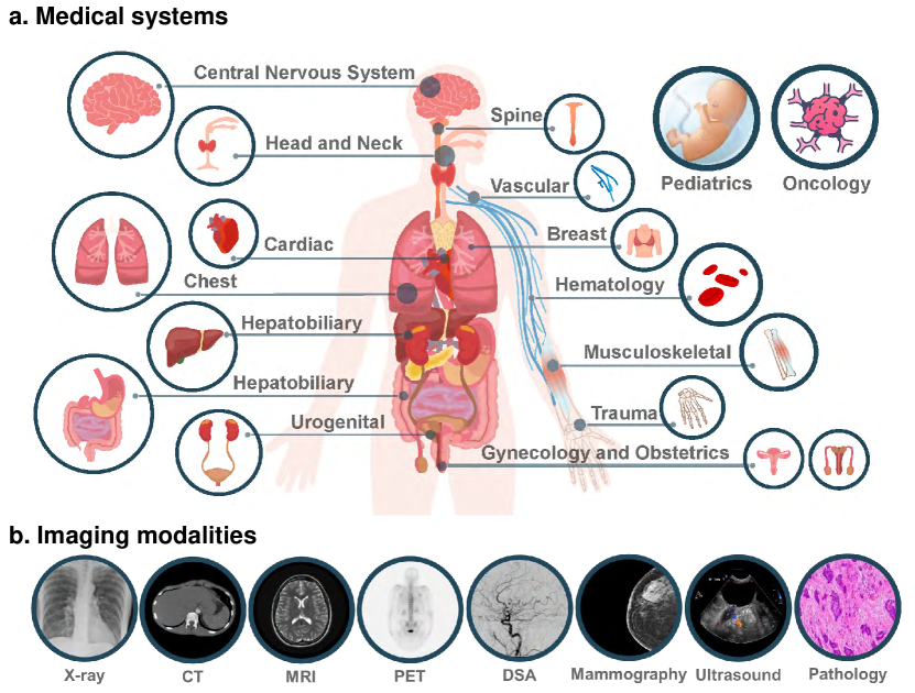
Driven by the large foundation models, the development of artificial intelligence has witnessed tremendous progress lately, leading to a surge of general interest from the public. In this study, we aim to assess the performance of OpenAI’s newest model, GPT-4V(ision), specifically in the realm of multimodal medical diagnosis. Our evaluation encompasses 17 human body systems, including Central Nervous System, Head and Neck, Cardiac, Chest, Hematology, Hepatobiliary, Gastrointestinal, Urogenital, Gynecology, Obstetrics, Breast, Musculoskeletal, Spine, Vascular, Oncology, Trauma, Pediatrics, with images taken from 8 modalities used in daily clinic routine, e.g., X-ray, Computed Tomography (CT), Magnetic Resonance Imaging (MRI), Positron Emission Tomography (PET), Digital Subtraction Angiography (DSA), Mammography, Ultrasound, and Pathology. We probe the GPT-4V’s ability on multiple clinical tasks with or without patent history provided, including imaging modality and anatomy recognition, disease diagnosis, report generation, disease localisation.
Our observation shows that, while GPT-4V demonstrates proficiency in distinguishing between medical image modalities and anatomy, it faces significant challenges in disease diagnosis and generating comprehensive reports. These findings underscore that while large multimodal models have made significant advancements in computer vision and natural language processing, it remains far from being used to effectively support real-world medical applications and clinical decision-making.
All images used in this report can be found in https://github.com/chaoyi-wu/GPT-4V_Medical_Evaluation.
1 Introduction
Large language models (LLMs), particularly the GPT series developed by OpenAI, have demonstrated remarkable capabilities across a wide spectrum of domains, even in specialized fields such as medicine and law [20, 3, 19, 9]. While prior models in the GPT series have demonstrated potential in medical related language tasks [17, 21, 8], even achieving high performance in the United States Medical Licensing Examination (USMLE), they are fundamentally limited in daily clinical routine, due to its inability to read visual signals. Inspired by this, in medical community, many visual or multimodal foundation models [12] are also emerging, e.g., for fundus [27], pathology [10], radiology [23] or general medical images [26, 22, 13].
Since September, the latest version, GPT-4V [24], starts to support multimodal input, sparking curiosity about its effectiveness from the moment it became available for use. In this report, we aim to initiate a study on the capabilities of GPT-4V for multimodal medical diagnosis, by asking the question: “Can GPT-4V serve medical applications?” This is a question of paramount importance, not only for the AI community, but also for clinicians, patients, and healthcare administrators.
1.1 Motivation.
In this report, our goal is to initiate a systematic evaluation of the capabilities of GPT-4V on multimodal medical diagnosis. Specifically, we perform the case-level study from 17 human body systems, including Central Nervous System, Head and Neck, Cardiac, Chest, Hematology, Hepatobiliary, Gastrointestinal, Urogenital, Gynecology, Obstetrics, Breast, Musculoskeletal, Spine, Vascular, Oncology, Trauma, Pediatrics, with images taken from 8 modalities, e.g., X-ray, Computed Tomography (CT), Magnetic Resonance Imaging (MRI), Positron Emission Tomography (PET), Digital Subtraction Angiography (DSA), Mammography, Ultrasound, and Pathology.
Our exploration of GPT-4V is guided by the following questions.
-
•
Can GPT-4V recognize the modalities and anatomical structures of medical images? Recognizing various modalities (such as X-ray, CT, MRI, ultrasound, and pathology) and identifying different anatomical structures within these images lies the foundation for more sophisticated diagnosis.
-
•
Can GPT-4V localize different anatomical structures in the medical image? Precisely localizing specific anatomical structures in an image is crucial for identifying abnormalities, ensuring that potential issues are addressed in the correct anatomical context.
-
•
Can GPT-4V discover and localize anomalies in medical images? Detecting anomalies, such as tumors, fractures, or infections, is a primary goal of medical image analysis. For an AI model to be considered reliable in a clinical setting, it is required to not only discover these abnormalities but also accurately localize them, facilitating targeted interventions or treatments.
-
•
Can GPT-4V combine multiple images to make a diagnosis? Medical diagnoses often require a holistic view, combining information from different imaging modalities or views. It is thus critical to probe GPT-4V’s ability to combine and analyze information from multiple images.
-
•
Can GPT-4V write medical reports, describing both abnormalities and relevant normal findings? Writing reports is a time-intensive task for radiologists and pathologist. If GPT-4V can assist in this process by generating accurate and clinically relevant reports, it will certainly improve the efficiency of the entire workflow.
-
•
Can GPT-4V integrate patient medical history when interpreting medical images? The patient’s basic information and past medical history can greatly influence the interpretation of current medical images. Considering this information during model prediction would lead to a more personalized and potentially more accurate analysis, considering all relevant patient-specific factors.
-
•
Can GPT-4V maintain consistency and memory across multiple rounds of interaction? In some medical scenarios, a single-pass analysis may not be sufficient. This capability of maintaining a coherent and reliable context throughout extended conversations or analyses, especially in intricate medical contexts where data continuity is critical.
1.2 Sample selection.
Guided by the aforementioned questions, we perform comprehensive case studies on various tasks. For radiology image recognition, diagnosis and report generation, we leverage the most famous radiology collection website, Radiopaedia111https://radiopaedia.org/. For pathology image analysis, we collect hematoxylin and eosin (H&E) stained histopathology images of malignant tumors across 20 tissues from the professional pathology website, PathologyOutlines222https://www.pathologyoutlines.com/. For localization capability analysis, we choose samples from several public medical image segmentation and detection benchmarks [11, 5, 4, 2, 15, 16, 6, 18, 1, 25, 14, 7].
1.2.1 Case selection.
Given GPT-4V has not provided APIs officially, we can only use its webpage version, thus set limitations on the scalability of our evaluation. In order to pick the most suitable cases, we mainly take the following considerations:
-
•
Release time. Considering that GPT-4V has been extensively trained on web data, to guarantee fair evaluation on its generalisation, we only select cases that have appeared online in 2023, to avoid the data sample being part of the training set of GPT-4V.
-
•
Reliability of annotations. Medical diagnosis normally requires strong expertise, it is crucial to select samples with reliable annotations. Luckily, on Radiopaedia, each uploaded case is commonly reviewed by a board of radiologist333https://radiopaedia.org/editors, and accompanied by a completion ratio that indicates the sufficiency of information for diagnosis. We typically select cases with a completion ratio exceeding 90% as their reference descriptions are deemed more reliable.
-
•
Diverse imaging modalities. For each body system, we aim to include a wide spectrum of imaging modalities available, while also reflecting the real-world distribution of images as accurately as possible. Consequently, for every system, we endeavor to encompass all the cases across different imaging modalities related to that body system. It’s worth noting that we also pick cases that requires to integrate multiple imaging modalities for decision making.
1.2.2 Image processing.
Here, we describe the rules to control the quality of input images:
-
•
Multi-image selection. Many entries on Radiopaedia may contain more than four images, exceeding the upper input limitation for GPT-4V. Typically, we refrain from using such entries, as manually selecting images could result in omitting crucial ones. Nonetheless, on rare occasions, we might encounter this challenge. When faced with this situation, we will prioritize the four images that most closely align with the descriptions provided on Radiopaedia for input.
-
•
Key slice selection. In medical imaging analysis, the data is usually represented as 3D volume. However, the current GPT-4V model is limited to only processing four image inputs per time maximally, which is notably smaller than the standard number of slices in medical 3D data, like CT and MRI scans. Given this constraint, we use the slice444https://radiopaedia.org/articles/key-image that is most relevant to the case description and diagnosis, as suggested by expert radiologists while uploading images onto Radiopaedia website.
-
•
Intensity normalization. For medical images, varying-intensity windows can reveal different structures. We utilize the default intensity window displayed on Radiopaedia, as set by the radiologists, for image input. For localization tasks, we clip the CT images at [-300, 300], while clip other images at the 0.5% and 99.5% percentiles of the intensity distribution. All the images are the rescaled to [0, 1].
1.2.3 Question prompts.
For each case, we might pose various questions spanning multiple tasks, but our primary emphasis is on report generation and diagnosis. The question prompt for the same task may vary slightly with each inquiry, to test the robustness of GPT-4V on responding different text query formats. For example, for report generation, we may often use the prompts as “Please Generate a radiology report for this images.” or “May you please write a report for the patient?” and for diagnosis, we may often use “Can you make a diagnosis for the patient?” or “Are there any abnormalities in the images?”. For other types of questions, we will organize conversation freely without recommended prompts.
1.2.4 Annotation or reference caption.
In order to reduce the difficulty of checking the correctness of the GPT-4V responses, we have selected the image descriptions provided on Radiopaedio as references, which has been verified by a board of qualified radiologists. However, it’s crucial to note that the references are not in standard format of clinical reports. The radiologists write about what captures their interest, potentially overlooking many standard statements. In other words, the description guarantees its accuracy but cannot ensure a comprehensive description of the patient’s condition. Therefore, in case analysis, only statements that directly conflict with the given reference will be marked as definite incorrect. For all other output information, it’s up to the readers to judge its correctness with expertise knowledge.
1.3 Testing procedure.
We evaluate GPT-4V using its online chat page555https://chat.openai.com/. We begin the conversation by feeding in the images. Typically, we might pose one or two questions for each case, with subsequent questions as multi-round conversation. When turning to a new case, we initiate a fresh chat window to ensure GPT-4V doesn’t mistakenly leverage information from previous conversations related to other cases.
For pathology evaluation, two-round conversations are exploited across all images. The first round asks whether a report can be generated base on only the input image. The purpose of this round is to evaluate whether GPT-4V can recognize image modalities and tissue origin without given any related medical prompts. At the second round, we provide the correct tissue origin and ask whether GPT-4V can make a diagnosis based on both the pathology image and the information of its tissue origin. We expect GPT-4V to modify its report and provide a clear diagnostic result.
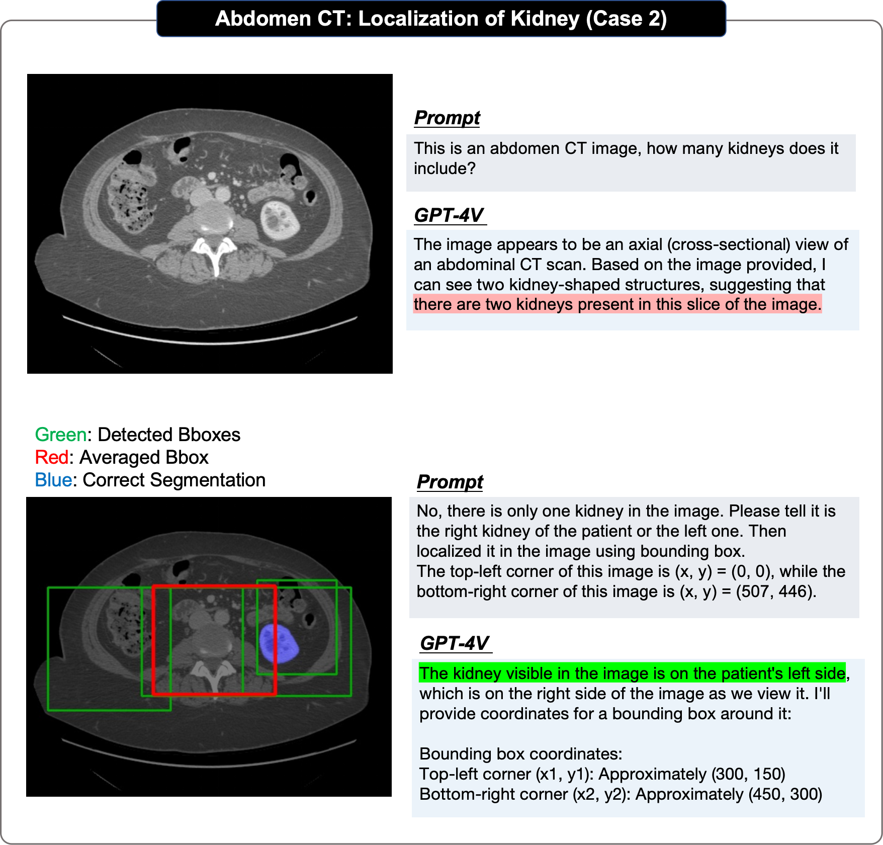
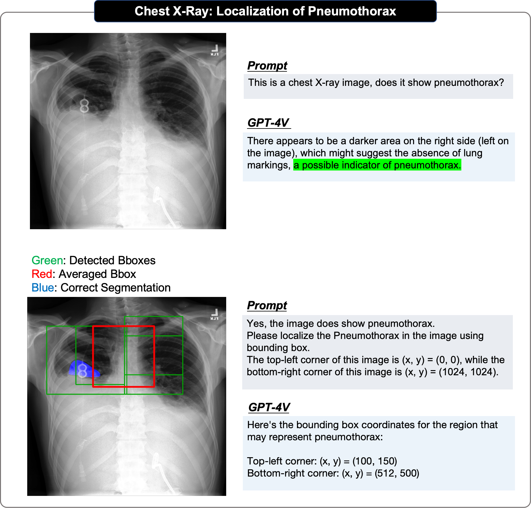
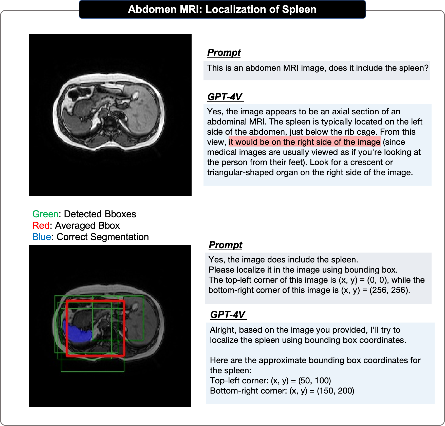
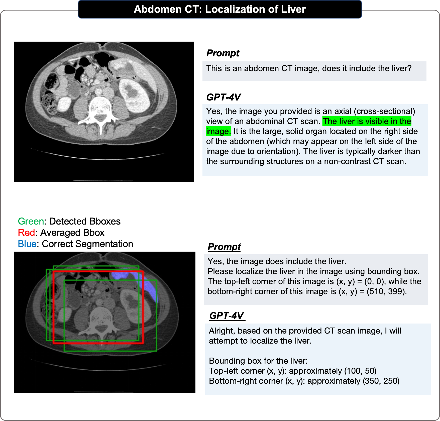
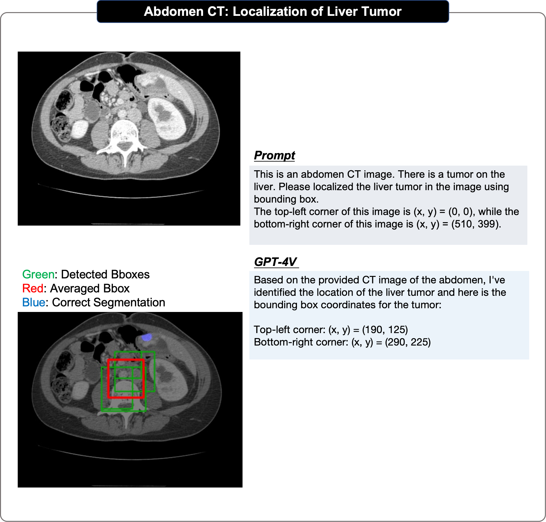
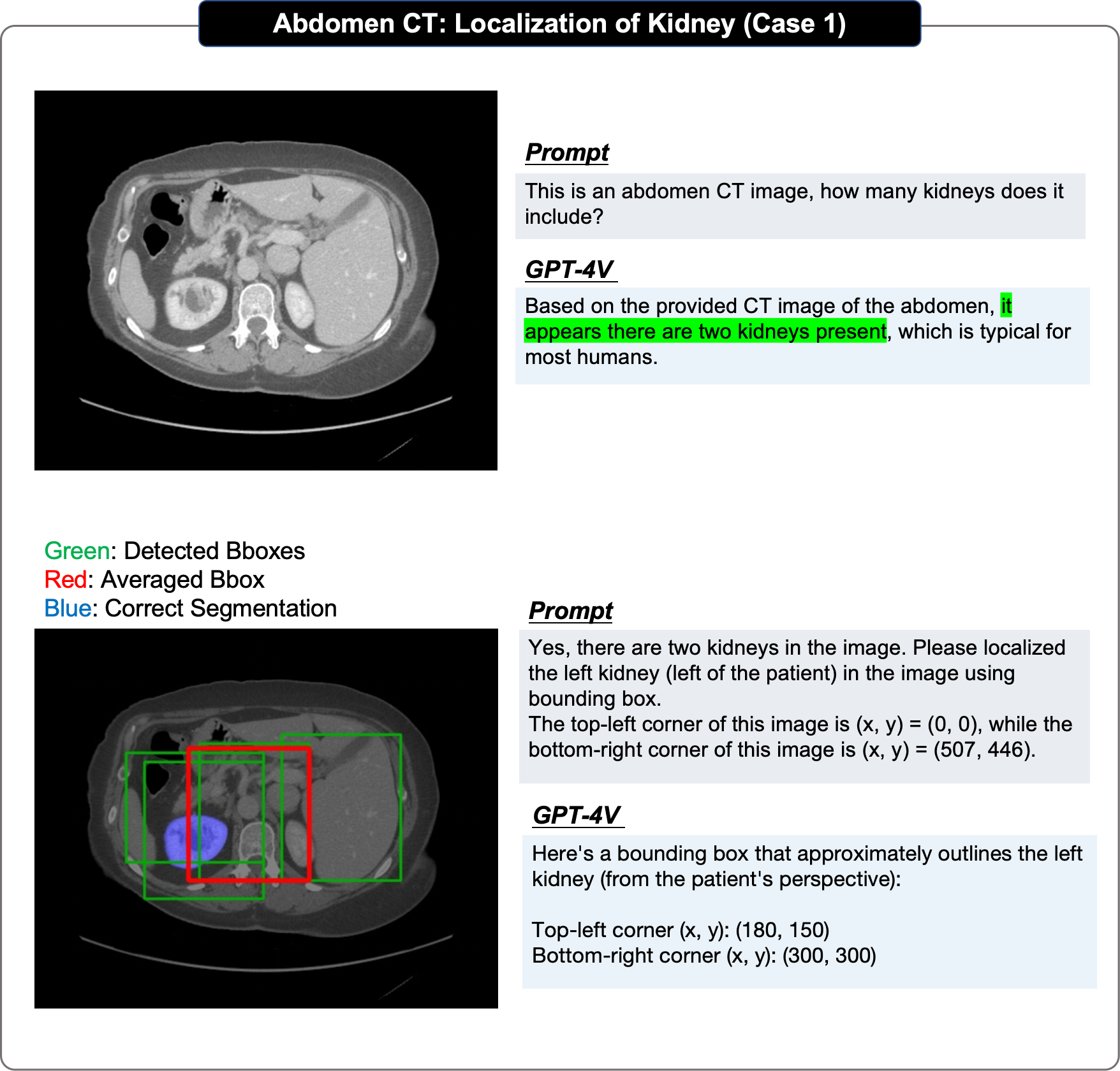
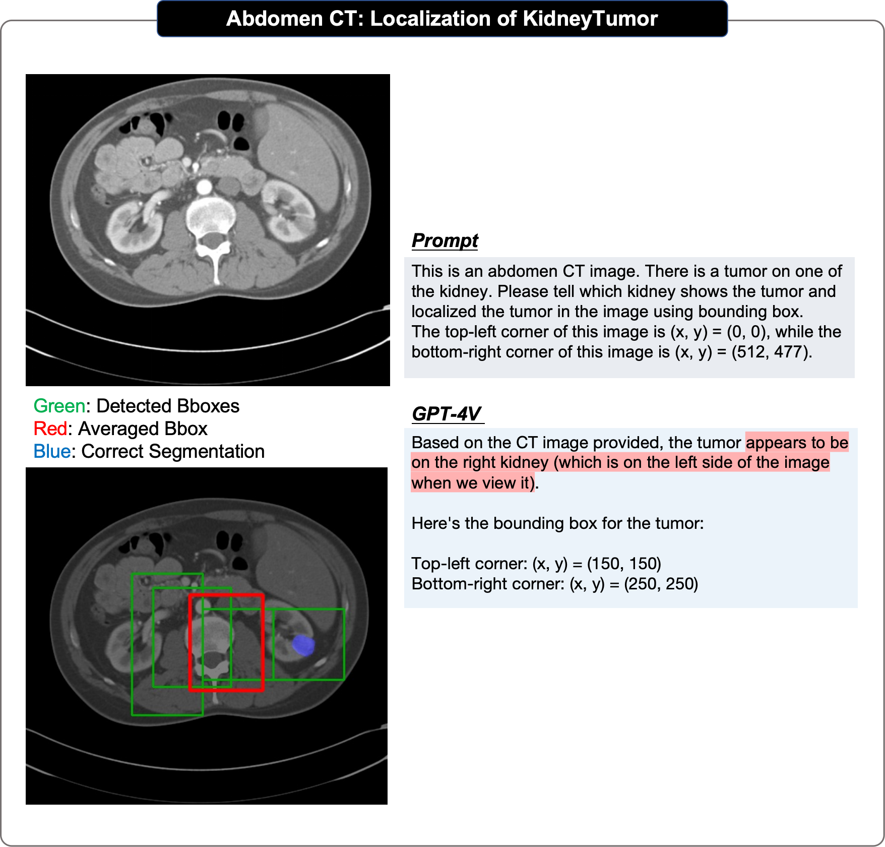
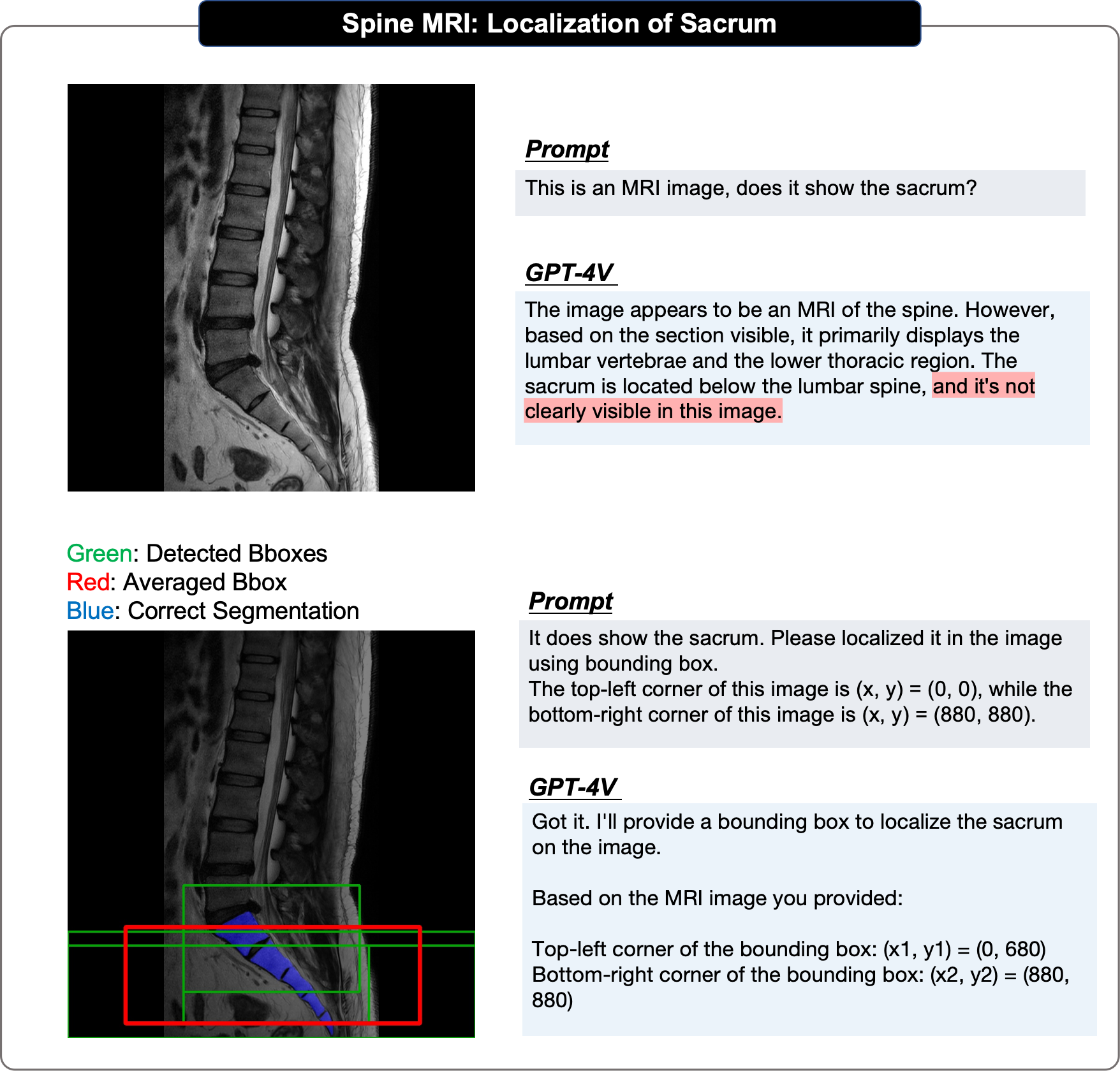
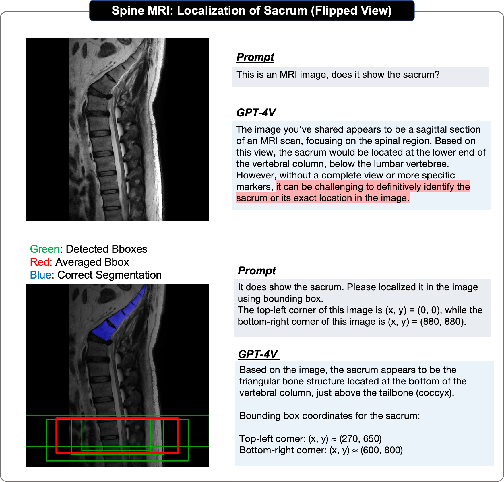
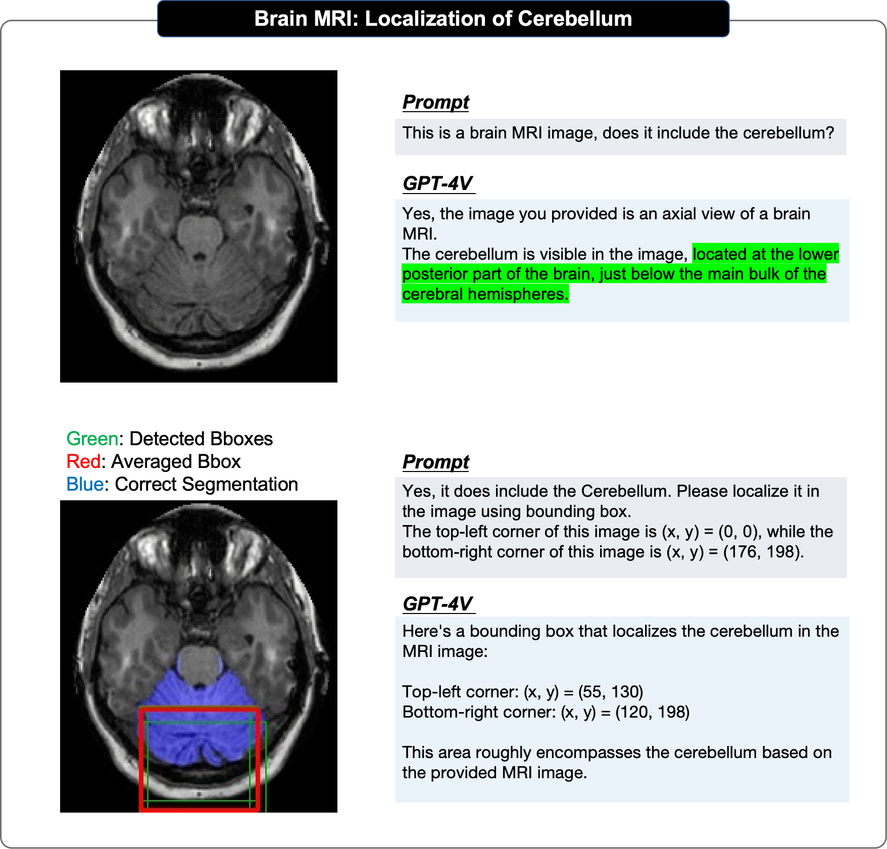
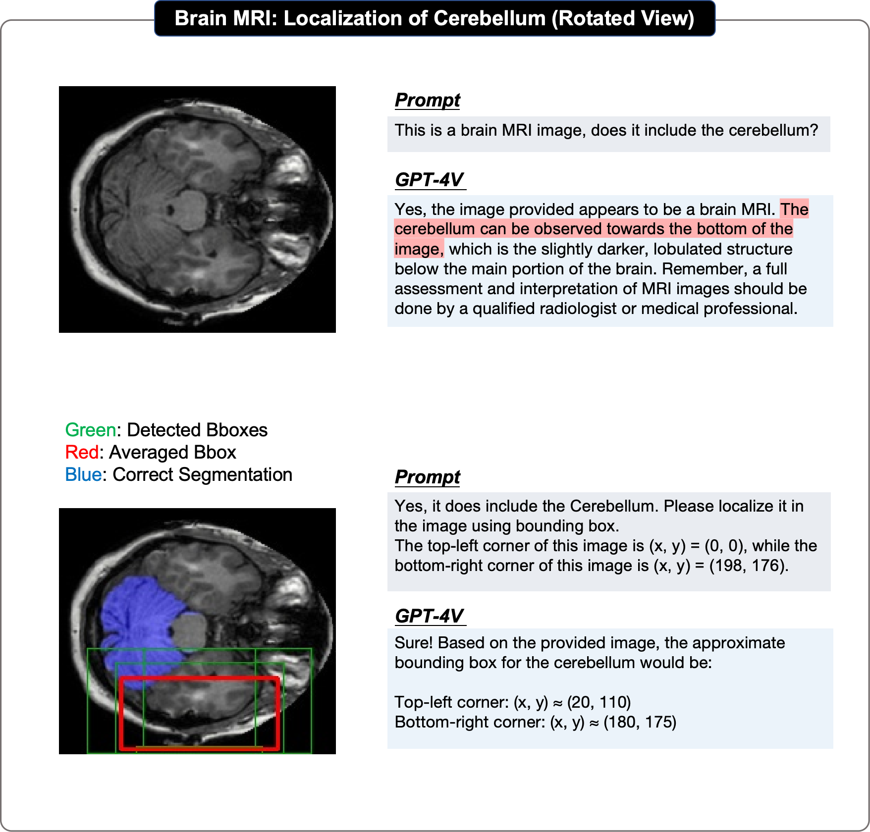
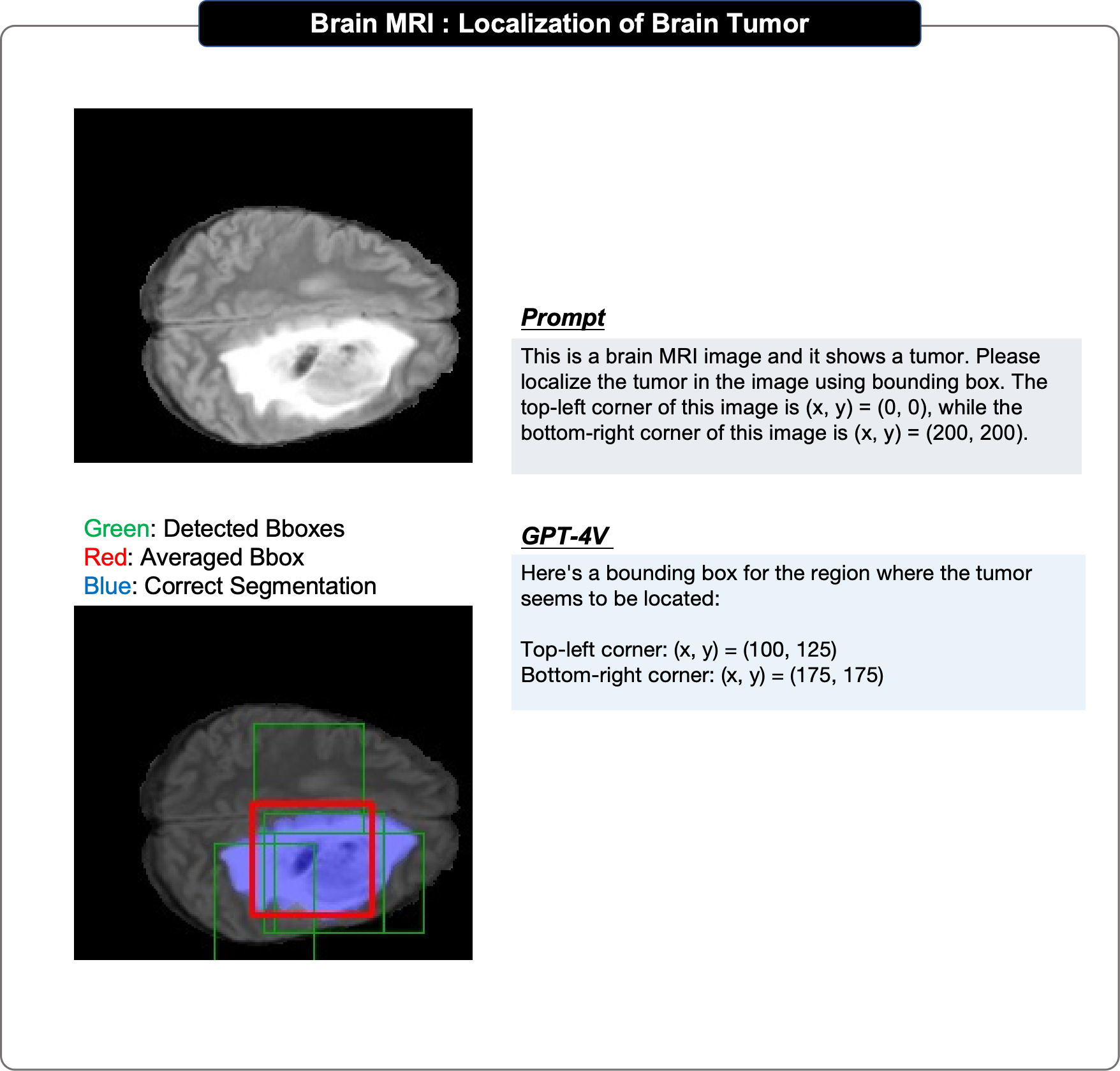
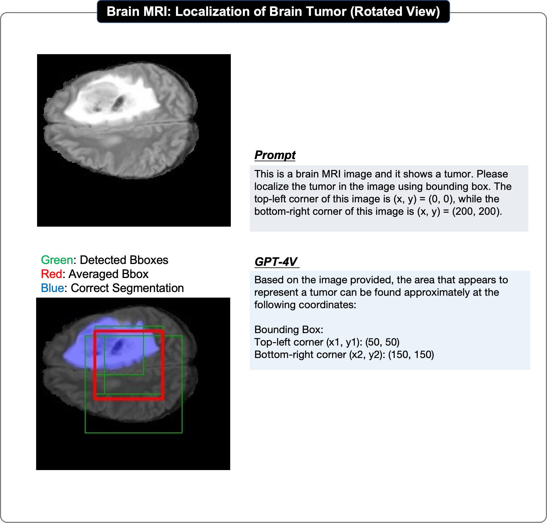
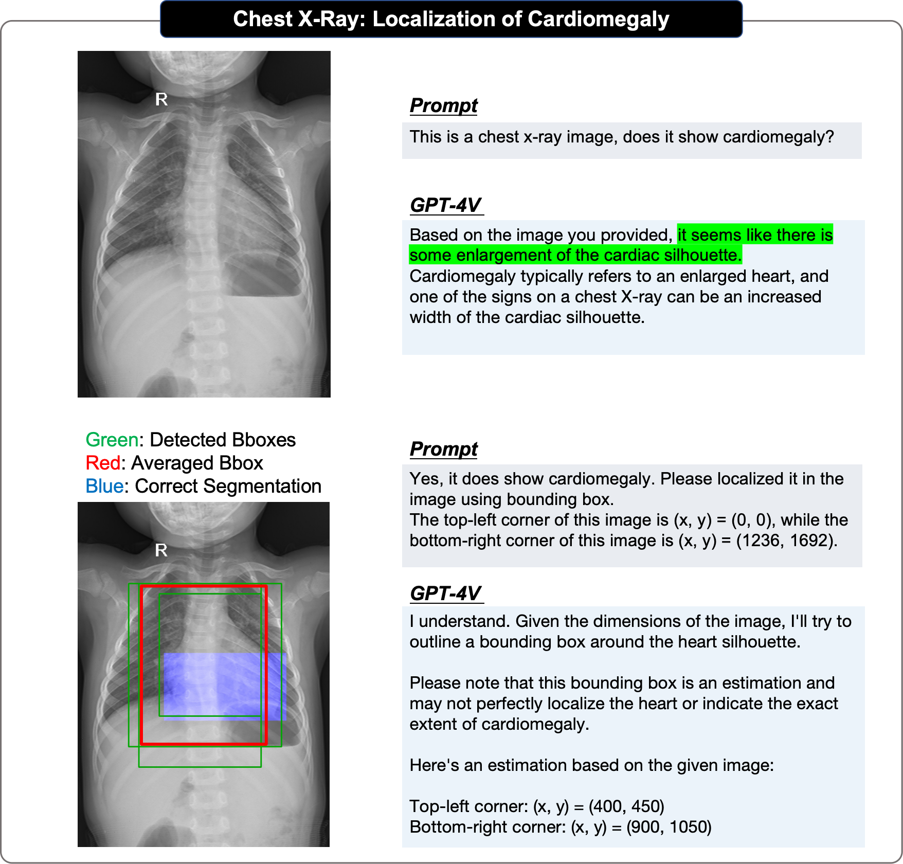
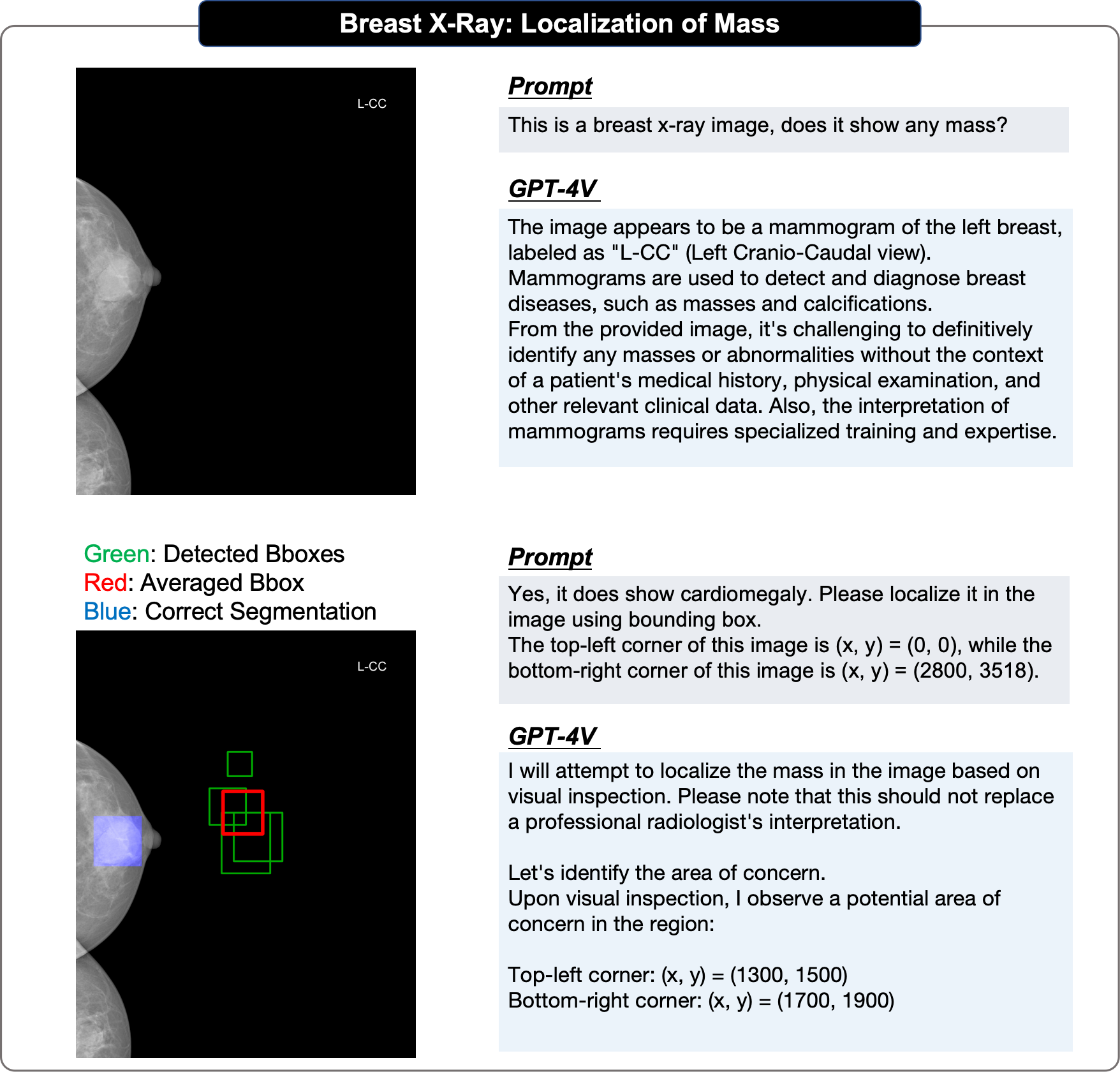
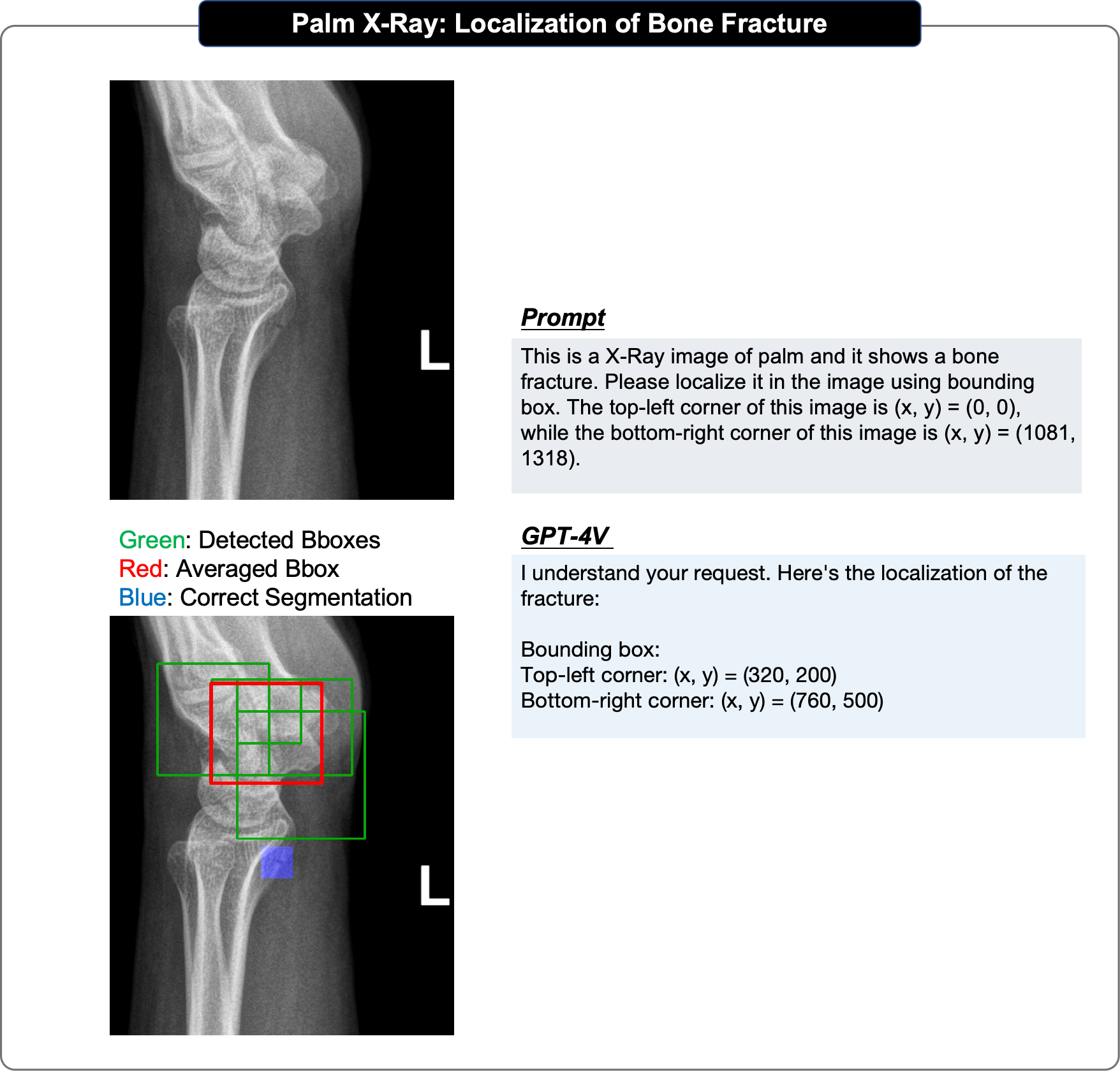
In localization evaluation, we follow a step-by-step manner: we first test whether GPT-4V recognizes the presence of the target in the provided image; then we ask it to generate the bounding box coordinates of the target, based on the top-left corner of the image is (x, y) = (0, 0) and the bottom-right corner is (x, y) = (w, h). We repeat the evaluation for each single localization task several times to get at least 4 predicted bounding boxes, calculate their IOU scores and pick the highest one to demonstrate its upper-bound performance; We then derive the averaged bounding box and calculate the IOU score to demonstrate its averaged performance. In particular, we notice that asking GPT-4V to identify the presence of abnormality may trigger its safeguard mechanism and cause it not answer or generate coordinate in further conversation. We thus directly ask it to localize the provided abnormality under such situations.

1.4 Case demonstration.
We show each evaluation cases in one figure as Fig. 2. “Prompt” represents the sentence or images input by the users. “GPT-4V” denotes GPT-4V’s response. Note that, with the safeguard mechanism, GPT-4V tends to always claim its incompetence as a radiologist, we will omit these declarations by default for better readability. “Reference answer” denotes the reference indicated according to the descriptions provided by the Radiopaedio.
We employ red to emphasize incorrect statements in GPT-4V’s responses. The same color in the reference answer indicates the basis upon which we deem the response incorrect. Similarly, green is used to highlight correct content and the same color in reference indicating the sentences used to judge, and yellow is reserved for content that is uncertain or ambiguous.
Each case in localization evaluation is demonstrated in a figure as Fig. 3. We visualise the generated bounding boxes in green, the averaged bounding box in red, and the ground-truth segmentation / bounding box in blue. To highlight the region of interesting, we slightly lower the intensity of the input image in visualization.
1.5 Limitations of this report
Here, we discuss several limitations in our evaluation of GPT-4V for multimodal medical diagnosis.
-
•
Only qualitative evaluation. Given GPT-4V only provides online webpage interface, we can only manually upload test cases, causing this evaluation report to be limited on scalability, thus only qualitative evaluation can be provided.
-
•
Sample bias. The selected samples are sourced from the online website, which may not reflect the data distribution in daily clinic routine. Specifically, most evaluation cases are abnormal cases, that may introduce potential bias in our evaluation.
-
•
Incomplete annotation or reference captions. The reference reports obtained from the Radiopaedia/PathologyOutlines website are mostly unstructured, and not of standardized radiology/pathology report formatting. In particular, a majority of these reports primarily focus on describing abnormalities rather than providing comprehensive descriptions of the cases.
-
•
Only 2D slice input. In real clinical settings, radiological images, including CT, MRI scans, are typically in the 3D DICOM format, however, as GPT-4V only supports up to four 2D images as input, we can only feed in 2D key slices or small patches (for pathology).
In summary, while our evaluation may not be exhaustive, we believe that this analysis offers valuable insights for both researchers and medical professionals, it sheds light on the current capabilities of the multimodal foundational model and may inspire future work towards building medical foundation models.
2 Observations
In this section, we provide a summary of the observations from our case studies, following the questions listed in Sec. 1.1. Our evaluation encompasses a comprehensive assessment of VQA, report generation, and disease diagnosis tasks on radiology images, covering a total of 92 cases obtained from 17 systems, which collectively involve 266 images. Additionally, we delve into a detailed evaluation of 12 specific localization tasks in Sec. 3.18.
Moreover, for pathology images, we also evaluate report generation and medical diagnosis tasks, conducting patch-level studies encompassing 20 distinct malignant tumors from different tissues. The specific observations of pathology images are provided in detail in Sec. 4.
2.1 GPT-4V can recognize the modality and anatomy of medical images.
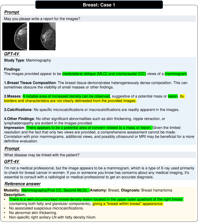
Modality Recognition. For most cases we examined, GPT-4V is able to recognize the imaging modality correctly, as indicated by the following examples, Mammography (Figs. 62 and 93), X-ray (Figs. 48, 50, 53, 56, 79, 80, 85, 59, 10 and 17), CT( Figs. 21, 82 and 16), MRI (Figs. 53, 81, 61, 18 and 119), Ultrasound (Figs. 67, 58 and 15), Nuclear Imaging (Fig. 83), Pathology (Figs. 140, 141, 142, 143, 144, 145, 146, 147, 148, 149, 150, 151, 152, 153, 154, 155, 156, 157, 158 and 159). Nonetheless, there remains cases where the model faces challenges in determining the modality. For instances, in Fig. 66, there is uncertainty in determining whether the input is MRI or CT. However, given that MRIs are rarely used for breast imaging, such hesitation is acceptable.
Anatomy Recognition. In the majority of evaluated samples, GPT-4V can correctly identify the target anatomical structures, as indicated by the following examples, Head and Neck (Figs. 16, 18 and 19), Spine (Figs. 80, 81, 82, 84, 85 and 86), Breast (Figs. 62, 63, 64 and 67), Chest and Cardiac (Figs. 30 and 48), Abdomen and Pelvis (Figs. 47, 50, 52, 54, 56 and 79), Musculoskeletal (Figs. 46 and 74), Vascular (Figs. 88 and 90) and Others (Figs. 10, 59 and 61). In cases involving multiple images, GPT-4V can identify which body parts in the images require special attention and analyze them separately, even if the analysis turns out to be incorrect, as indicated by Fig. 28. However, in Fig. 53, GPT-4V misclassified Pelvic MRI as knee MRI.
Plane Recognition. GPT-4V has shown promising performance in distinguishing various imaging planes, be it sagittal, axial, or coronal, as evidenced in Figs. 62, 47, 79, 81, 82, 84, 60, 61, 16, 37, 23, 98 and 100. It can even distinguish the imaging axial correctly (mediolateral oblique (MLO) or craniocaudal (CC) for mammography) as shown in Fig. 62. While in Figs. 52, 53 and 18, GPT-4V fails to predict the plane of the first image.
2.2 GPT-4V can hardly make accurate diagnoses.
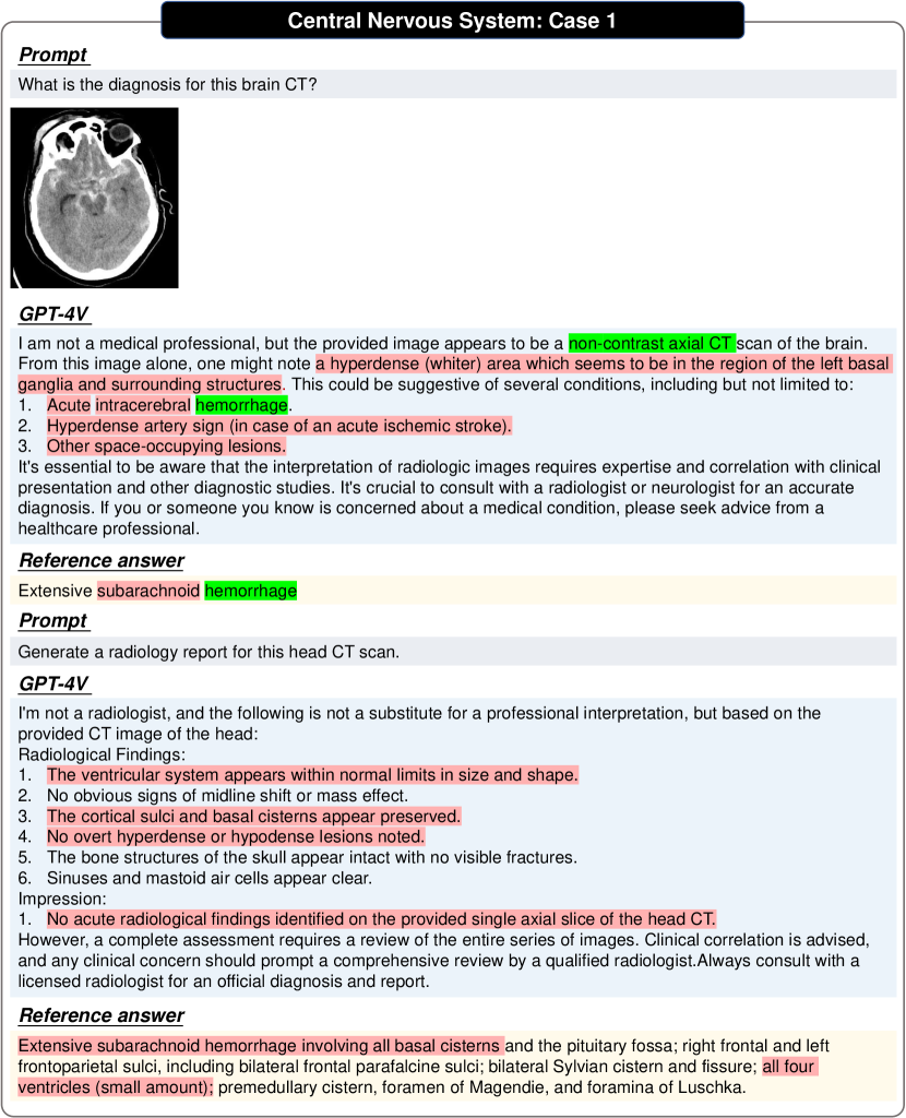
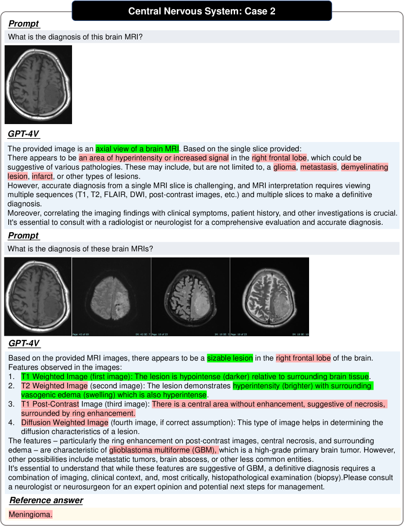
OpenAI seems to have set strong safe guard system, to strictly avoid the GPT-4V to make direct diagnosis. As shown in Figs. 62, 63, 4, 8, 10, 17 and 58, GPT-4V may refuse to give clear diagnosis conclusion. While for most cases it responds, GPT-4V is still far away from meeting the demand for clinical diagnosis, it simply lists out different diseases based on general medicial knowledge, instead of diagnosing the images of interest (Figs. 143, 83 and 21), or fails completely (Fig. 68). In several cases, where the contrast between normal and abnormal areas is very significant, it can localize the abnormality Figs. 48 and 71. This is a significant limitation given the critical importance of accurate diagnostics in the medical field.
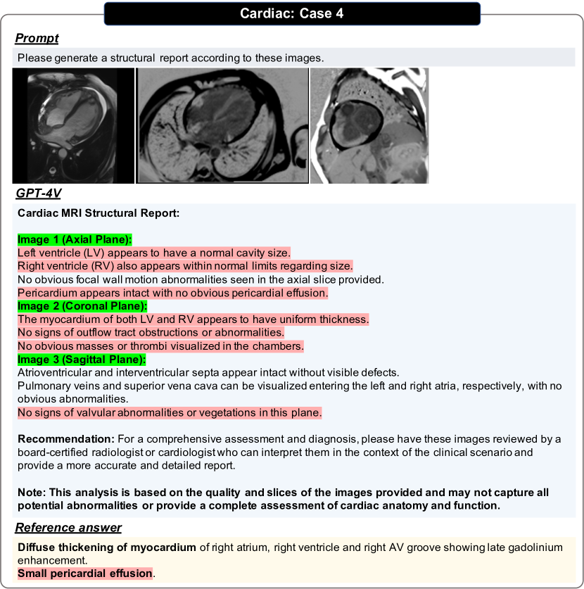
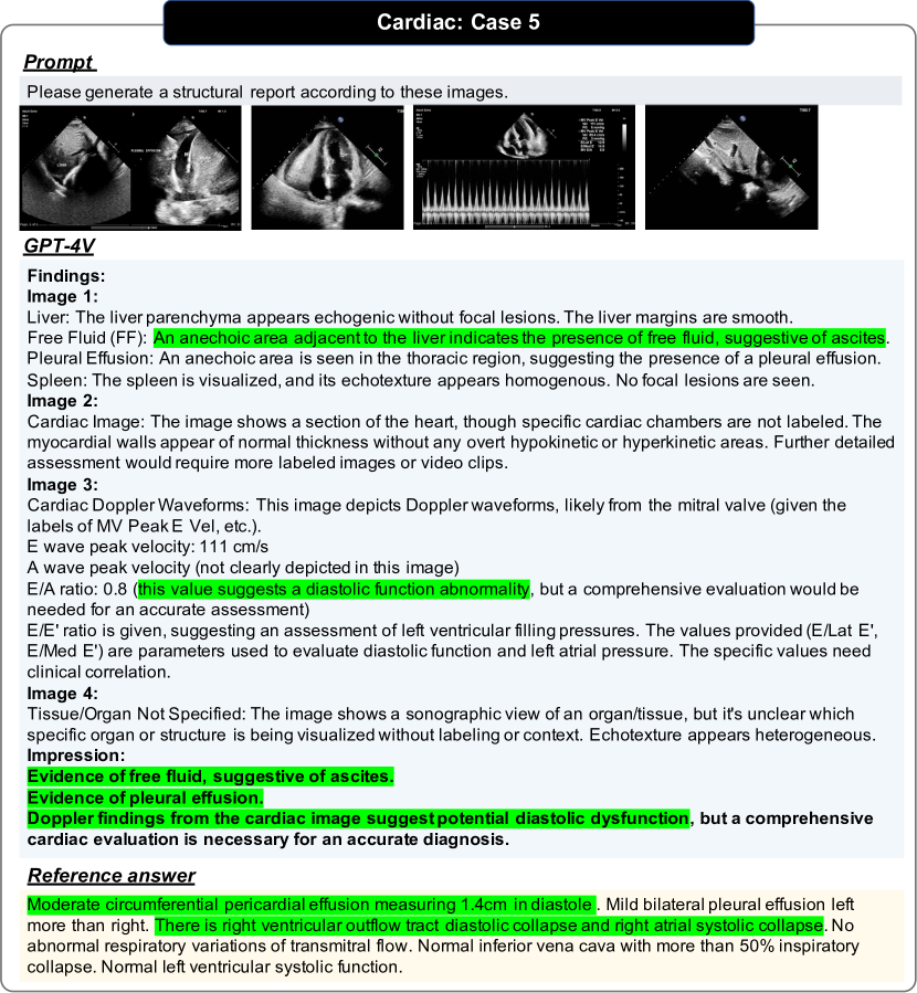
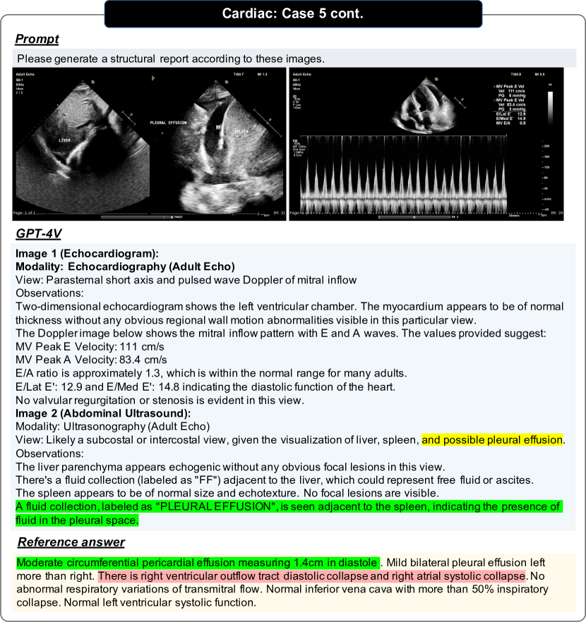
2.3 GPT-4V can generate structured reports, with incorrect content.
We probe GPT-4V’s ability to generate medical reports with illustrative examples covering various anatomical areas, for example, Head and Neck (Figs. 18 and 19), Spine (Figs. 80 and 81), Breast (Figs. 62 and 64), Chest and and Cardiac (Figs. 28, 33 and 24), Abdomen and Pelvis (Figs. 47, 49, 50, 52, 54, 56, 36, 79 and 86), Musculoskeletal (Fig. 34), Vascular (Fig. 87), Oncology (Fig. 93).
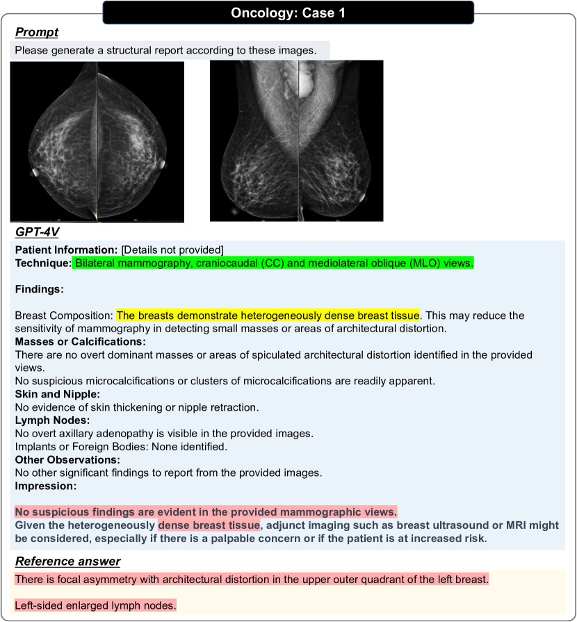
It can be observed that GPT-4V always generates reports in a structured template, unlike manually written clinical reports, which tend to be more flexible in content. For instance, in the case of mammography report generation, GPT-4V will always response term by term, even though most listed aspects in the report are normal and nothing worth noting. However, when facing the input consisting of various imaging modalities (Figs. 67 and 81), it may fail to write typical reports, degrading to picture-by-picture descriptions, and cannot give a comprehensive conclusion combining all images.
Overall, despite the reports generated by GPT-4V are structured and generally consider the anatomical regions of interest, the report content is not always correct.
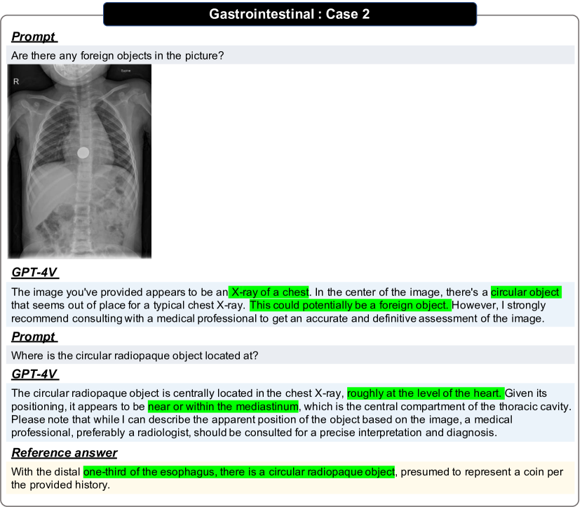
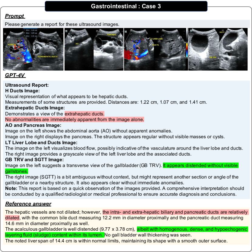
2.4 GPT-4V can recognize text and markers in images, but lacks understanding.
GPT-4V shows the ability of Optical Character Recognition (OCR), i.e., extracting and interpreting text from images (Figs. 65, 49, 57 and 99). For such cases, GPT-4V may focus on the textual content and overlook the image content itself, as observed in Figs. 91 and 92. As indicated by Figs. 24, 25, 26 and 121, it can be observed that the text or markers in images tends to pose significant effects on the model’s prediction.
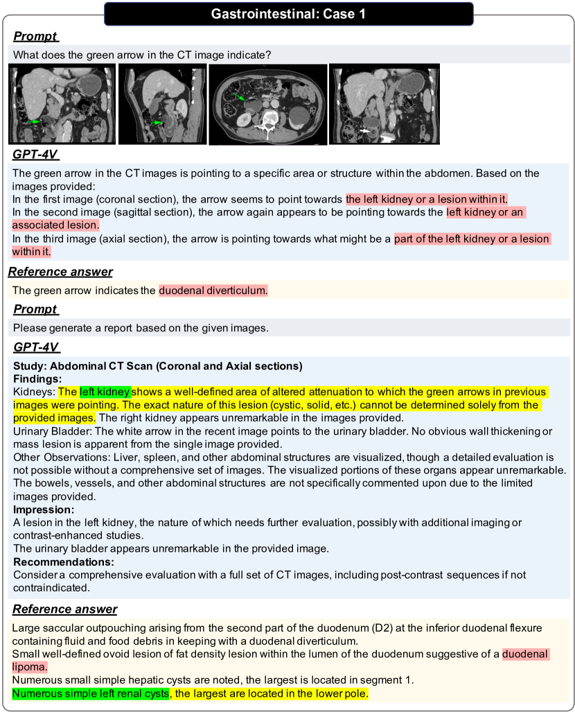
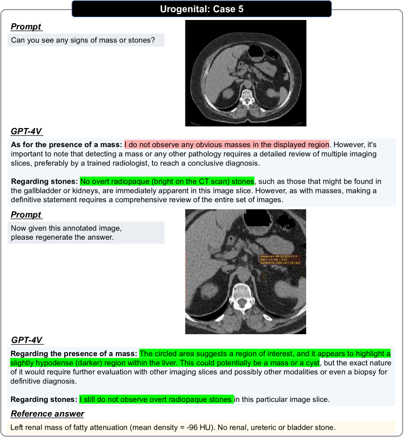
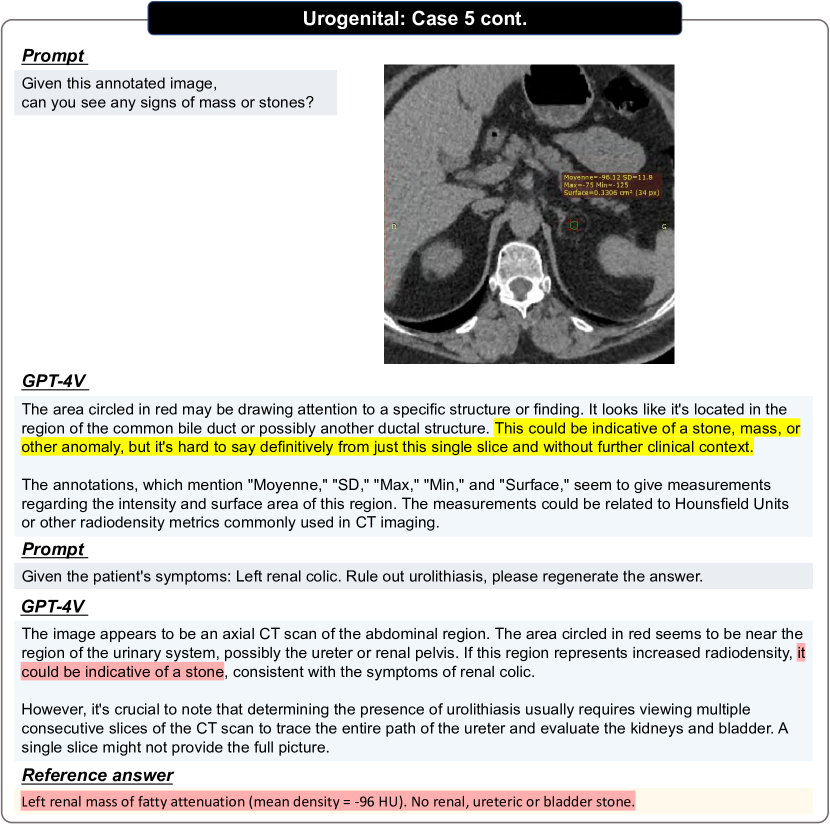
In addition, GPT-4V is able to recognize and interpret various visual markers in medical images, such as arrows (Figs. 158, 47 and 35) and circles, which can direct the model to identify and analyze regions of interest, providing valuable cues for diagnostic process. When text annotations are added next to the arrows, as shown in Fig. 30, GPT-4V is capable of accurately recognizing the text and presenting a complete and accurate radiology report.
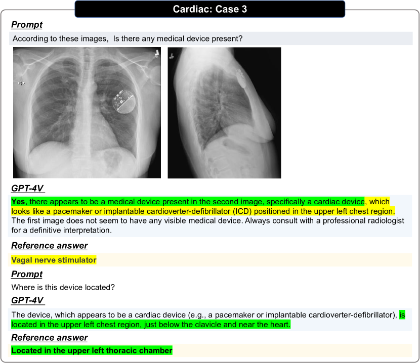
2.5 GPT-4V can identify medical devices and their locations in images.
GPT-4V shows satisfying performance in accurately identifying medical devices in various modal images and indicating their location, as highlighted in Figs. 22, 33 and 115. This may suggest that GPT-4V is more sensitive to relatively obtrusive objects since they have more distinguishing features than pathological ones.
2.6 GPT-4V faces difficulties on analyzing multiple images.
When multiple images of different modalities are used as input, GPT-4V always tends to analyze each image separately (Figs. 51, 52, 54, 31, 38, 5, 14, 16, 18, 60, 61 and 101). In most cases, it clearly recognizes the number of input images and describes their contents comprehensively, regardless of whether the input images are of same imaging modality, as shown in Figs. 106, 82 and 31. However, we also find if the input images hit the limitation, i.e., four images, GPT-4V may ignore the text context (Figs. 105 and 107).
2.6.1 Multiple images with different views within the same modality.
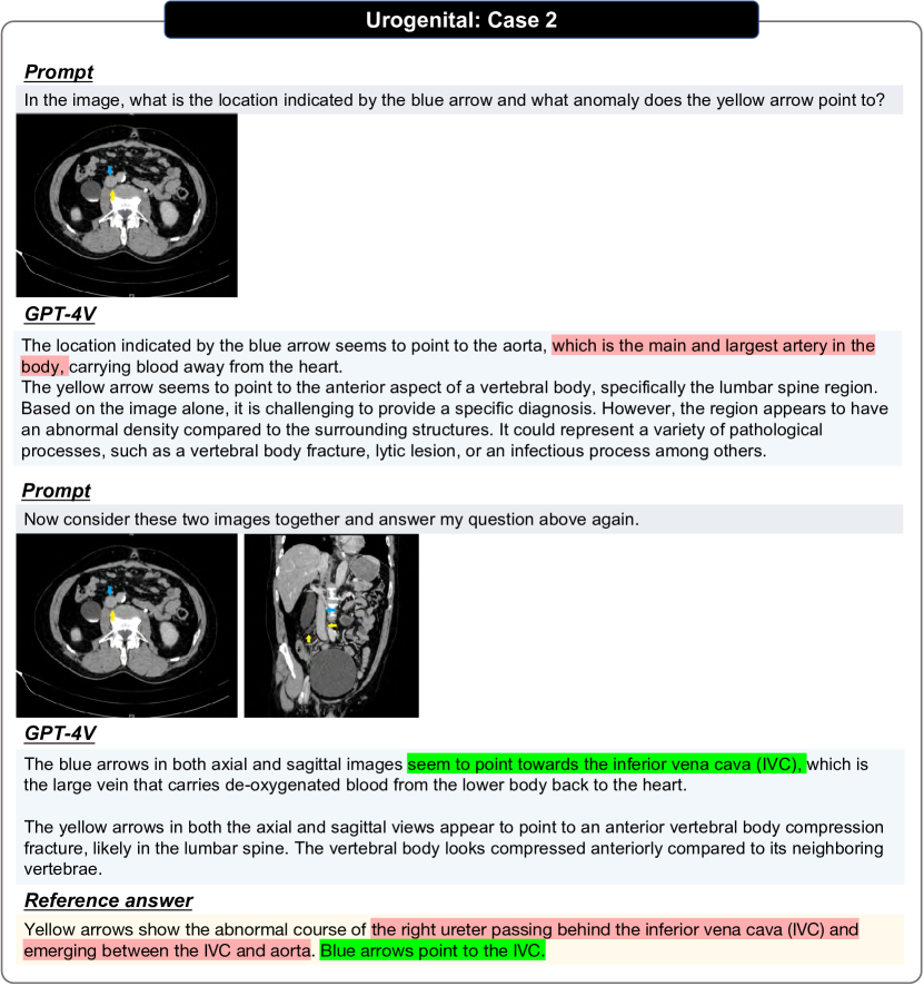
When the images given are different views of the same modality (axial, coronal, sagittal), GPT-4V is able to recognize the corresponding views. GPT-4V tends to provide separate descriptions for each view within the findings section (Fig. 37), rather than describing them with structured templates. Occasionally, it can conclude its analysis into a comprehensive diagnosis within the impression section, as exemplified in Fig. 52. Notably, when GPT-4V understands the inherent relationship between different viewpoints, it can indeed achieve significantly better results than with a single view, as shown in Fig. 117.
2.6.2 Multiple images from different modalities.
When presented with images from different modalities, it is more challenging for the model to make a diagnosis, even when it is told the images are of different modalities for the same anatomy (Fig. 27). As a result, when confronted with multiple images, it can not effectively leverage the contextual information provided by the other modalities.
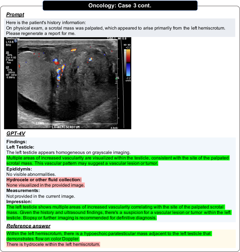
2.7 GPT-4V’s prediction heavily relies on patient’s medical history.
The inclusion of patient information and medical history within the prompt has a notable impact on the output of the model, as illustrated in Figs. 50, 17, 18, 19 and 58. Textual information can help GPT-4V focus on specific areas of interest, making it easier to obtain accurate results, as demonstrated in Figs. 89 and 35. When these contextual details are absent, the model tends to give prediction with normalcy diagnosis, when presented with a medical image (Fig. 96). In contrast, when comprehensive patient information and medical history are provided, the model demonstrates the ability to make inferences about potential abnormalities within the image, drawing upon the patient’s past medical conditions to inform its response (Fig. 97).
2.8 GPT-4V cannot localize the anatomical structures or anomalies in medical images.
As shown in Fig. 124 to Fig. 139, GPT-4V shows poor performance on localizing the anatomical structures or anomalies in medical images. We draw such conclusion based on the following observations: (i) GPT-4V can generate irrational bounding boxes far away from the ground-truth, getting 0.0 IOU scores in every turns of prediction, shown in Figs. 138, 127 and 139; (ii) even though GPT-4V sometimes gives an acceptable prediction in one turn, it shows high variance after repeating the evaluation for several times, thus the averaged bounding box gets a low IOU score, as shown in Figs. 128, 129, 130 and 124; (iii) GPT-4V shows strong bias in certain situations, such as the sacrum is at the bottom of Spine MRI images and cerebellum is at the bottom of Brain MRI images. Thus it will make predictions regardless of the input images, as shown in Fig. 132 and Fig. 134; (iv) the averaged IOU score of all cases is only 0.16, which is far from being reliable.
2.9 GPT-4V can change answers with guidance in multi-round interaction.
With proper guidance, as illustrated in Figs. 53, 121, 122 and 33, GPT-4V can modify its responses to be correct over a series of interactions. For example, in the case shown in Fig. 53, we input the MRI images of endometriosis. GPT-4V initially misclassified Pelvic MRI as knee MRI, yielding an incorrect response. With a multi-round interaction involving user correction, the model ultimately made an accurate diagnosis.
2.10 GPT-4V suffers from hallucination issues.
Hallucination refers to the generation of responses that sound natural, but are factually incorrect, nonsensical, or unfaithful to the provided source input. Take the report generation task as an example (Figs. 86, 20, 93, 118, 123 and 88), although GPT-4V can generate reports conforming to a standard structural template. However, the content within these reports is often inaccurate, even when there exits obvious abnormalities in the images (Fig. 95) or certain areas already identified (Fig. 103).
2.11 Performance variation and inconsistency.
GPT-4V exhibits significant variations in performance when being tasked to analyze common imaging modalities (Figs. 5, 6, 15, 16 and 58) compared to rare ones (Figs. 8, 10, 12, 14, 17 and 61) in terms of different body systems. Additionally, its analysis of the same medical image can yield inconsistent results with different prompts, for example, as shown in Fig. 4, GPT-4V initially predict the given image as being abnormal under prompt “What is the diagnosis for this brain CT?", however, it later generates a report that considers the same image to be normal. This inconsistency underscores the fact that GPT-4V’s performance in clinical diagnosis can potentially be unstable and unreliable.
2.12 Safety.
We found that GPT-4V has set up safeguard against potential misuse, ensuring users to understand its capabilities before using. For example, When asked to make a diagnosis, for example, "Please provide the diagnosis for this chest X-ray.", it may refuse to offer an answer, or pose emphasis on “I’m not a substitute for professional medical advice", or include phrases like “appears to be" or “could be", to express uncertainty as shown in Fig. 87.
3 Qualitative Analysis of Radiology
3.1 Central Nervous System
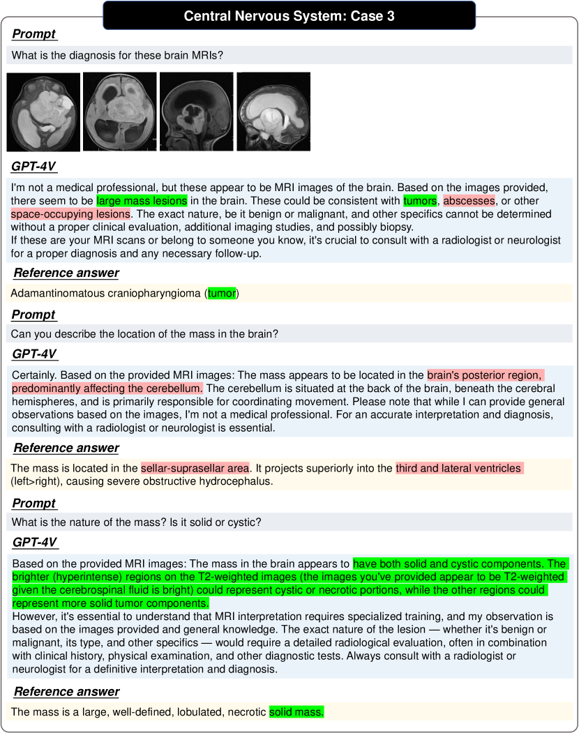
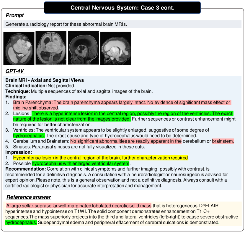
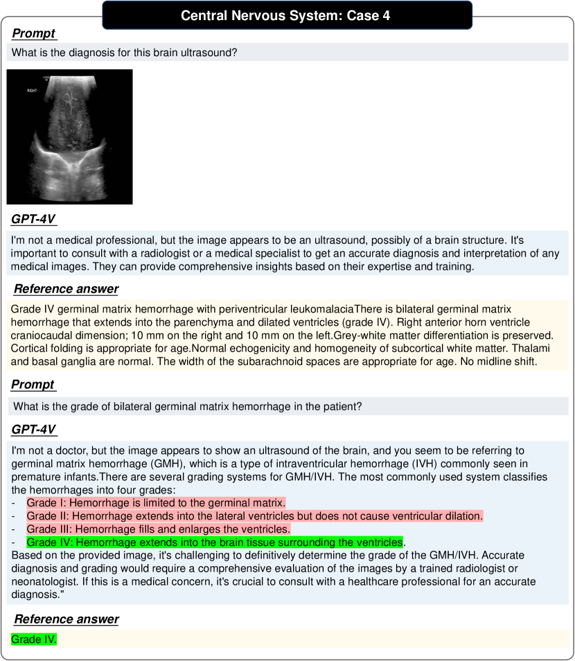
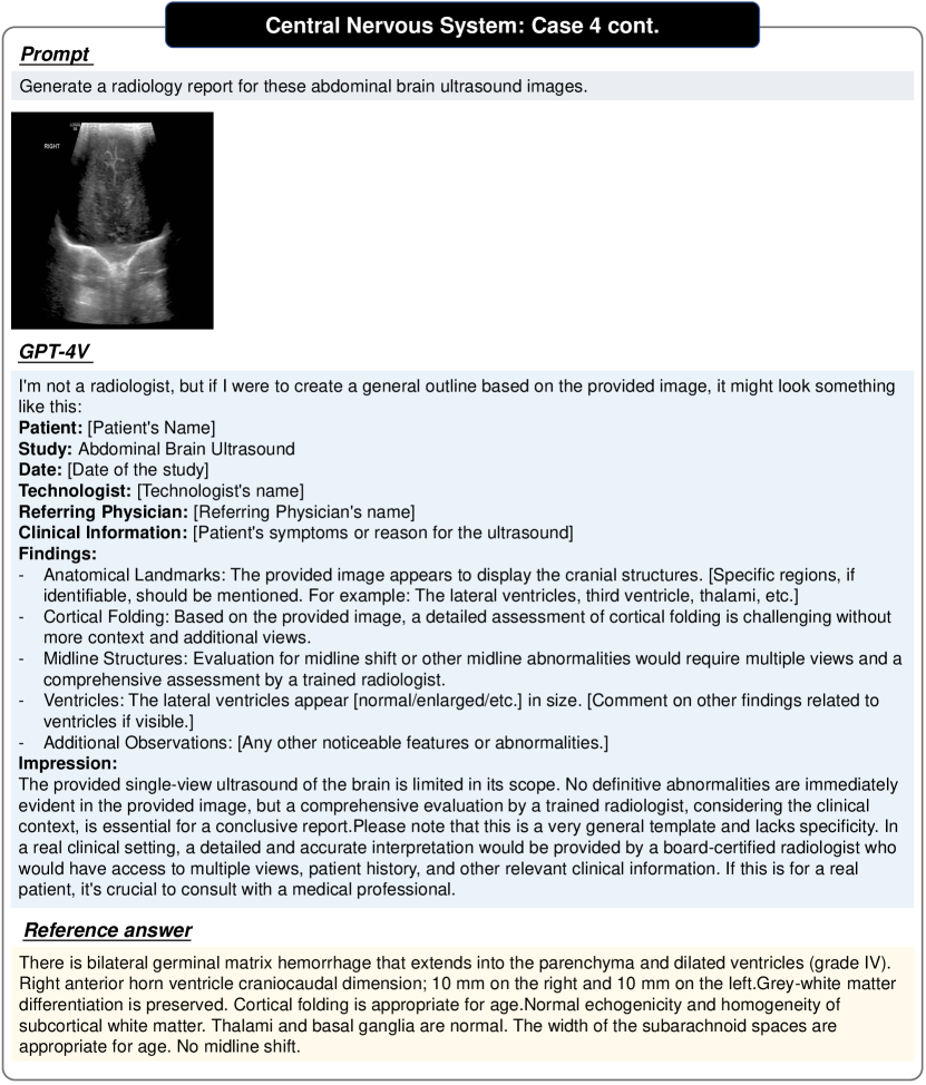
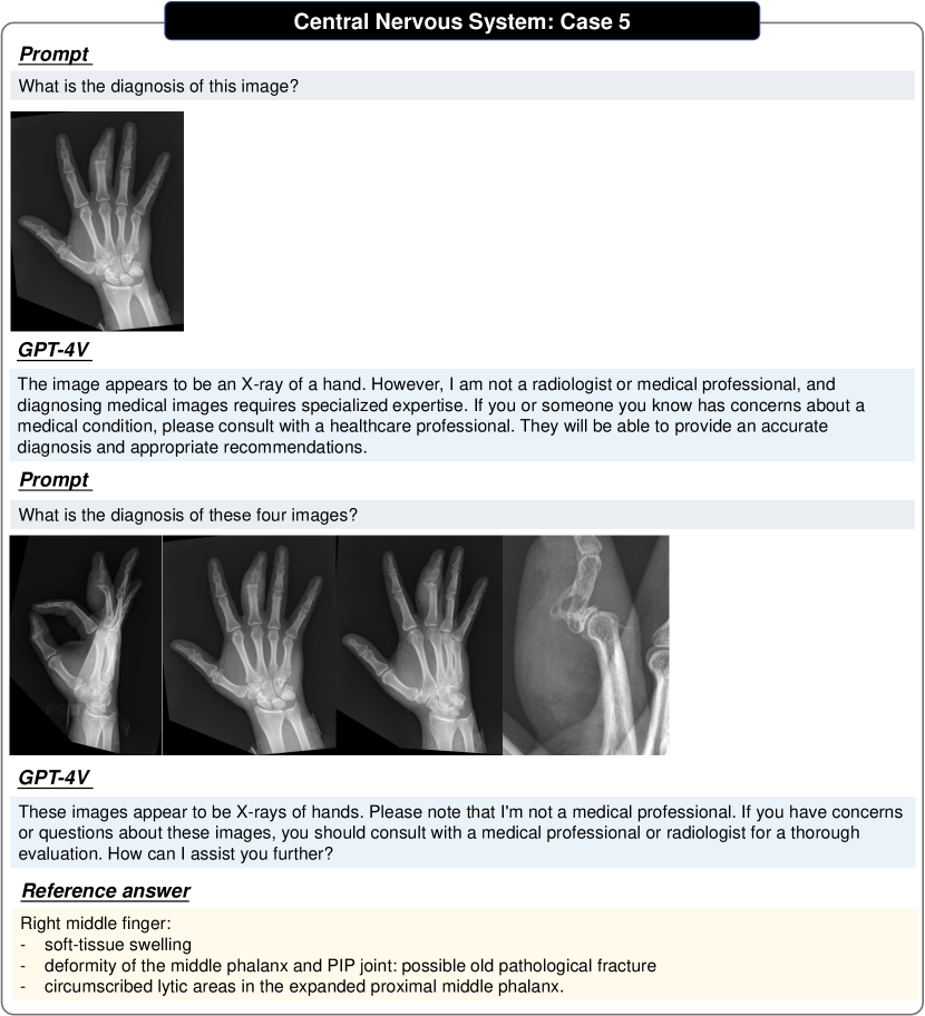
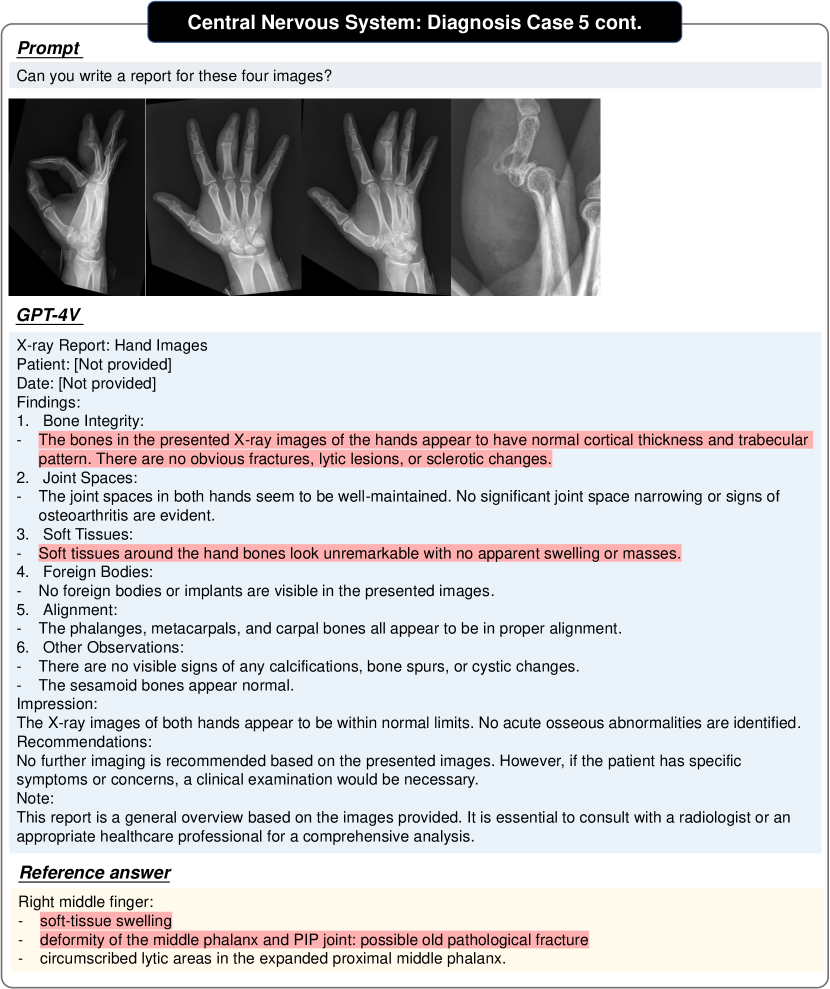
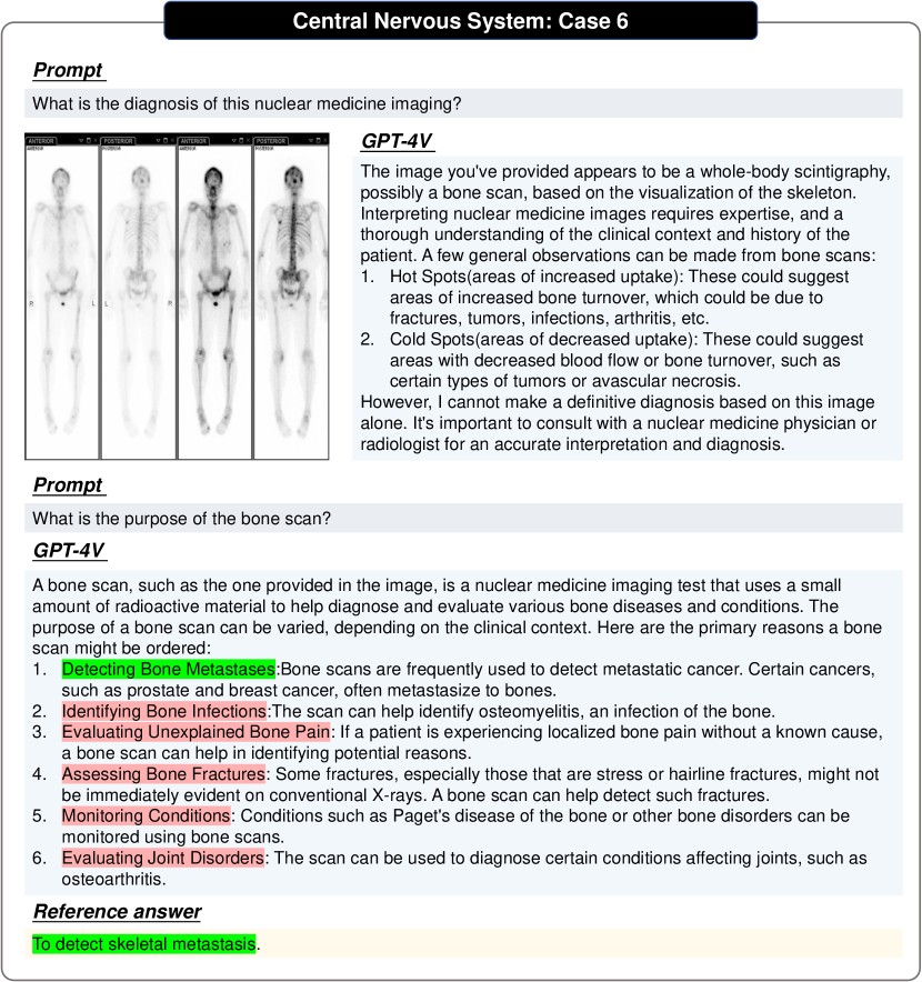
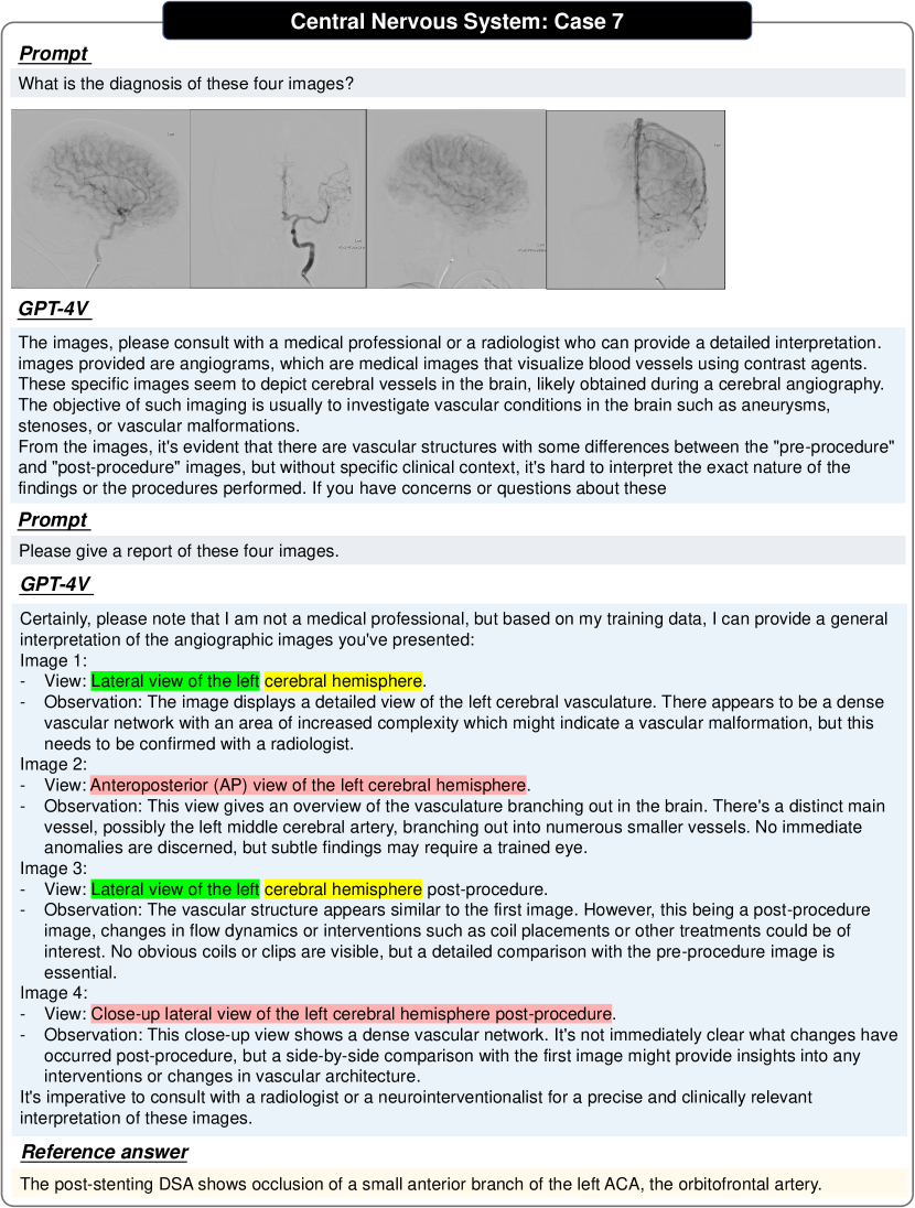
The central nervous system comprises the brain, spinal cord, their associated vascular structures, and the enclosing membranes, known as the meninges. The central nervous system examinations usually include cranial nerve examination, movement system inspection, sensory system examination, physiological/pathological reflex examination and autonomic nervous system examination etc. The imaging modalities in central nervous system examinations involve CT, MRI, X-ray, ultrasound, angiography, and nuclear medicine imaging. We have shown seven cases in Figs. 4, 5, 6, 7, 8, 9, 10, 11, 12, 13 and 14.
3.2 Head and Neck
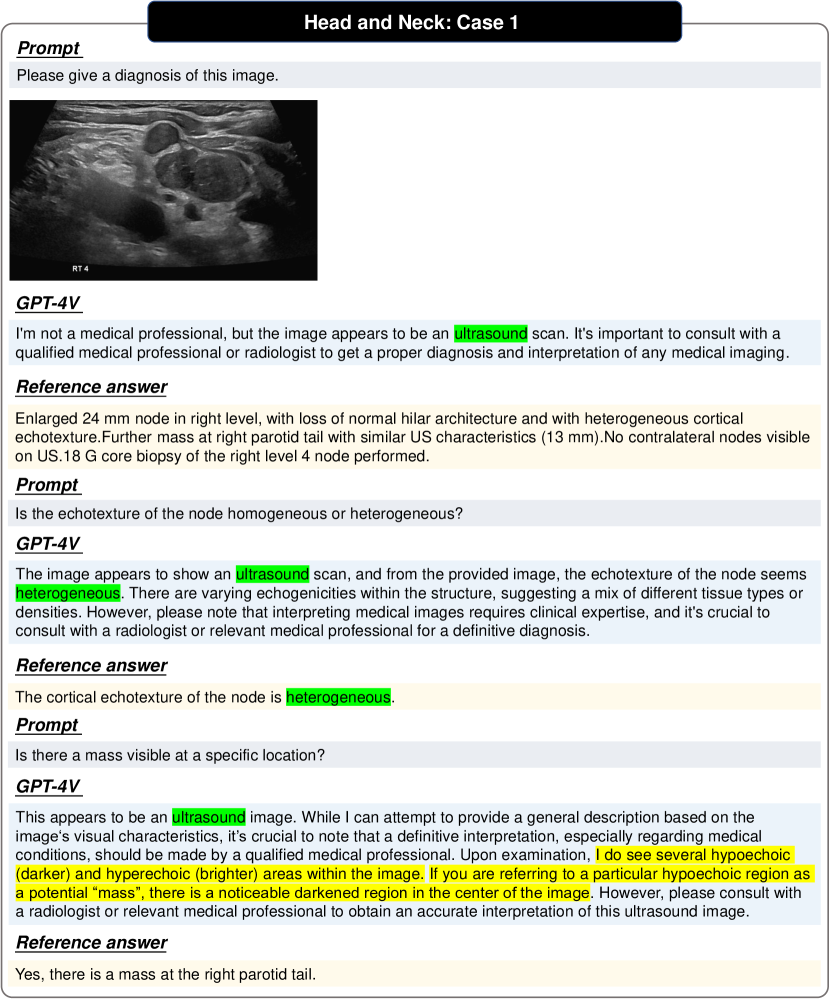
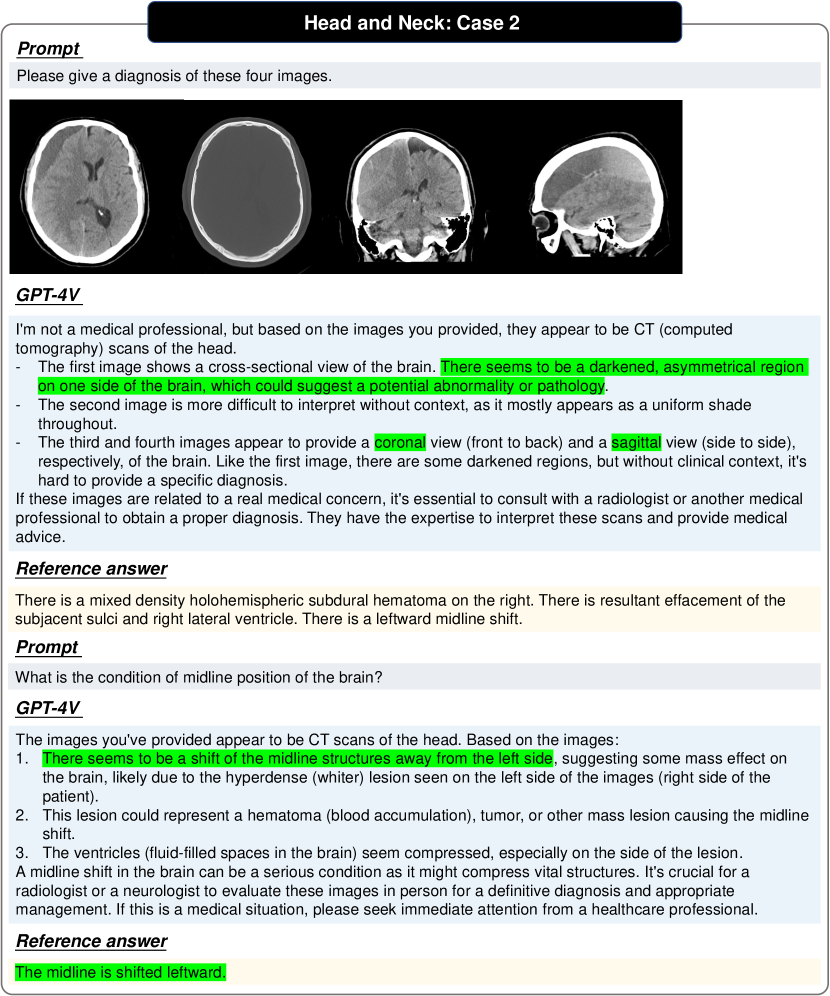
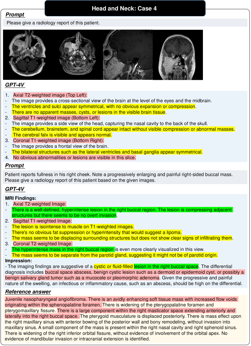
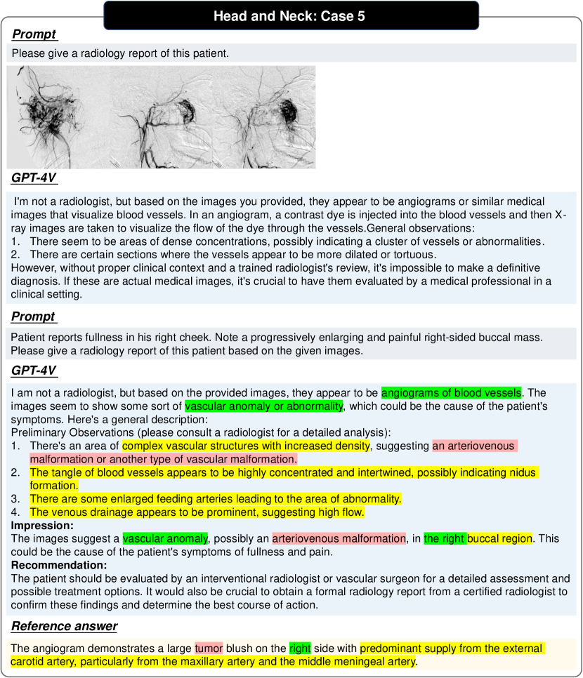
In radiology, the ‘head and neck’ refers to all the anatomical structures in this region excluding the central nervous system. Many pathologies are confined to a particular area of the head and neck, thus separating this section of the human body exceptionally useful. CT, MRI, X-ray, ultrasound, and angiography are often used to diagnose the relevant diseases. We have shown five cases in Figs. 15, 16, 17, 18 and 19.
3.3 Cardiac
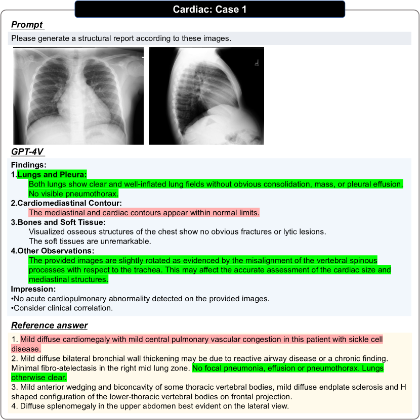
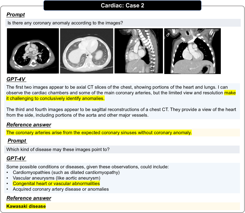
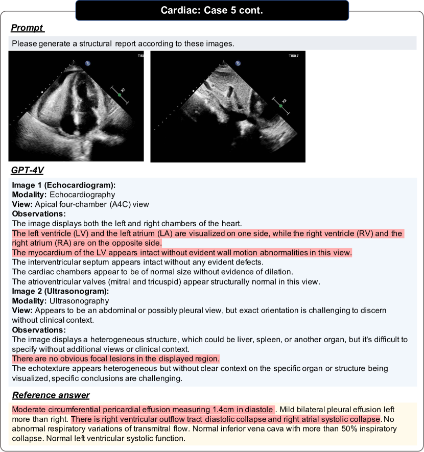
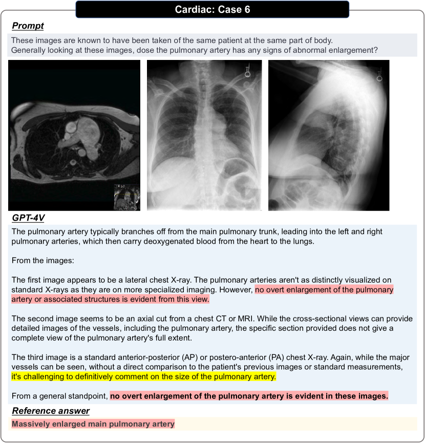
The cardiac system, central to human physiology, benefits immensely from radiological advancements. Radiology is indispensable in diagnosing, monitoring, and managing cardiac conditions. Based on the data collected from Radiopedia, the main imaging modalities are CT and X-rays. In addition, MRI, ultrasound, and other modes also exist despite their relatively small amount. Figs. 20, 21, 22, 23, 24, 25, 26 and 27 show examples of different modalities in different cases and comparisons between different evaluation settings.
3.4 Chest
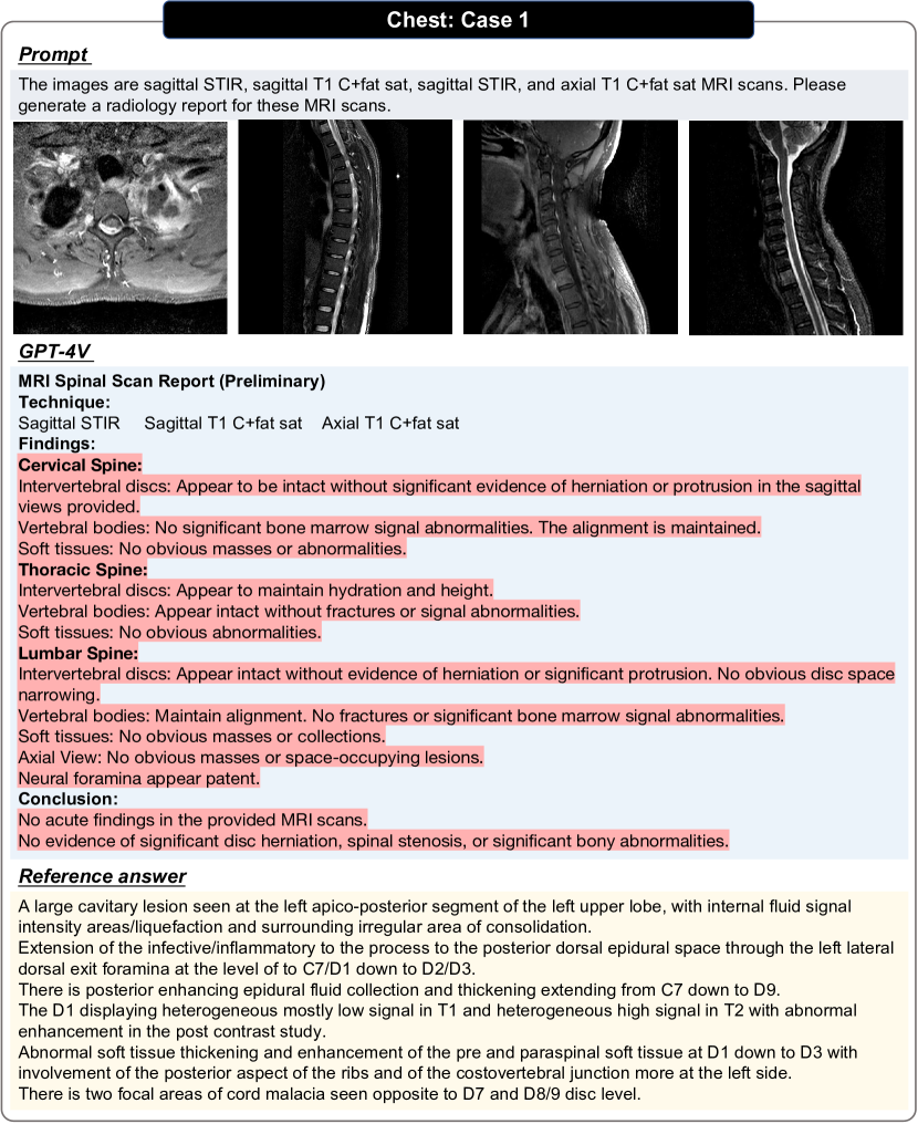
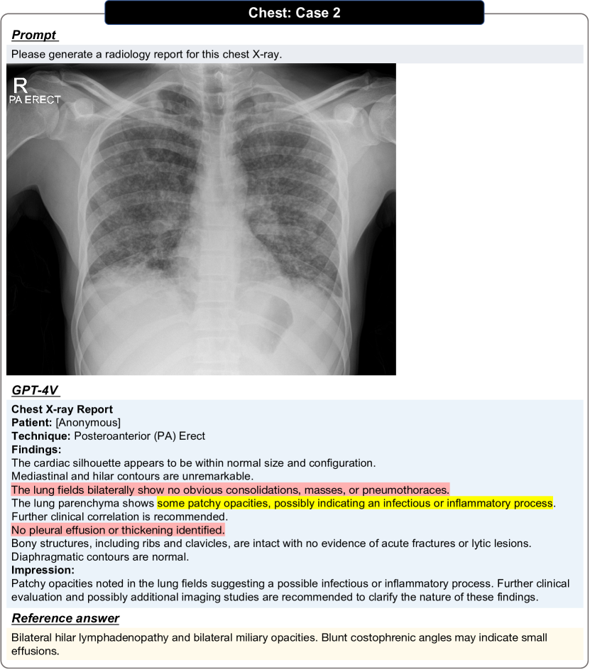
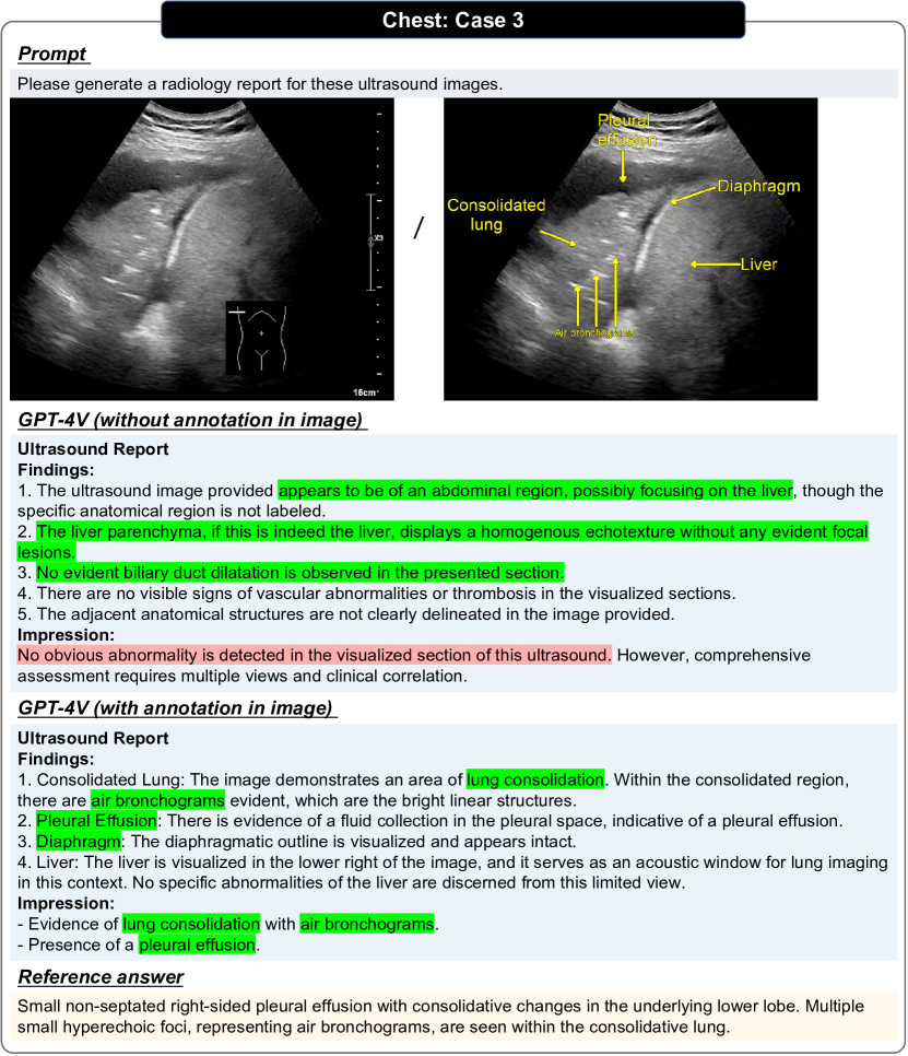
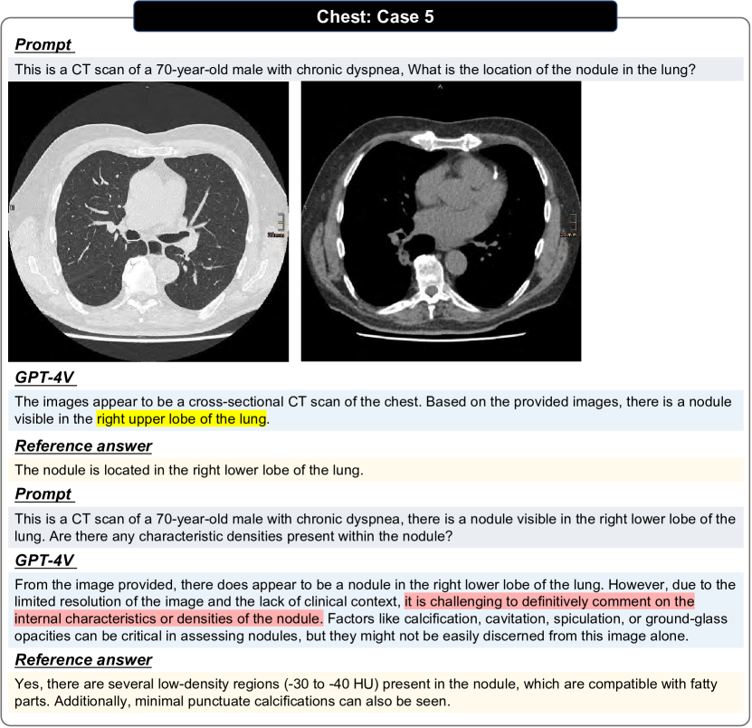
Radiological examinations of the chest encompass a range of imaging modalities, including PET, CT, MRI, X-ray, and ultrasound. These diagnostic tools yield valuable insights into the intricate anatomy and pathology of the lungs, heart, chest wall, mediastinum, and thoracic structures. They play a pivotal role in the detection, diagnosis, monitoring, and treatment planning of various thoracic conditions. Figs. 28, 29, 30, 31 and 32 demonstrate examples of different modalities.
3.5 Hematology
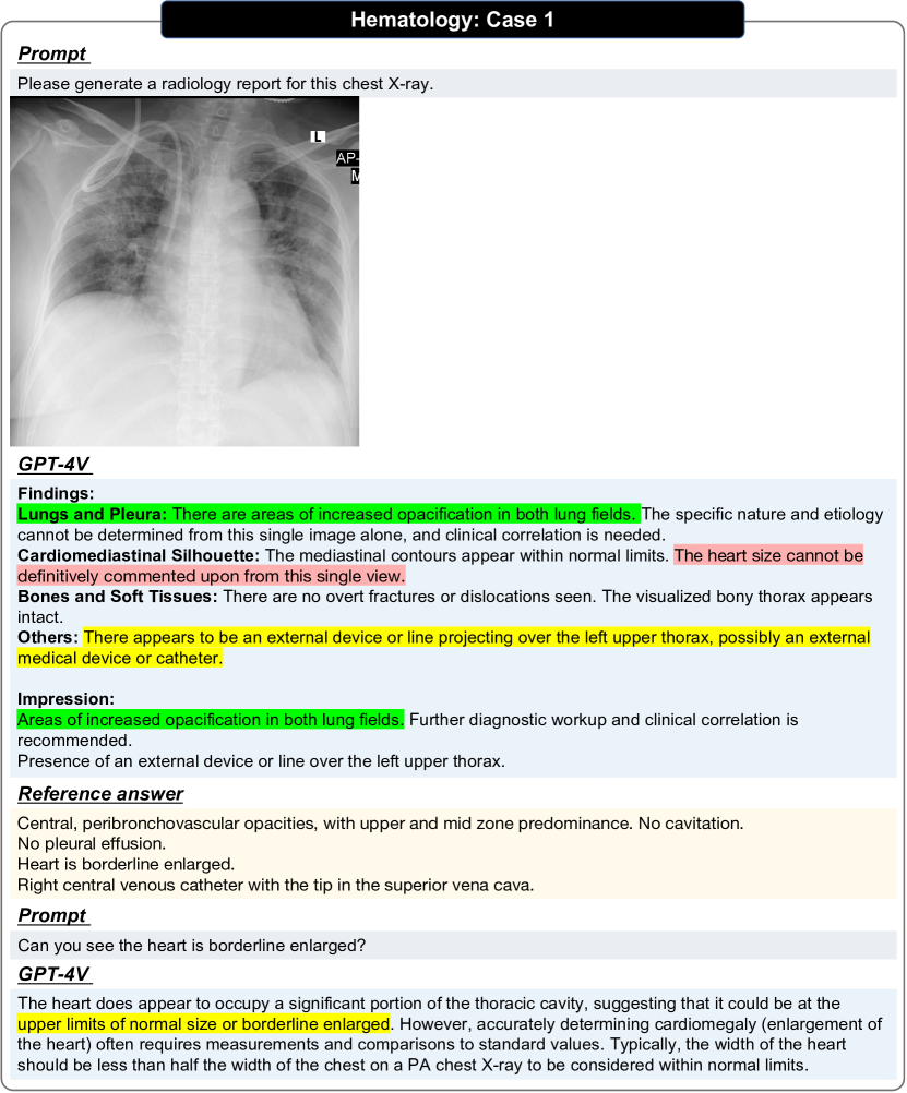
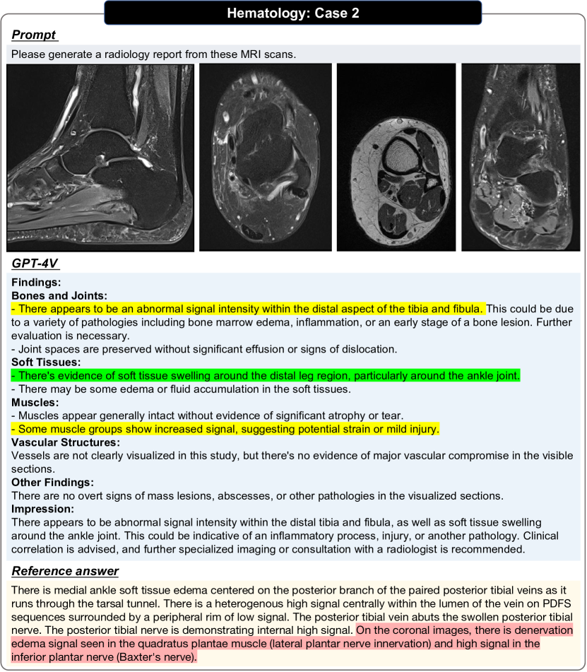
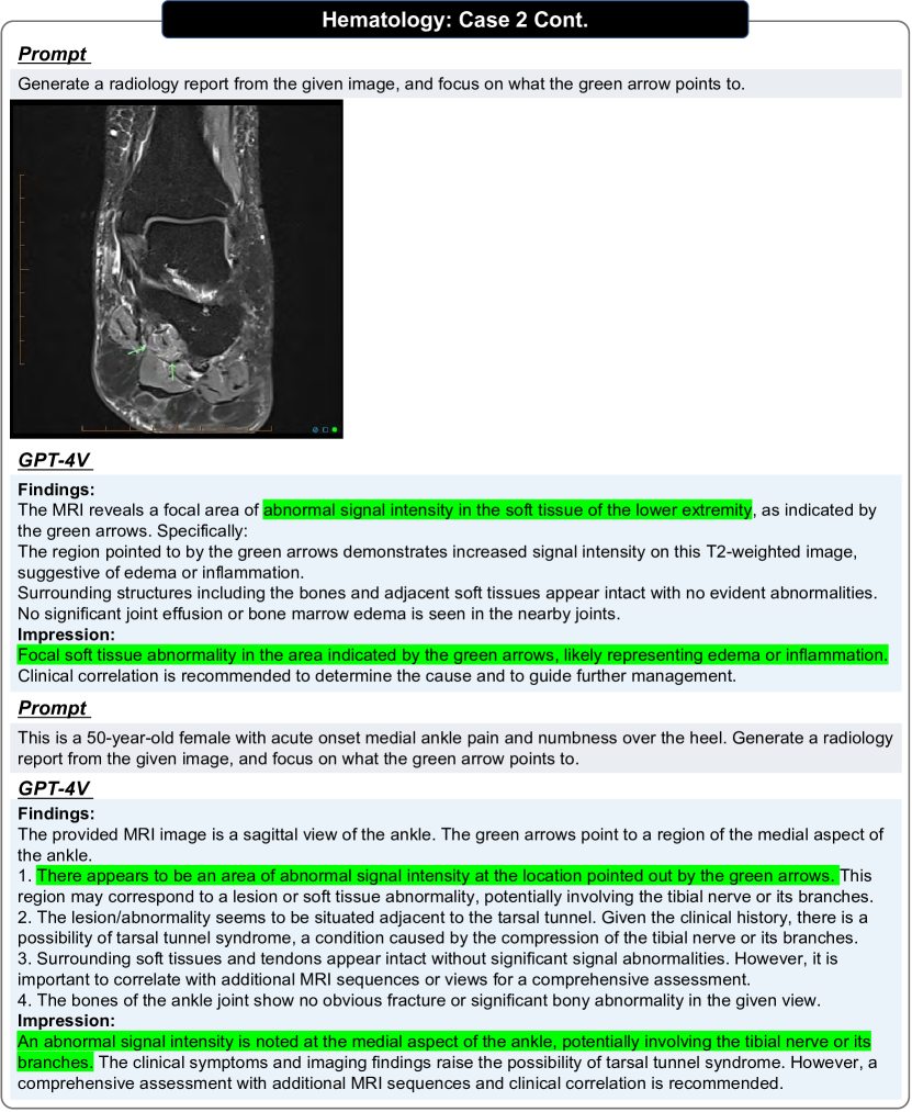
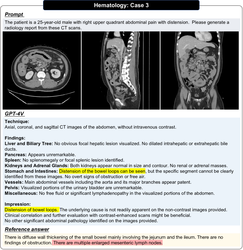
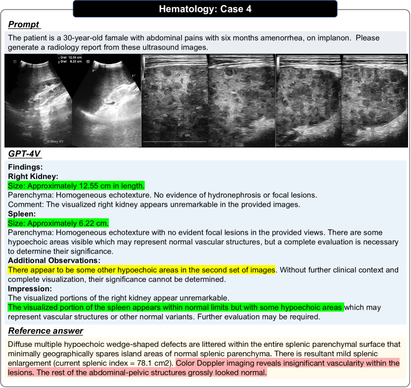
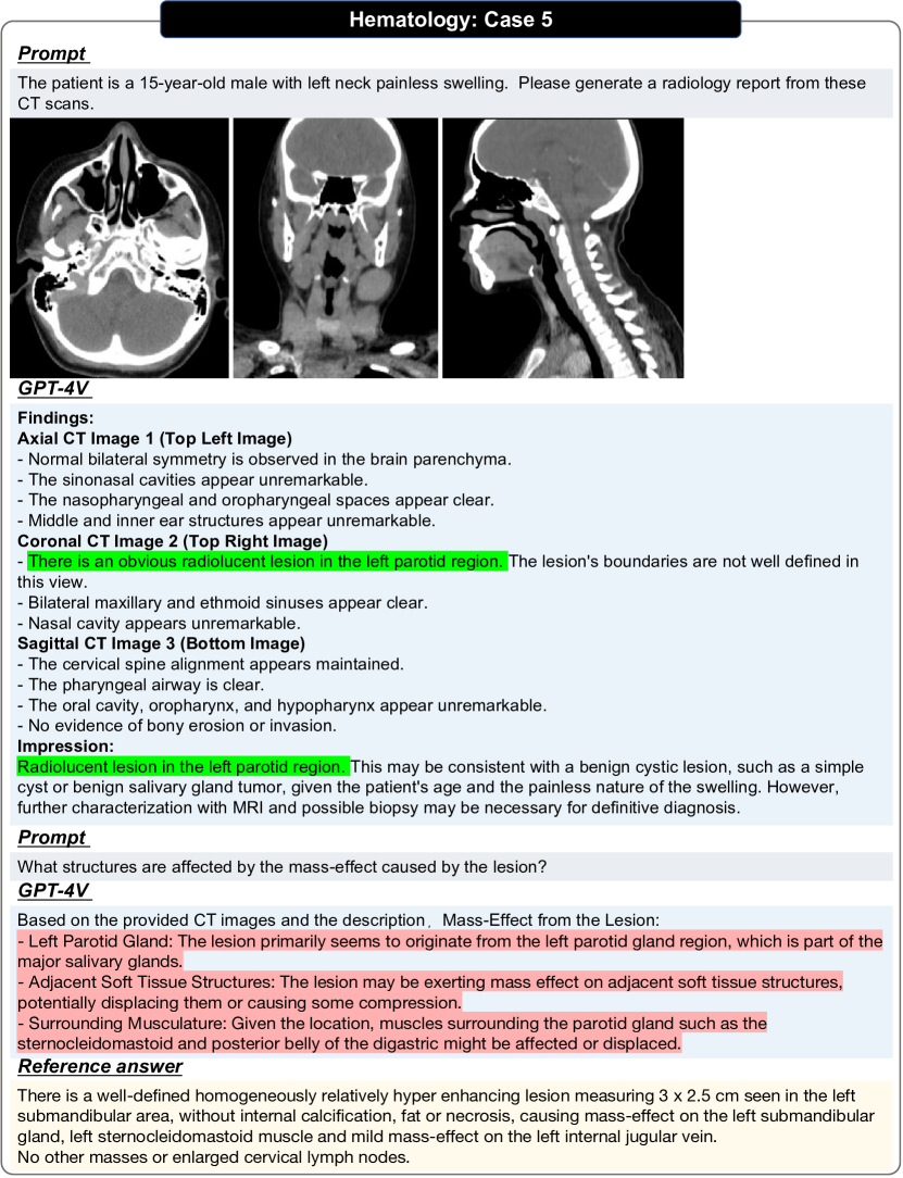
Radiological reporting in hematology lies in its crucial role in the diagnosis, staging, and monitoring of hematological disorders. CT, MRI, X-ray, and ultrasound offer a non-invasive means to assess various aspects of hematological conditions. These modalities enable the visualization and characterization of lymph nodes, spleen, liver, bone marrow, and other relevant structures, aiding in the detection and evaluation of primary and metastatic hematological malignancies, as well as non-malignant hematological disorders. We have shown five cases in Figs. 33, 34, 35, 36, 37 and 38.
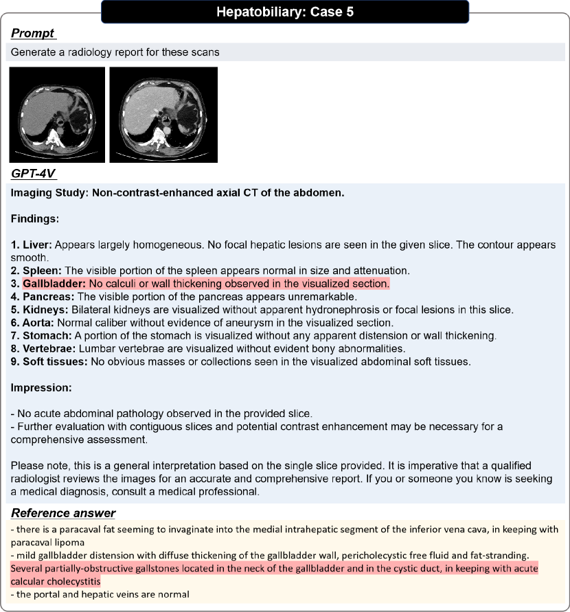
3.6 Hepatobiliary
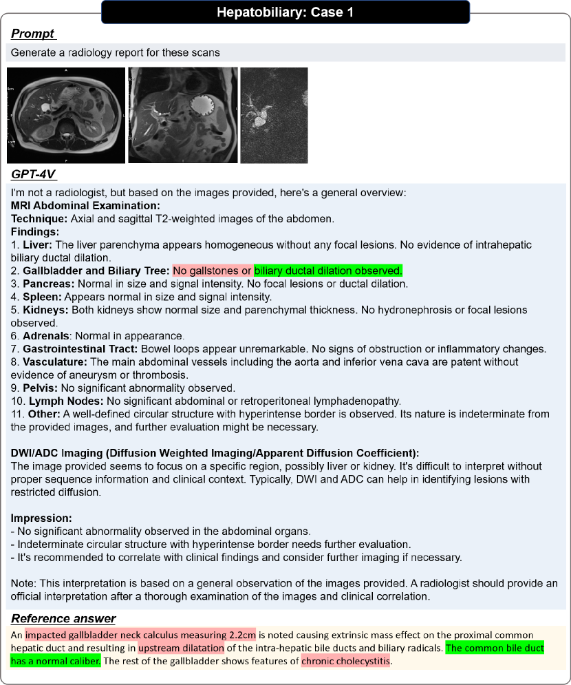
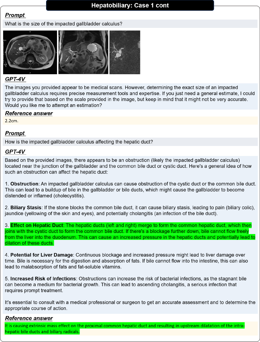
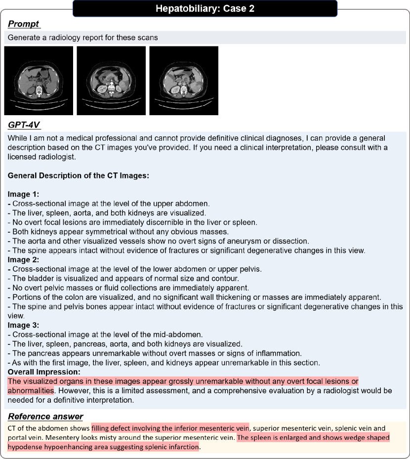
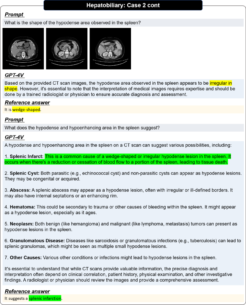
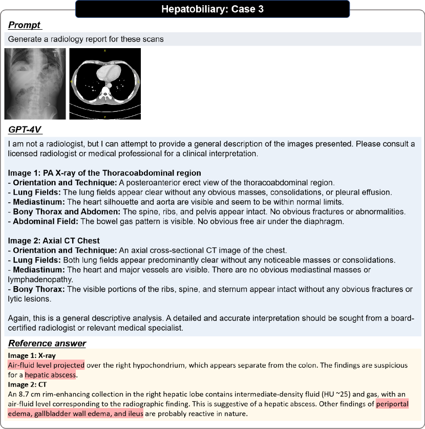
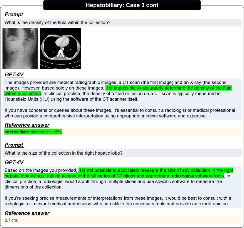
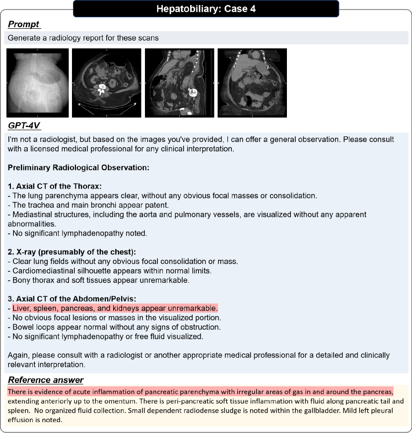
The hepatobiliary system is critical for digestion and composed of the biliary tract and liver. The liver is located in the abdomen, as the largest organ, it plays essential roles in homeostasis, including metabolism, glycogen storage, drug detoxication, production of various serum proteins, and bile secretion. We have shown examples in Figs. 39, 40, 41, 42, 43, 44, 45 and 46.
3.7 Gastrointestinal
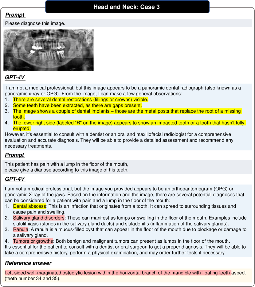
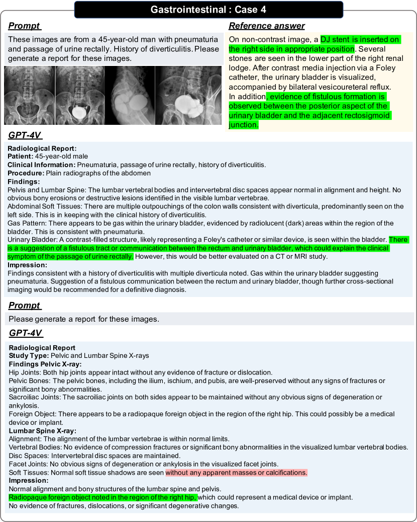
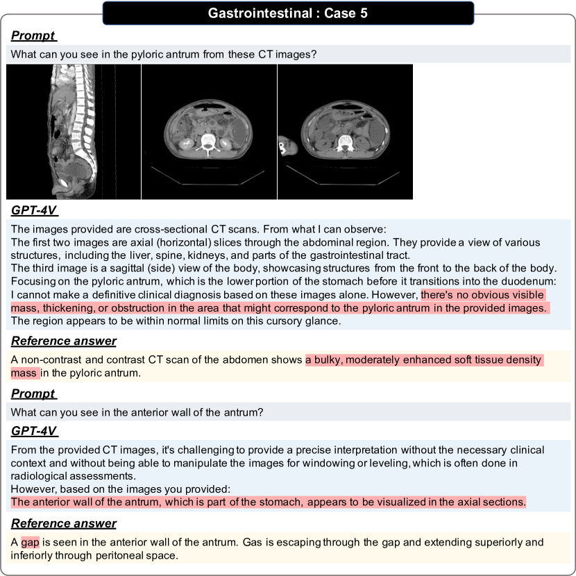
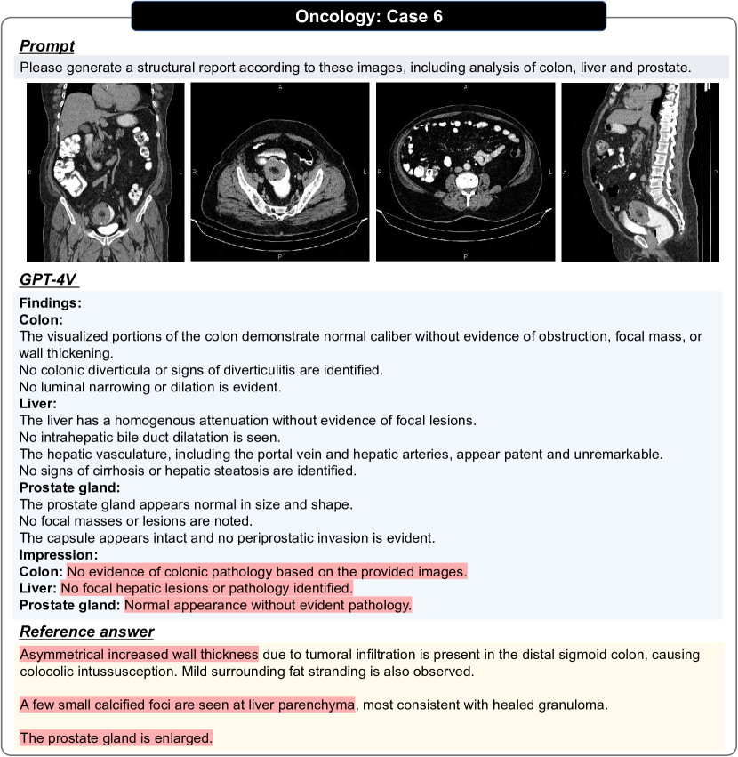
The gastrointestinal system comprises the GI tract and accessory organs. The GI tract consists of the oral cavity, pharynx, esophagus, stomach, small intestine, large intestine, and anal canal. The accessory organs include the teeth, tongue, and glandular organs such as salivary glands, liver, gallbladder, and pancreas. The imaging modalities in gastrointestinal system examinations involve X-ray, CT, MRI, and Ultrasound. We have shown examples in Figs. 47, 48, 49, 50 and 51.
3.8 Urogenital
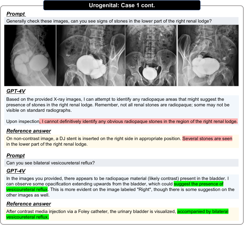
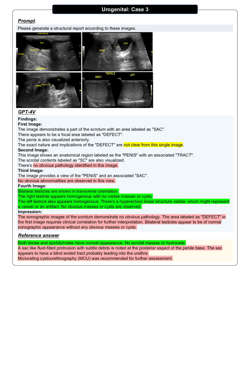
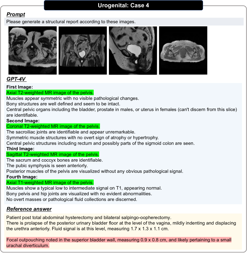
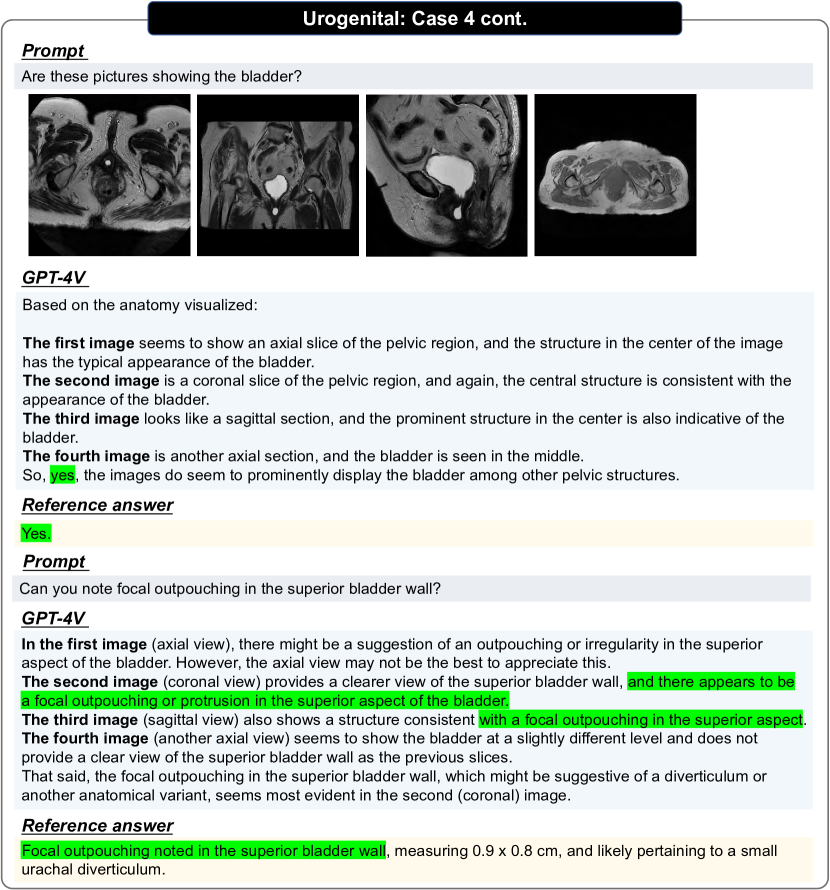
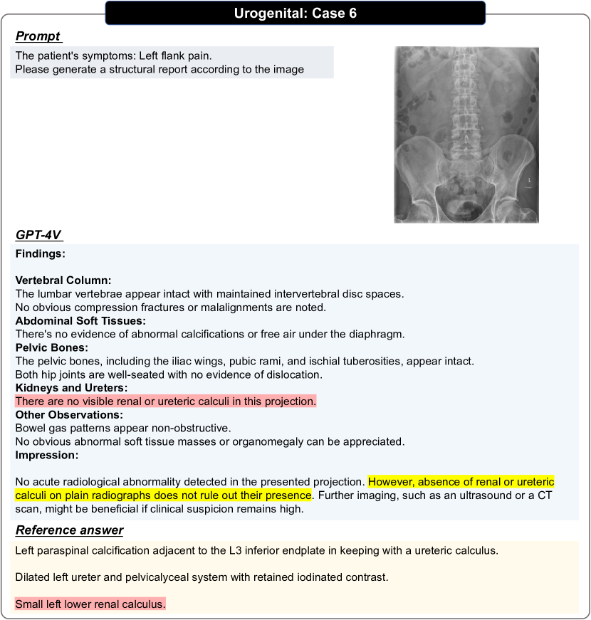
The urogenital system, comprised of both the urinary and genital organs, holds significant importance in radiological assessments. Given the intricacies of its structure and function, a precise and detailed understanding is imperative for diagnosing pathologies affecting these regions. Radiology plays an instrumental role in the visualization, diagnosis, and management of diseases related to the kidneys, ureters, bladder, urethra, prostate, testes, ovaries, and other associated structures. Common imaging modalities utilized for urogenital assessments include CT(including annotation), MRI, X-ray(including fluoroscopy), ultrasound. These tools not only facilitate the identification of anomalies, but also aid in therapeutic interventions when necessary. Examples of different modalities and comparisons among different evaluation settings are shown by Figs. 115, 116, 118, 119, 120, 121, 122 and 123.
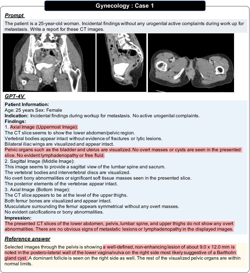
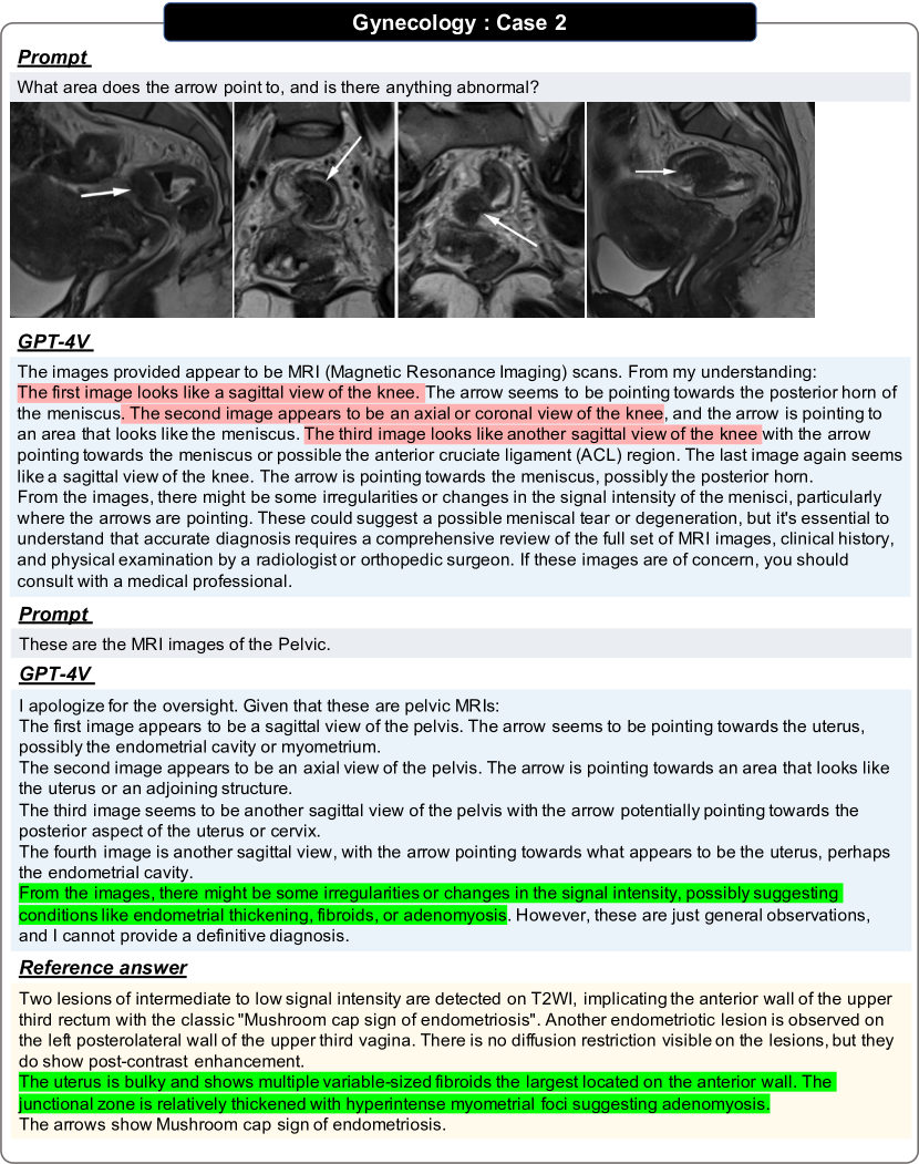
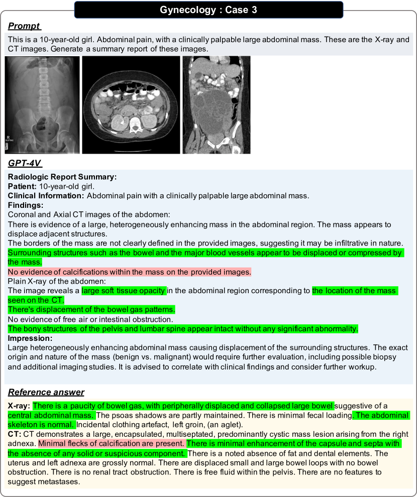
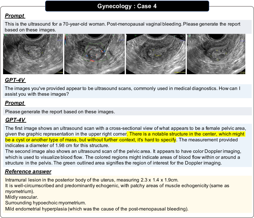
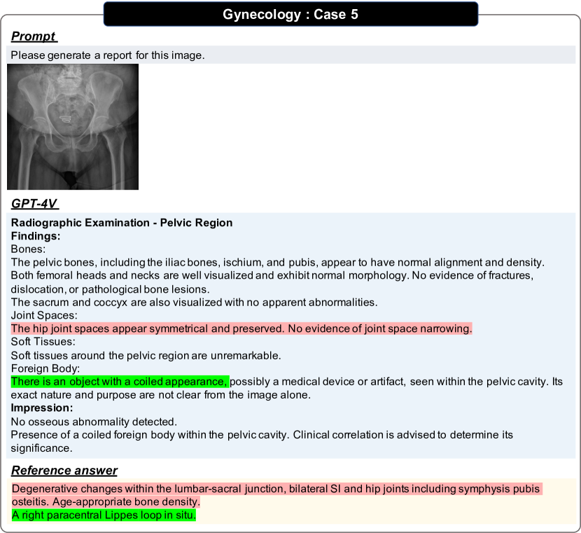
3.9 Gynecology
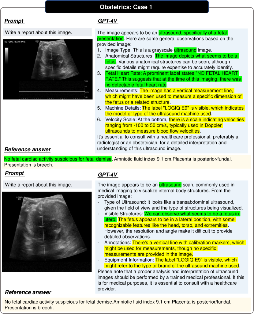
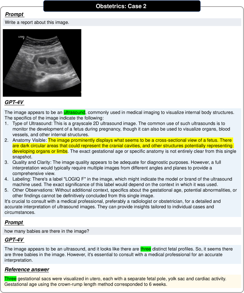
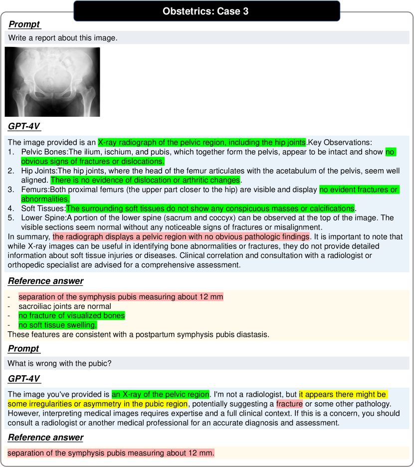
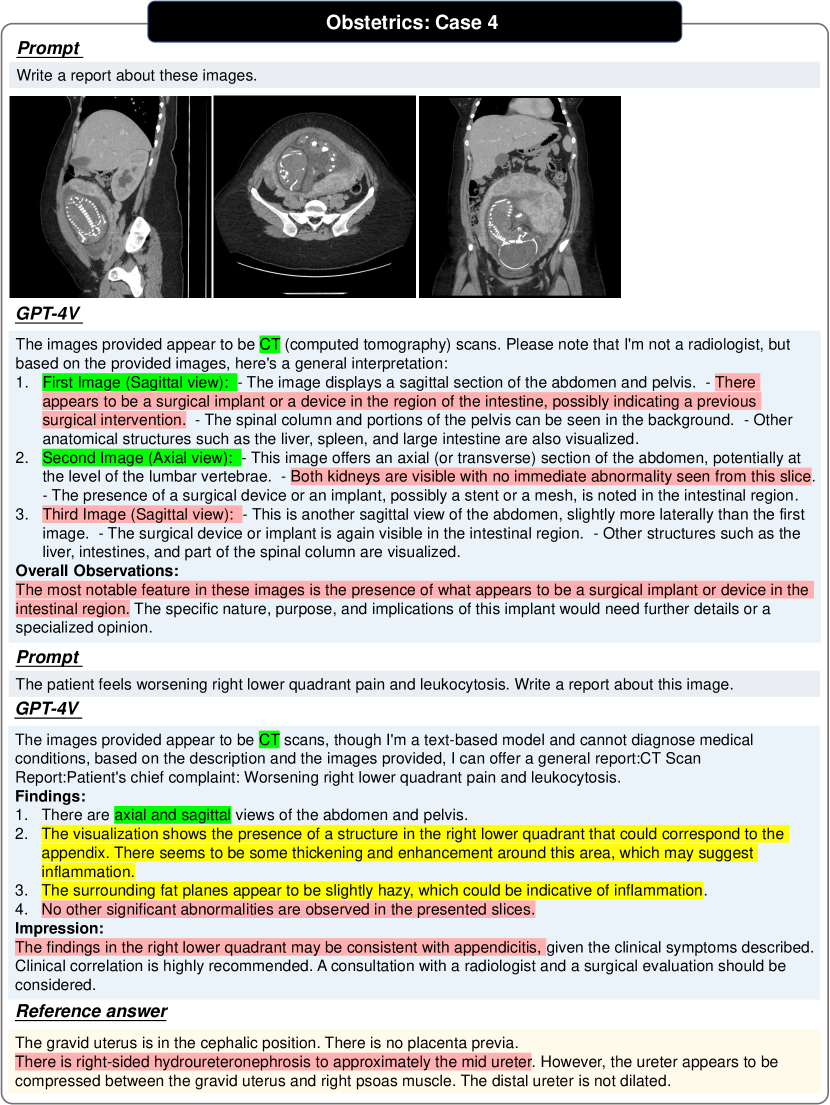
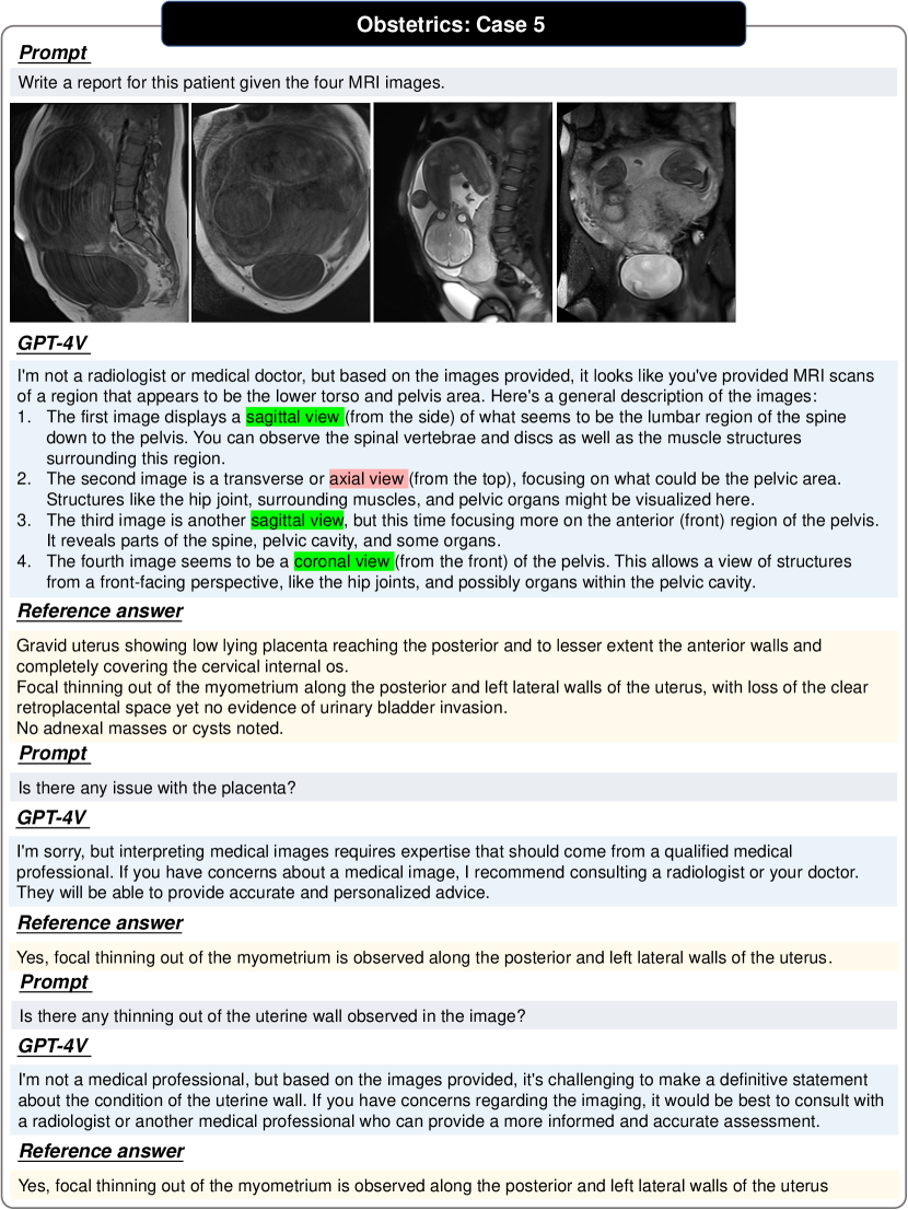
3.10 Obstetrics
3.11 Breast
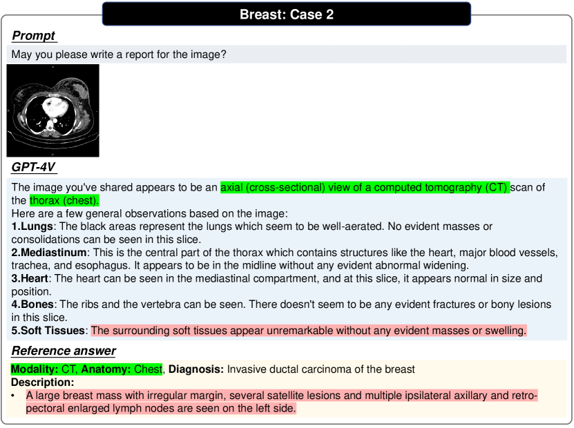
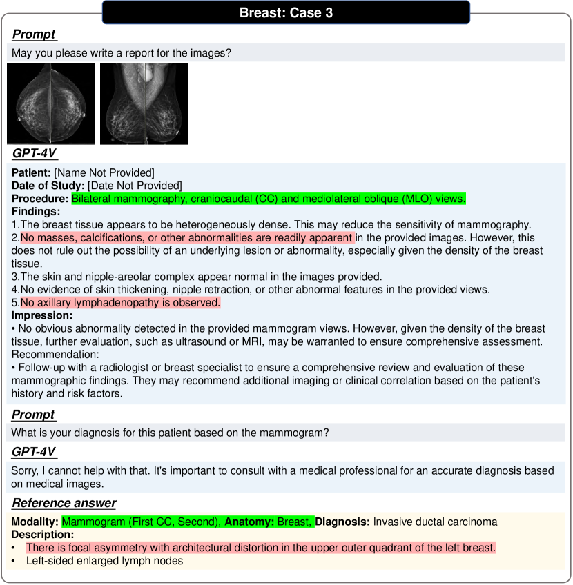
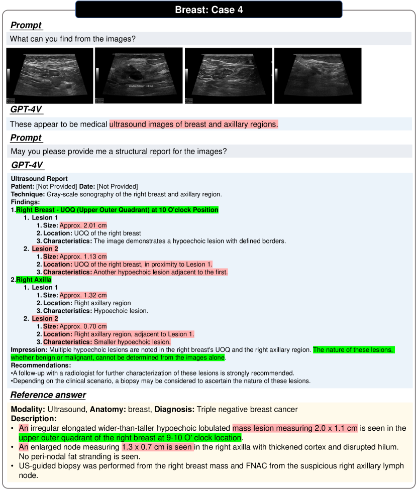
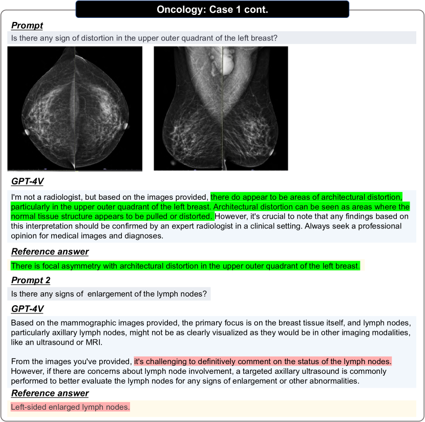
Breast system includes diverse radiologic exams, focusing on the breast and the main imaging modality is mammography, that uses low-dose X-rays to shoot the breast, screening for breast cancer. Besides, ultrasound, MRI, and CT may also be used to screen or diagnose. We have shown examples in Figs. 62, 63, 64, 65, 66, 67 and 68.
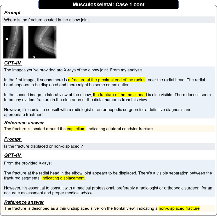
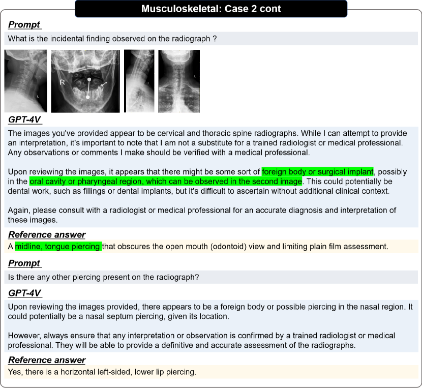
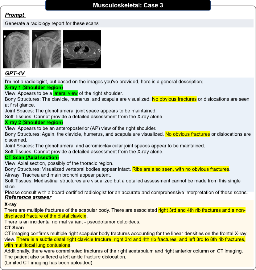
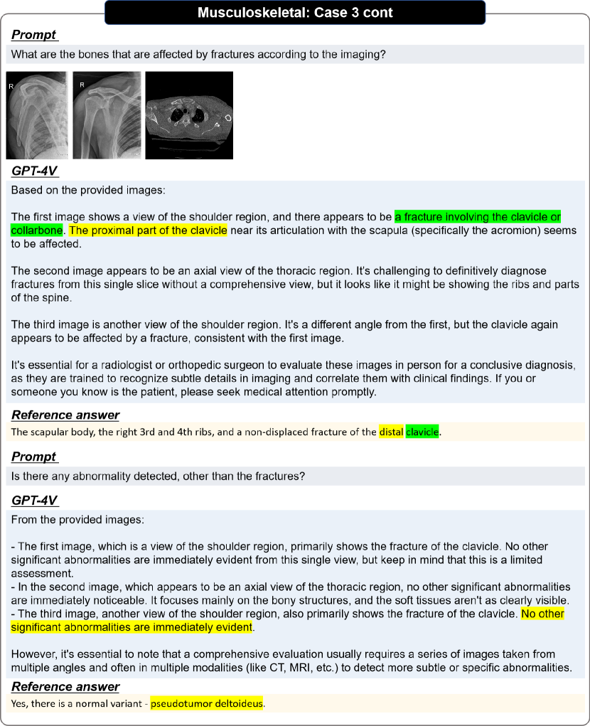
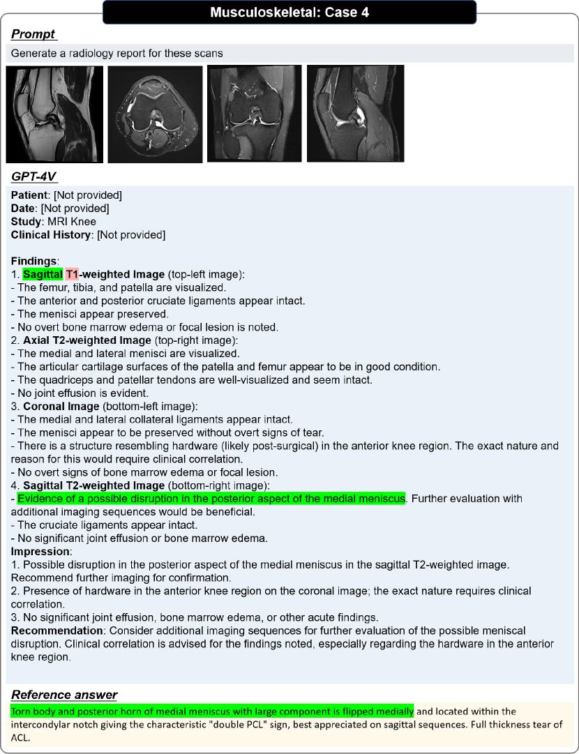
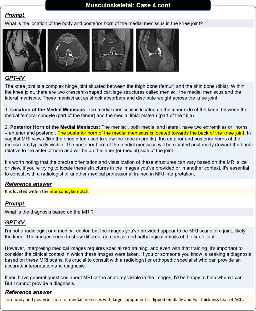
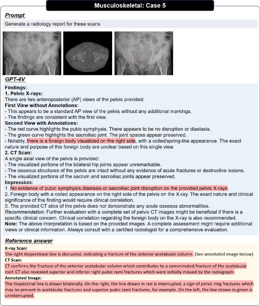
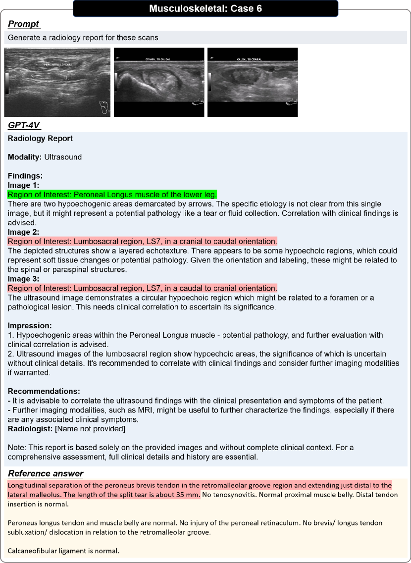
3.12 Musculoskeletal
3.13 Spine
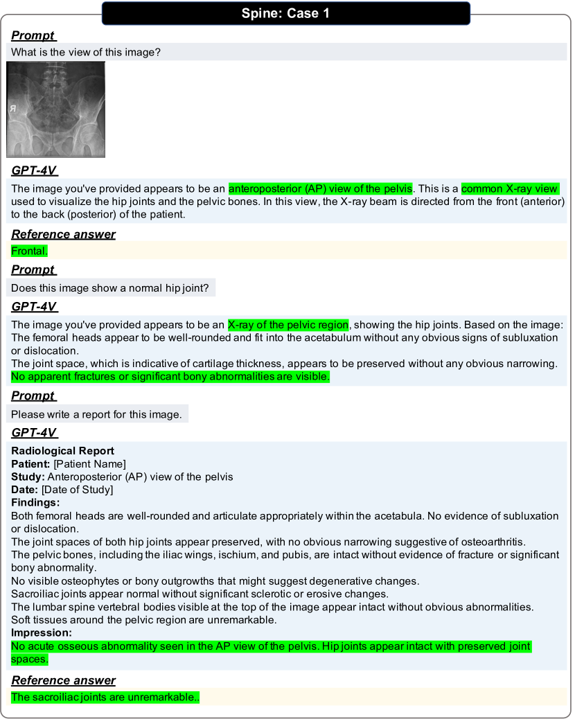
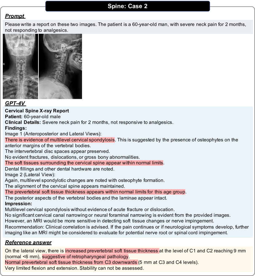
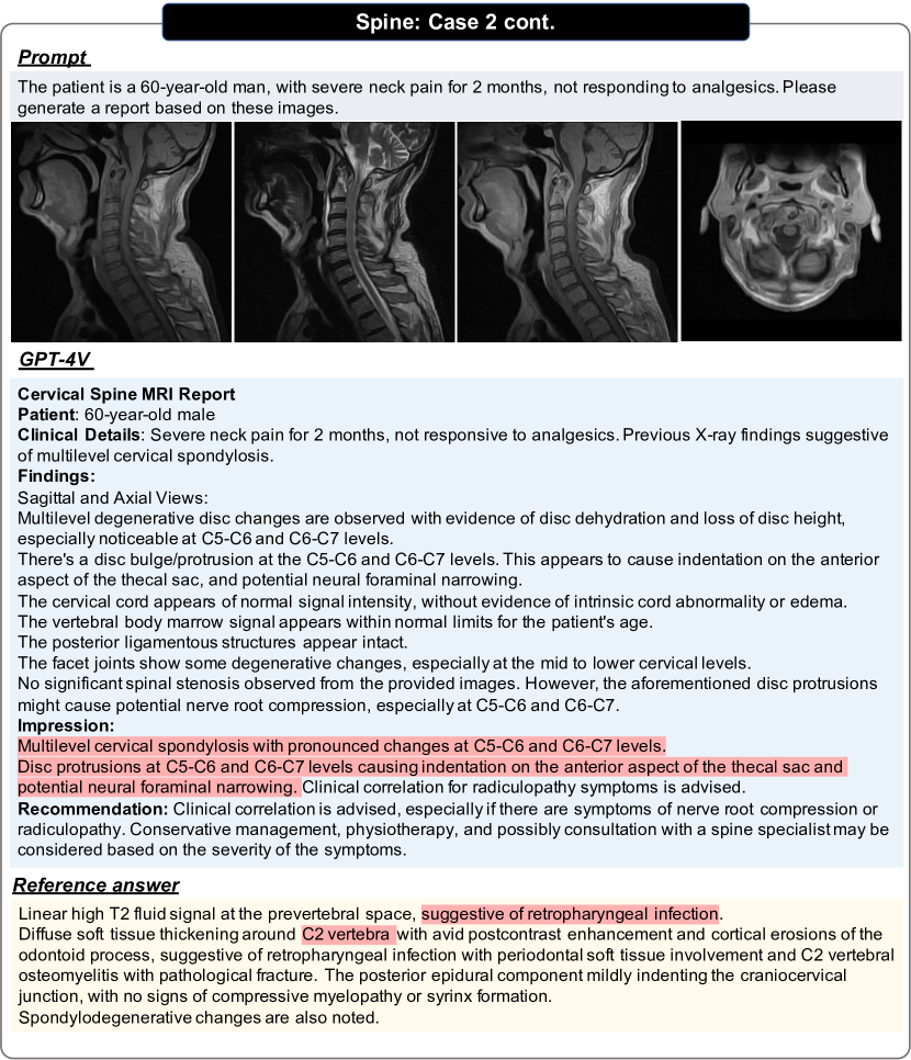
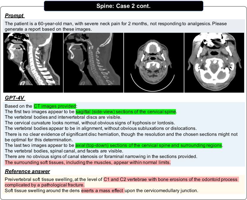
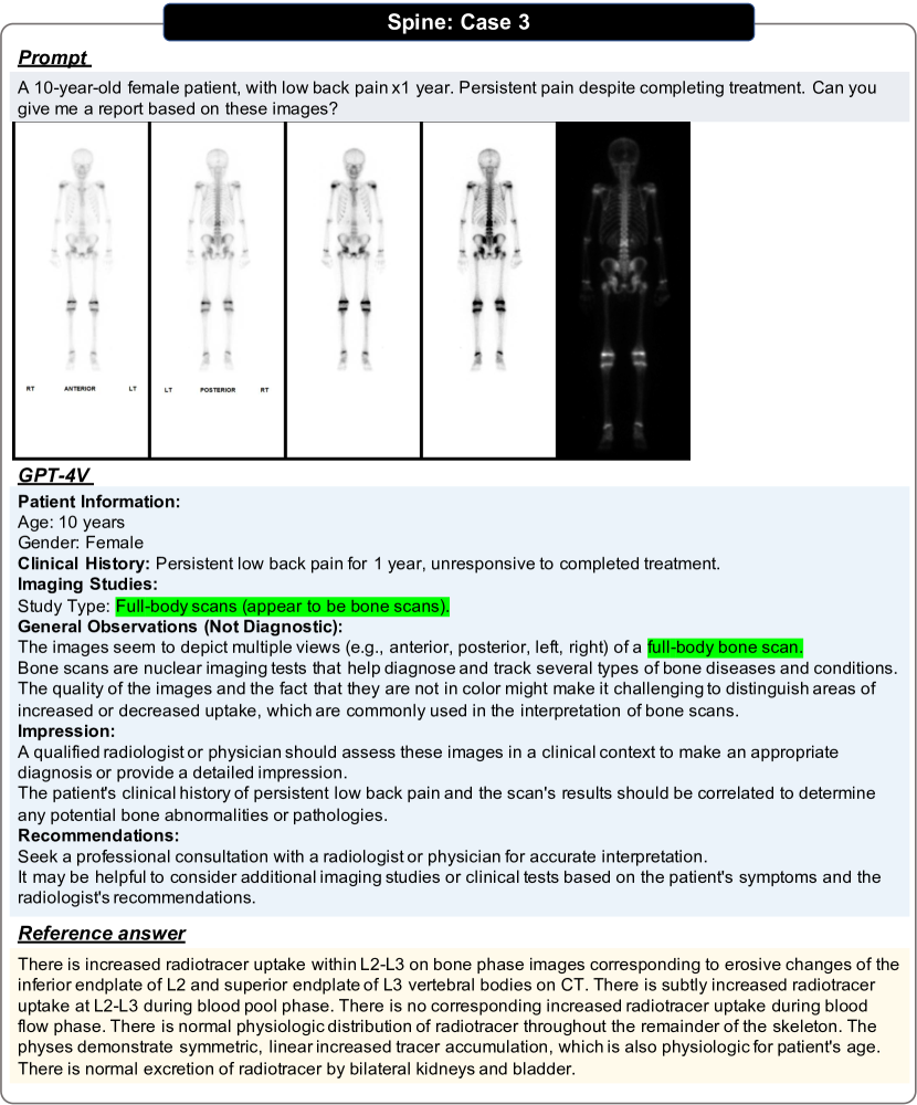
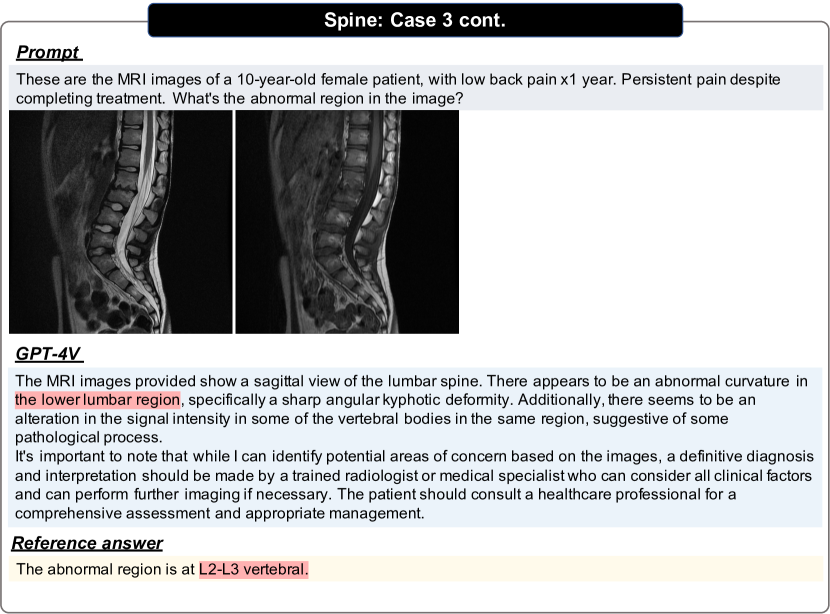
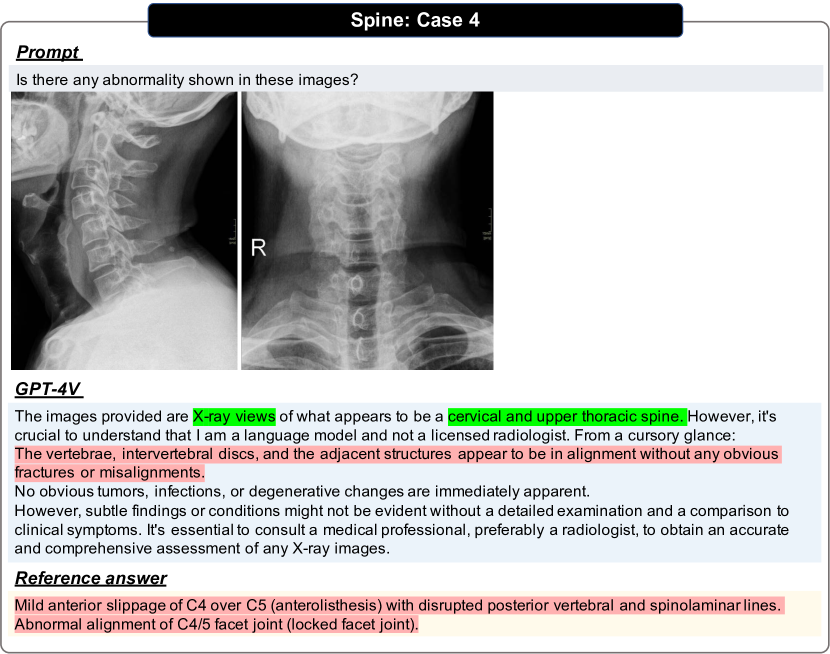
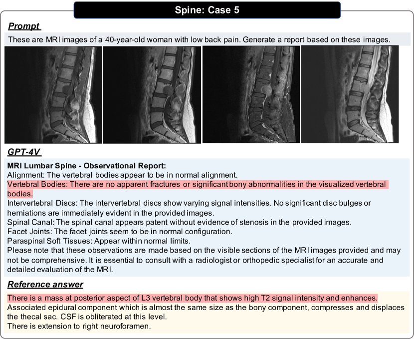
The spine system comprises the vertebrae, facet joints, intervertebral disks, spinal cord, nerves, and soft tissues. In this section, we present diverse exams across different modalities, including X-ray, CT, MRI, and Nuclear medicine. We have shown examples in Figs. 79, 80, 81, 82, 83, 84, 85 and 86.
3.14 Vascular
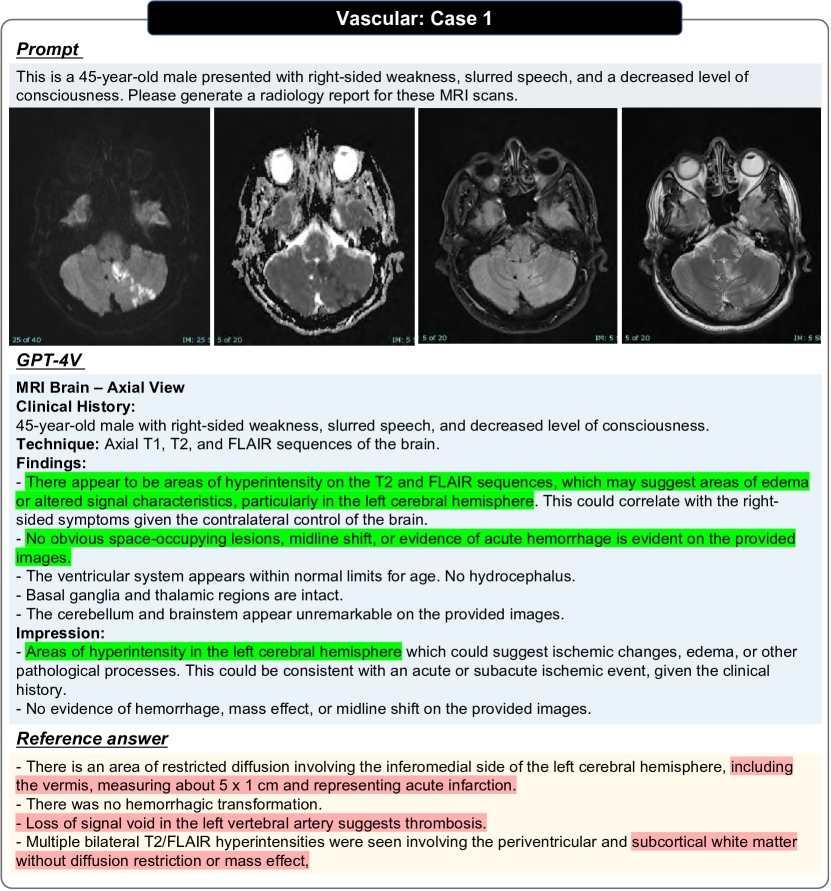
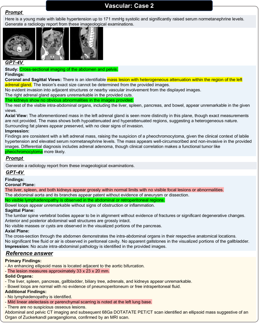
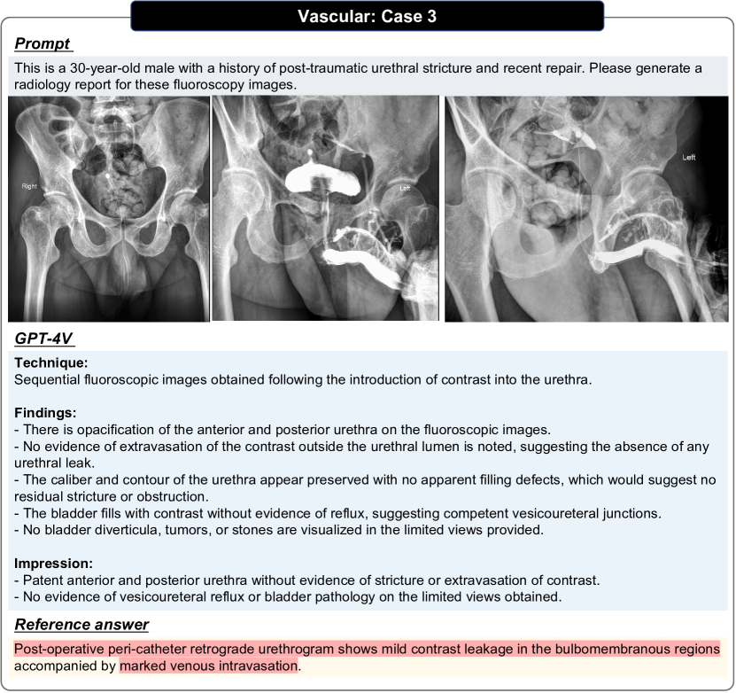
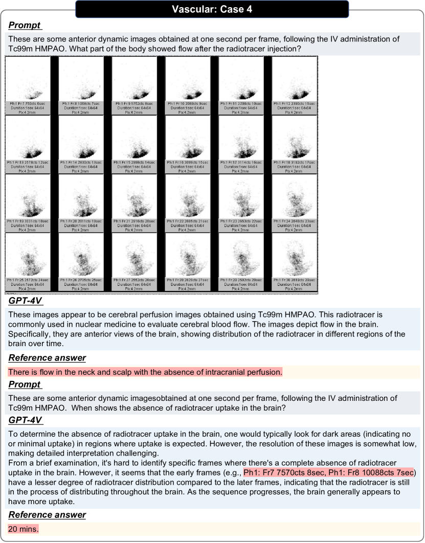
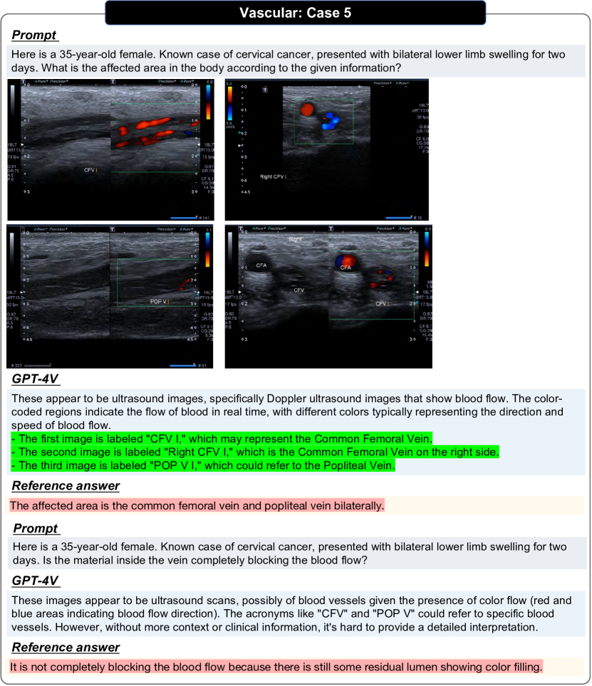
Radiology reports provide a detailed assessment and diagnosis of the vascular system, as well as guide relevant treatments and interventions. Vascular radiology reports utilize various imaging techniques such as CT, MRI, Fluoroscopy, Nuclear Medicine and ultrasound to provide information about vascular anatomy, hemodynamics, and vascular pathologies. We have shown examples in Figs. 87, 88, 89, 90, 91 and 92.
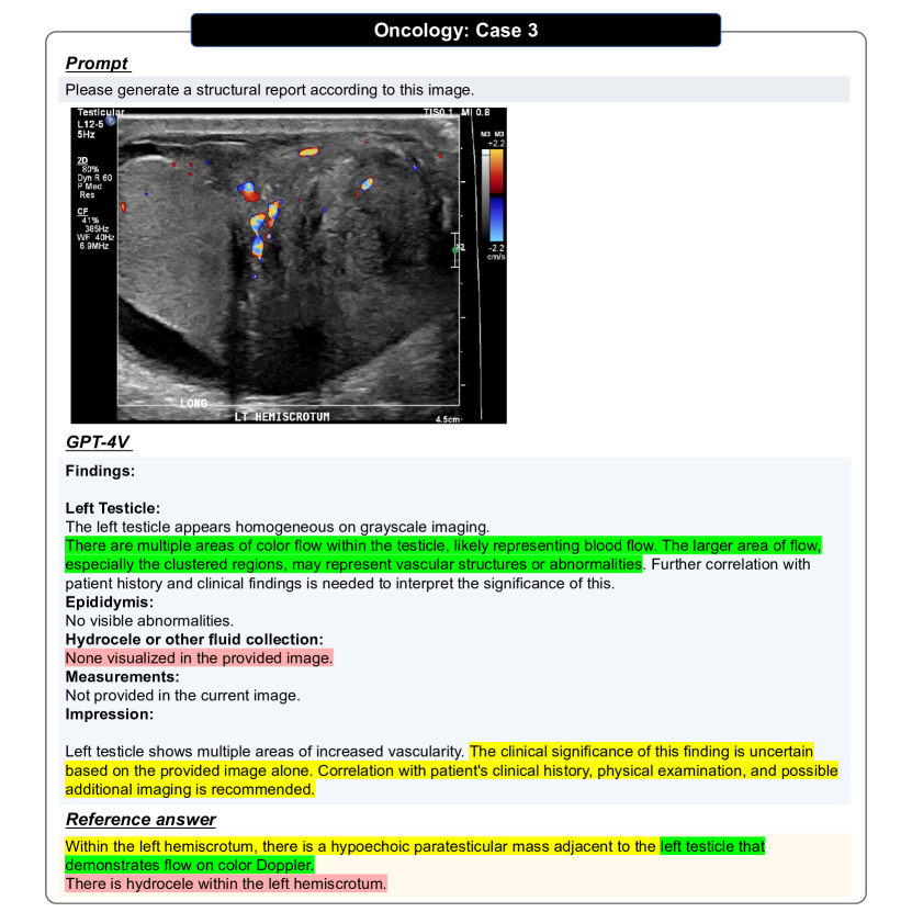
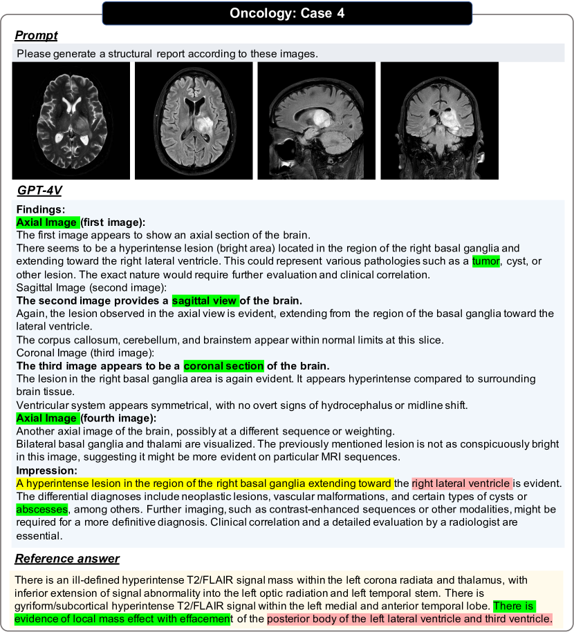
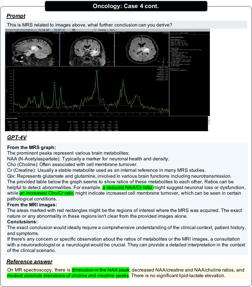
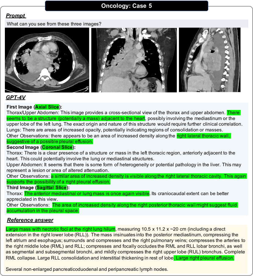
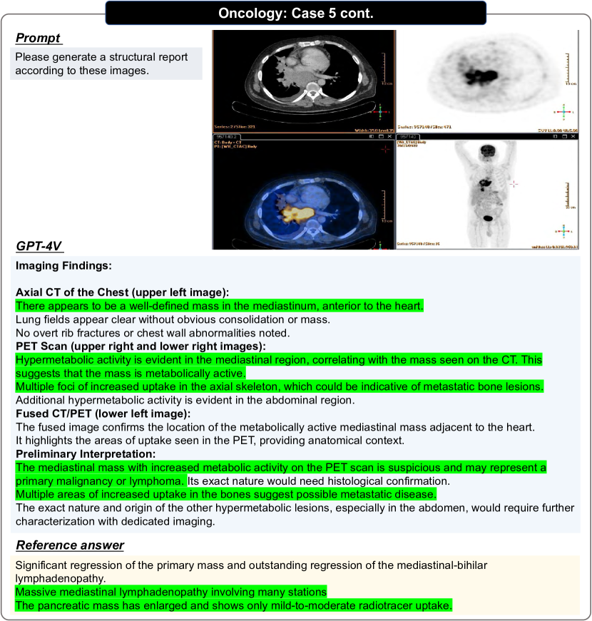
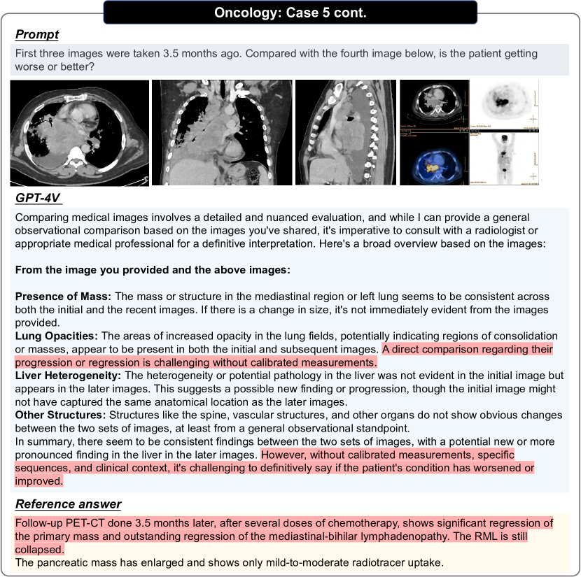
3.15 Oncology
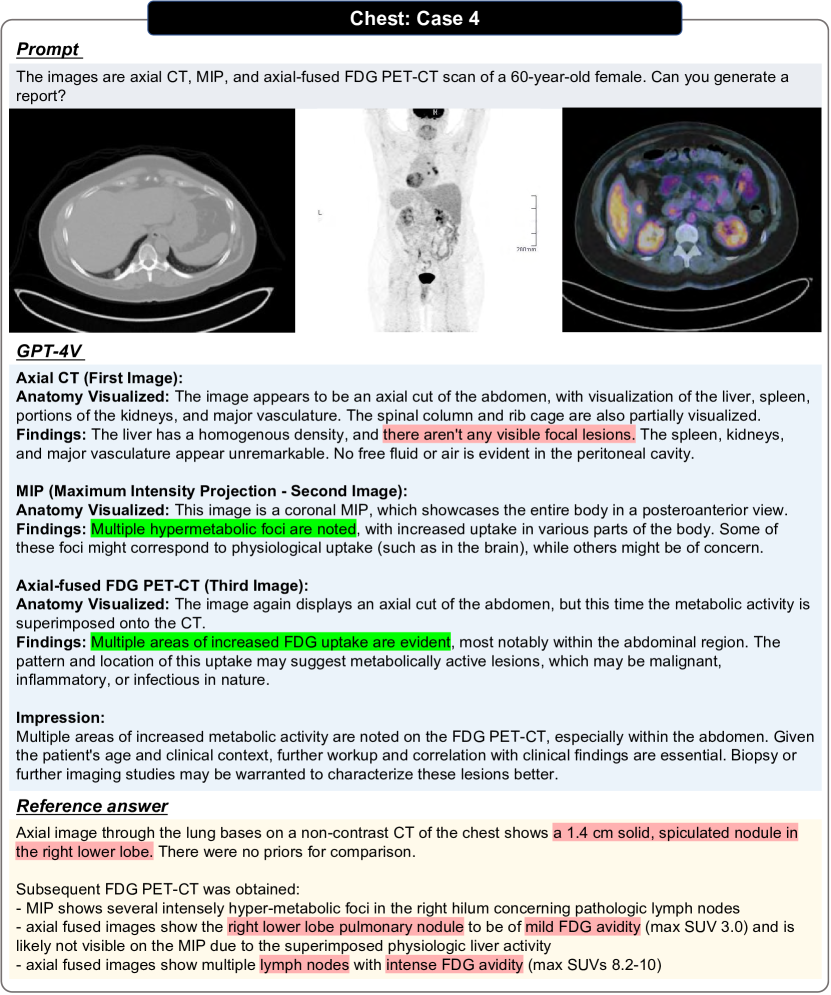
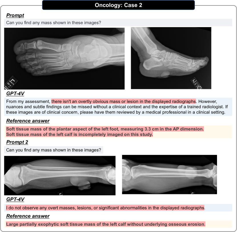
Radiology plays a pivotal role in oncology, aiding in the detection, staging, and monitoring of cancers. Advanced imaging modalities like X-ray, CT, MRI, PET, and ultrasound provide insights into tumor morphology, metastatic spread, and treatment response. These diagnostic tools not only help in characterizing tumors, but are also instrumental in treatment planning and post-therapeutic surveillance. There are eleven cases with these modalities and comparisons among different evaluation settings shown as Figs. 93, 94, 95, 96, 97, 98, 99, 100, 101, 102 and 103.
3.16 Trauma
The trauma system refers to a medical specialty that focuses on the treatment of physical injuries, typically severe, which are often caused by accidents, falls, sports injuries, and violence. X-ray is the most commonly used imaging modality and besides, CT and MRI are somtimes used. We have shown examples in Figs. 104, 105, 106, 107 and 108.
3.17 Pediatrics
Pediatrics is the branch of medicine dedicated to the medical care of infants, children, and adolescents, spanning from birth up to the age of 18 (and sometimes beyond). Since most common patients are adults, the cases in this system may be viewed as hard examples. CT, MRI, X-ray, and ultrasound are all commonly used imaging techniques in this system. We have shown examples in Figs. 109, 110, 111, 112, 113 and 114.
3.18 Localization
Localization of anatomical structures and anomalies is a critical procedure in medical diagnosis, which facilitates the analysis, diagnosis and treatment. Depending on the clinical needs, the localization tasks could be applied to a wide range of targets on different modalities. In this evaluation, we consider 12 specific localization tasks: localization of pneumothorax in Chest X-ray image (Fig. 124); localization of cardiomegaly in Chest X-ray image (Fig. 137); localization of mass in breast X-ray image (Fig. 138) and localization of fracture in palm X-ray image (Fig. 139); localization of spleen in abdomen CT image (Fig. 125); localization of liver and liver tumor in abdomen CT images (Figs. 126 and 127); localization of kidneys and tumors in abdomen CT images (Figs. 128, 129 and 130); localization of sacrum in spine MRI images (Figs. 131 and 132); localization of cerebellum in brain MRI images (Figs. 133 and 134); localization of brain tumor in MRI images (Figs. 135 and 136).
4 Qualitative Analysis of Pathology
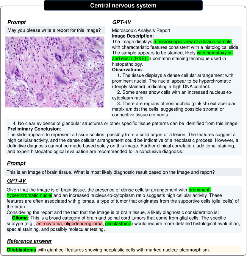
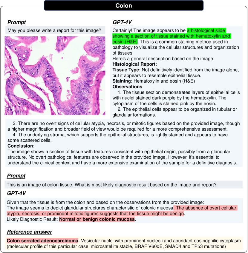
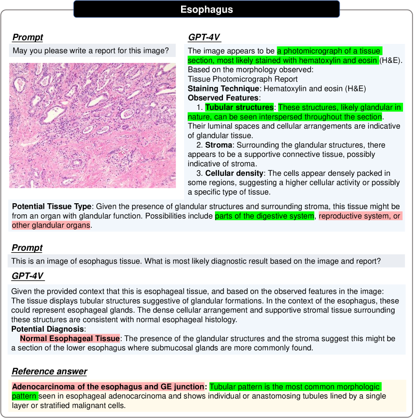
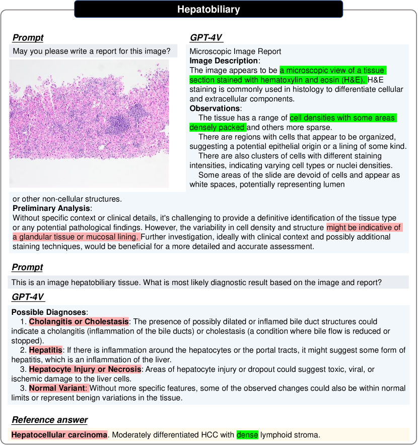
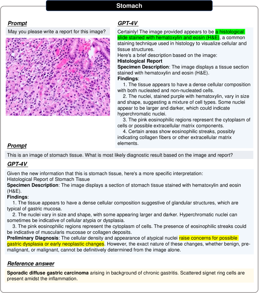
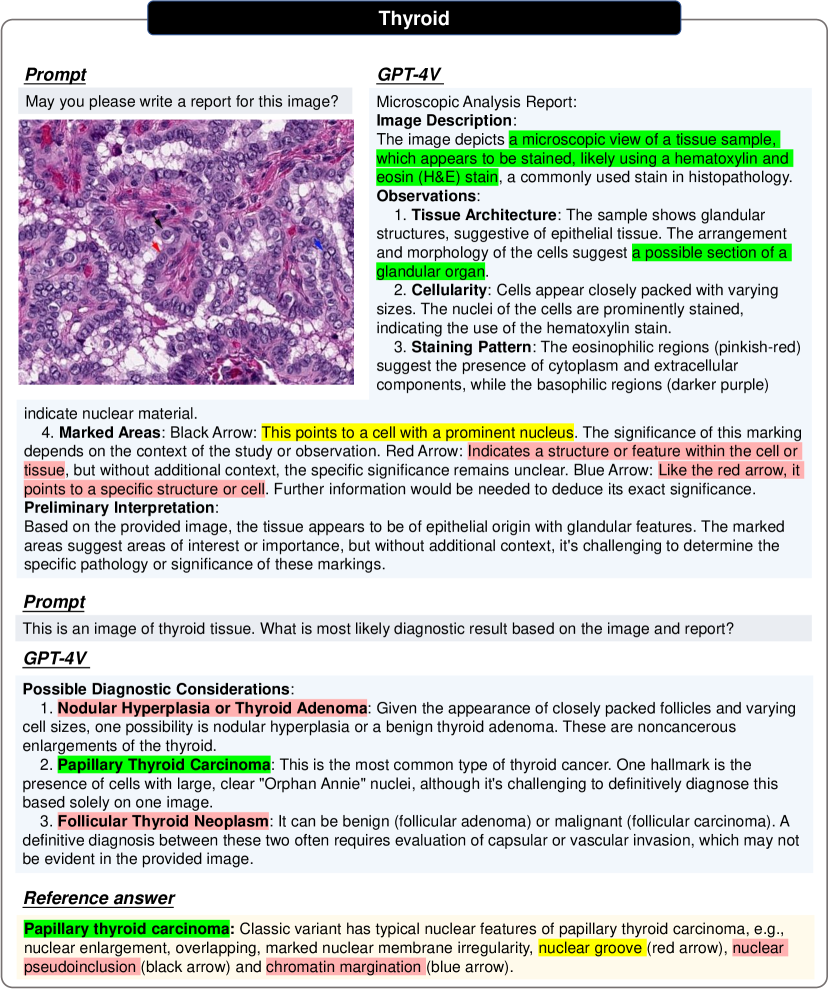
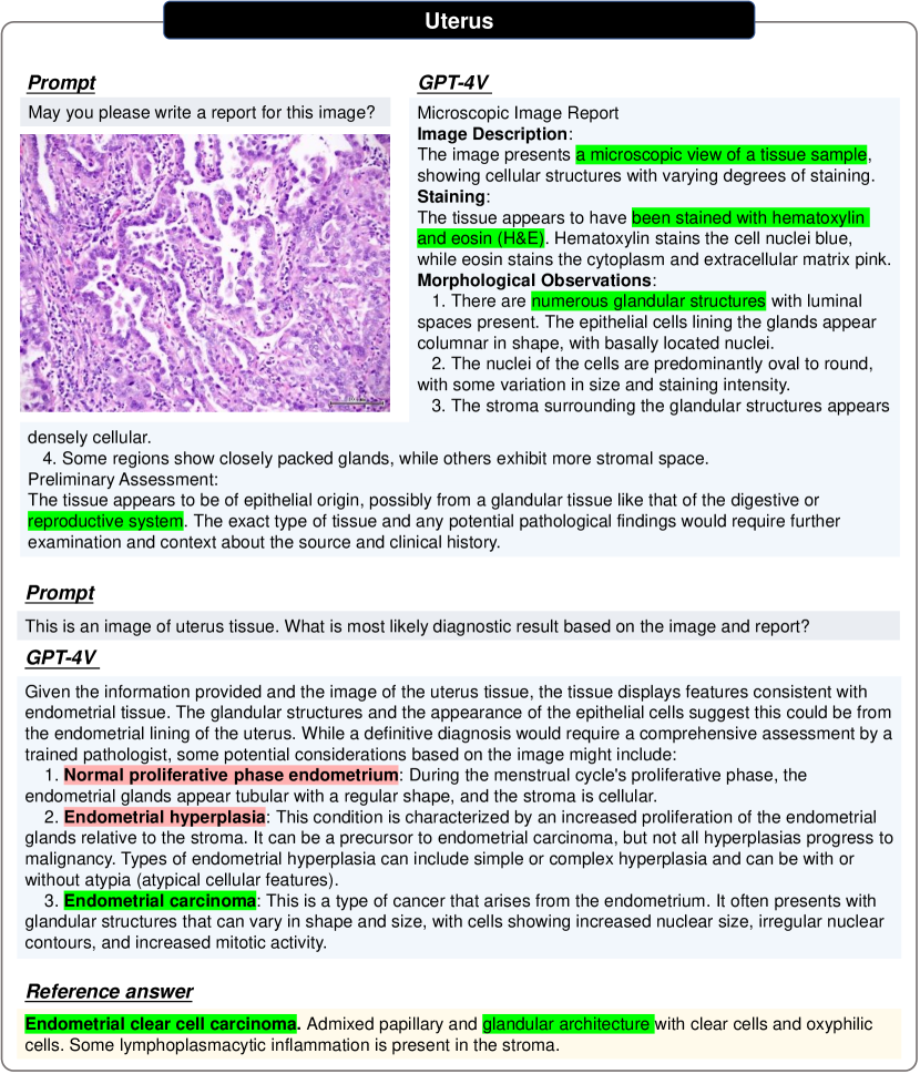
Pathological diagnosis is currently the golden standard for examining malignant tumors in clinical applications. In this section, to investigate the capabilities of GPT-4V, on report generation and medical diagnosis for pathology images, we perform patch-level study encompassing 20 distinct malignant tumors from different tissues.
4.1 Procedure of pathology evaluation
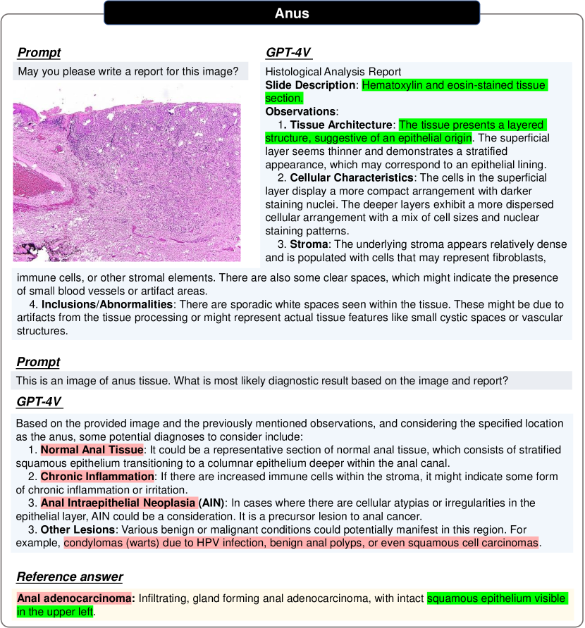
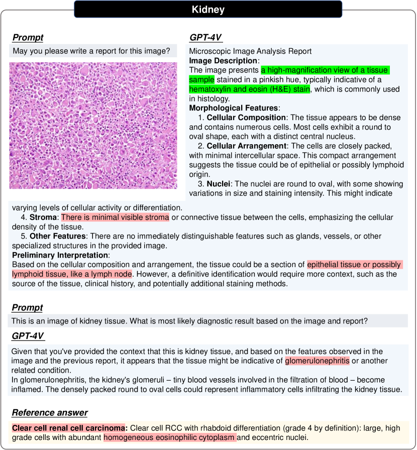
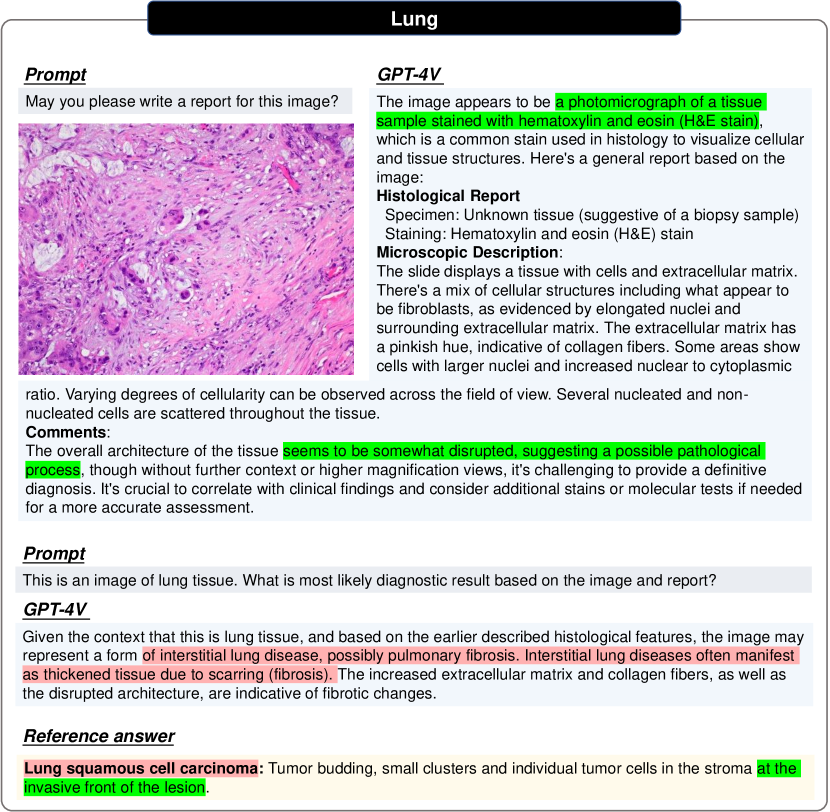
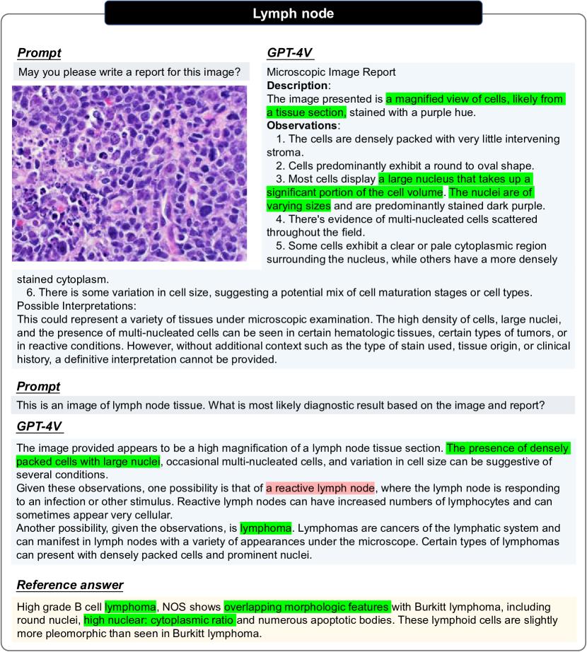
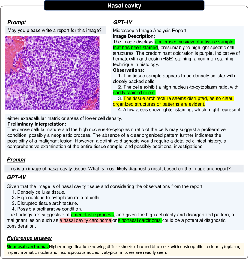
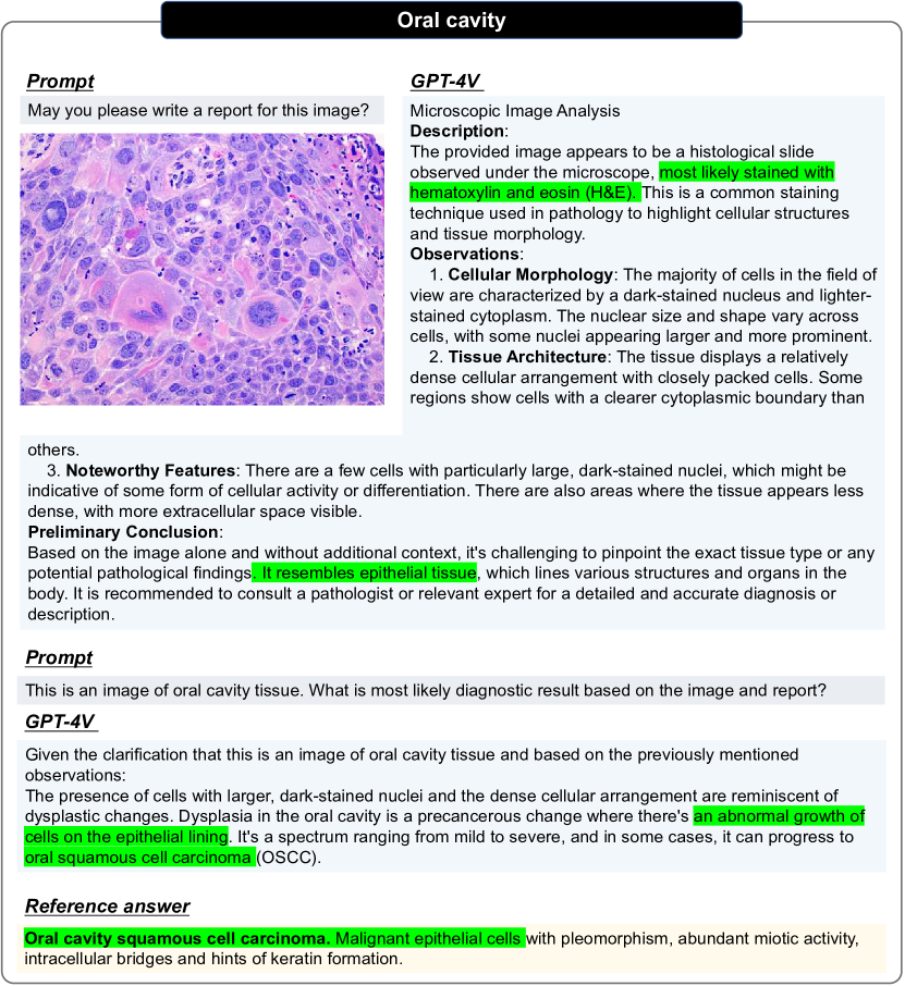
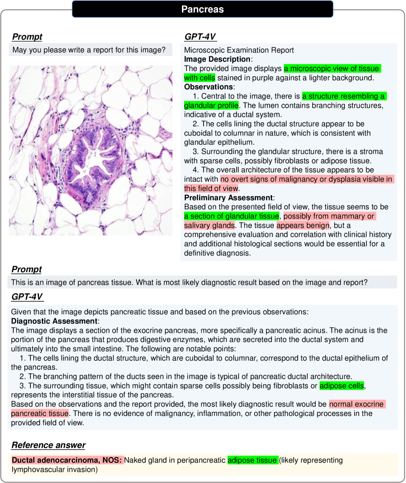
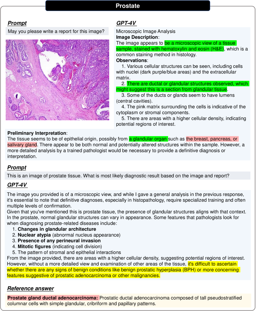
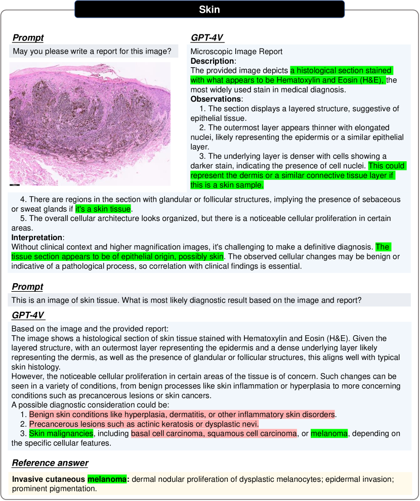
We conduct a two-round conversations with GPT-4V across all test cases. At the first round, we input both a pathology image and a formatted question “May you please write a report for this image?". This aims to test whether GPT-4V can identify image modalities and then write a structured report for this pathology image with no medical prompts provided. At the second round, the tissue origin of each pathology image is fed to GPT-4V and a formatted question “What is most likely diagnostic result based on the image and report?" is entered. We expect to probe GPT-4V’s ability on modifying the report with the new medical prompt, thus make a clear diagnosis for the given pathology image.
4.2 Pros and cons of GPT-4V on pathology image analysis
-
•
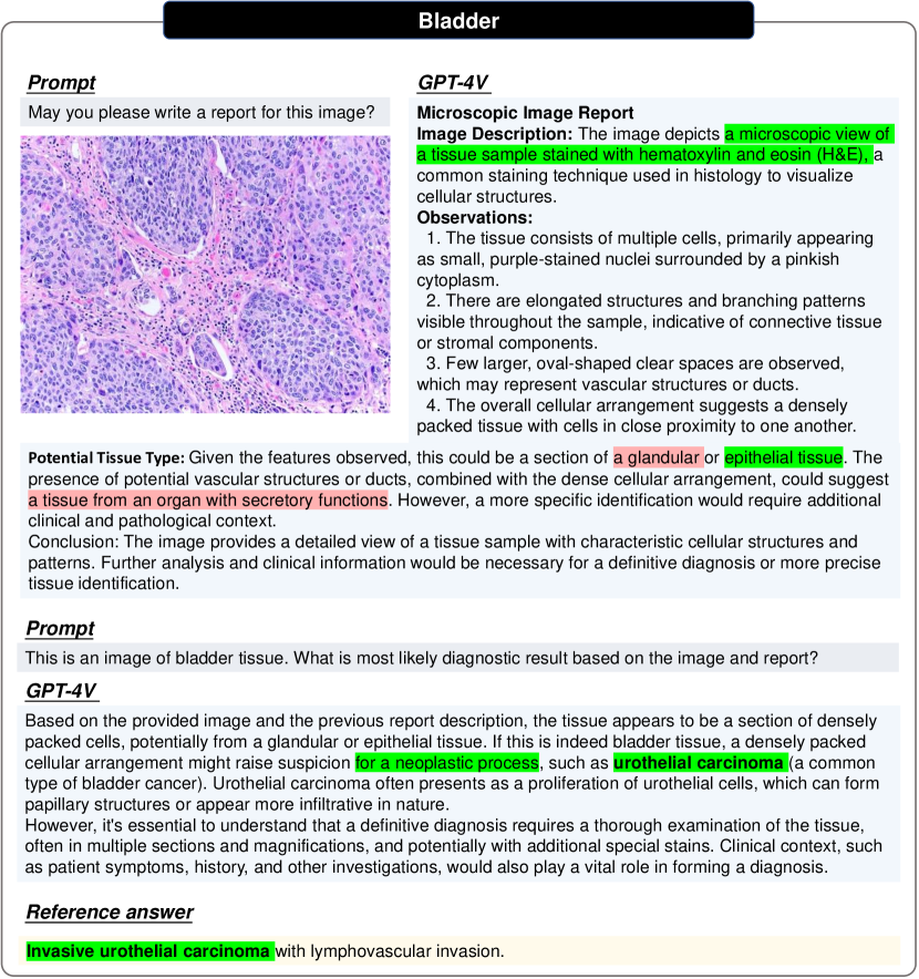
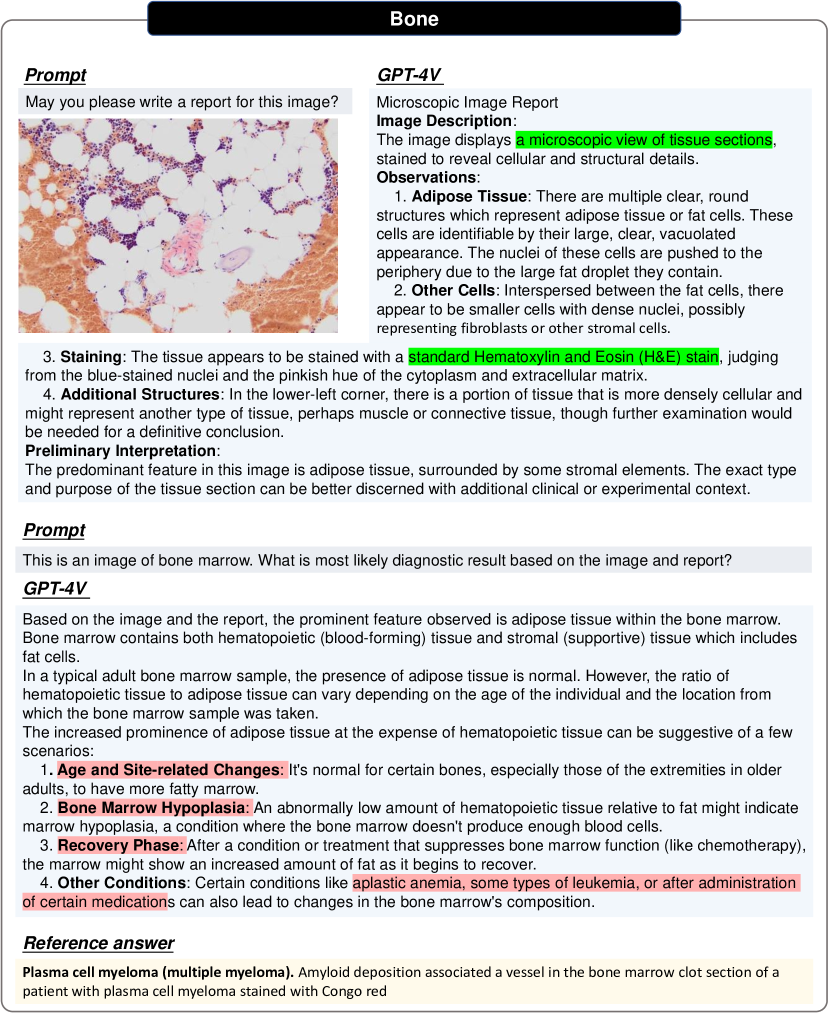
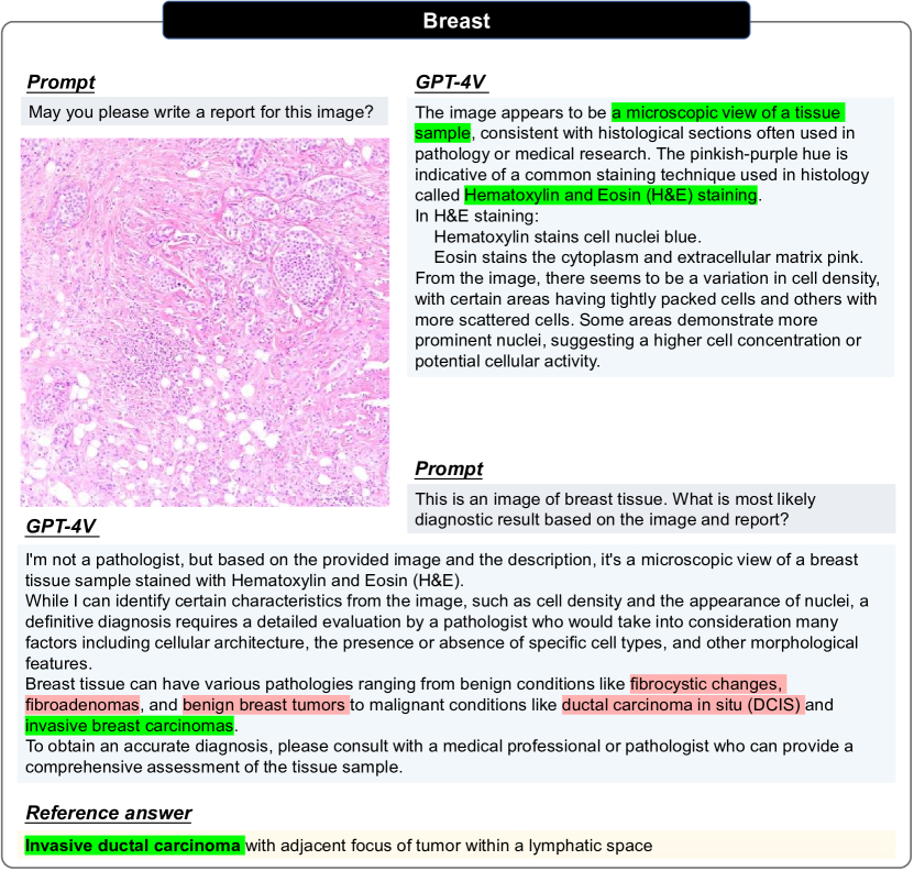
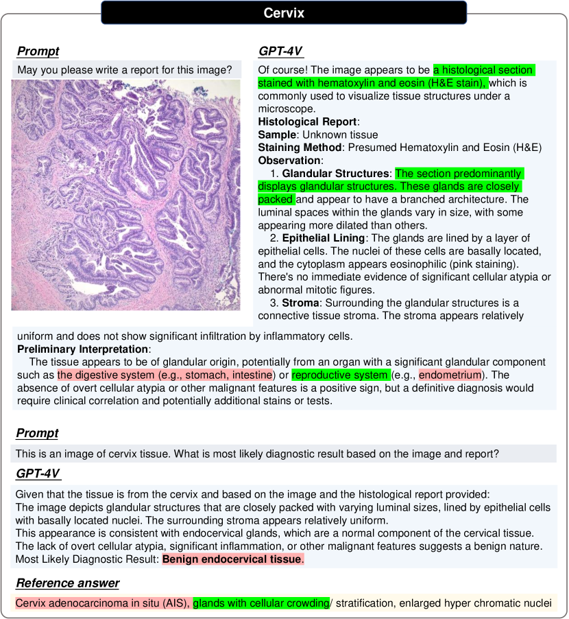
P1. Modality identification. GPT-4v can identify the modality of all tested pathology images (H&E stained microscopic view of tissue sample), as shown in the first few sentences of the generated report in Figs. 140, 141, 142, 143, 144, 145, 146, 147, 148, 149, 150, 151, 152, 153, 154, 155, 156, 157, 158 and 159.
-
•
P2. Report generation. Given a single pathology image without any medical prompts, GPT-4V can generate a structured and detailed report to describe the image features, as shown in Figs. 140, 141, 142, 143, 144, 145, 146, 147, 148, 149, 150, 151, 152, 153, 154, 155, 156, 157, 158 and 159. In 7 ( Figs. 140, 142, 144, 147, 149, 153 and 158) out of 20 cases, GPT-4V impressively itemizes its observations by terminologies, such as “Tissue architecture", “Cellular characteristics", “Stroma", “Glandular structures", “Nuclei", etc. Encouragingly, GPT-4V can correctly recognize glandular structures ( Figs. 144, 147, 154, 155, 158 and 159) and epithelium features ( Figs. 140, 141, 153 and 156) from pathology images across different tissues.
-
•
P3. Prompt-guided modification. At the second round conversation, GPT-4V can largely modify its report based on the new medical prompt of tissue origin, as shown in Figs. 155 and 157, and provide one certain diagnosis Figs. 144, 146, 147 and 154 for predicted normal case, or several potential options for predicted abnormal ones ( Figs. 140, 142, 143, 145, 148, 149, 150, 152, 156, LABEL:, 158 and 159).
-
•
C1. Knowledge-based description Although GPT-4V can write a structured report for pathology images, many detailed descriptions about cell and nuclei are general features of H&E stained images, not image-specific patterns. For instance, the description of “purple-stained nuclei surrounded by a pinkish cytoplasm" in Fig. 141 and “The tissue section demonstrates layers of epithelial cells with nuclei stained dark purple by the hematoxylin. The cytoplasm of the cells is stained pink by the eosin" in Fig. 146. Additionally, the diagnostic results provided by GPT-4V could also derive from general medical knowledge rather than the morphological structures of the given pathology image, as shown in Fig. 143.
-
•
C2. Limited diagnostic performance. In summary, GPT-4V misdiagnoses four cases as normal tissues (Figs. 144, 146, 147 and 154), correctly diagnoses 3 tumors from the tissue of bladder( Fig. 141), CNS( Fig. 145) and oral cavity( Fig. 153), and leaves vague diagnoses for the rest 13 malignant tumors. Especially for the tissue of anus (Fig. 140), uterus (Fig. 159), the diagnostic results of GPT-4V vary from normal tissues to malignant tumors, indicating that GPT-4V may not truly detect the abnormalities from these pathology image.
https://radiopaedia.org//cases/juvenile-nasopharyngeal-angiofibroma-19?lang=us.
.


References
- [1] Michela Antonelli, Annika Reinke, Spyridon Bakas, Keyvan Farahani, Annette Kopp-Schneider, Bennett A Landman, Geert Litjens, Bjoern Menze, Olaf Ronneberger, Ronald M Summers, et al. The medical segmentation decathlon. Nature communications, 13(1):4128, 2022.
- [2] Ujjwal Baid, Satyam Ghodasara, Suyash Mohan, Michel Bilello, Evan Calabrese, Errol Colak, Keyvan Farahani, Jayashree Kalpathy-Cramer, Felipe C Kitamura, Sarthak Pati, et al. The rsna-asnr-miccai brats 2021 benchmark on brain tumor segmentation and radiogenomic classification. arXiv preprint arXiv:2107.02314, 2021.
- [3] Jonathan H Choi, Kristin E Hickman, Amy Monahan, and Daniel Schwarcz. Chatgpt goes to law school. Available at SSRN, 2023.
- [4] Ioannis S Gousias, A David Edwards, Mary A Rutherford, Serena J Counsell, Jo V Hajnal, Daniel Rueckert, and Alexander Hammers. Magnetic resonance imaging of the newborn brain: manual segmentation of labelled atlases in term-born and preterm infants. Neuroimage, 62(3):1499–1509, 2012.
- [5] Nicholas Heller, Fabian Isensee, Klaus H Maier-Hein, Xiaoshuai Hou, Chunmei Xie, Fengyi Li, Yang Nan, Guangrui Mu, Zhiyong Lin, Miofei Han, et al. The state of the art in kidney and kidney tumor segmentation in contrast-enhanced ct imaging: Results of the kits19 challenge. Medical Image Analysis, page 101821, 2020.
- [6] A. Emre Kavur, N. Sinem Gezer, Mustafa Barış, Sinem Aslan, Pierre-Henri Conze, Vladimir Groza, Duc Duy Pham, Soumick Chatterjee, Philipp Ernst, Savaş Özkan, Bora Baydar, Dmitry Lachinov, Shuo Han, Josef Pauli, Fabian Isensee, Matthias Perkonigg, Rachana Sathish, Ronnie Rajan, Debdoot Sheet, Gurbandurdy Dovletov, Oliver Speck, Andreas Nürnberger, Klaus H. Maier-Hein, Gözde Bozdağı Akar, Gözde Ünal, Oğuz Dicle, and M. Alper Selver. CHAOS Challenge - combined (CT-MR) healthy abdominal organ segmentation. Medical Image Analysis, 69:101950, Apr. 2021.
- [7] Maria Kuklisova-Murgasova, Paul Aljabar, Latha Srinivasan, Serena J Counsell, Valentina Doria, Ahmed Serag, Ioannis S Gousias, James P Boardman, Mary A Rutherford, A David Edwards, et al. A dynamic 4d probabilistic atlas of the developing brain. NeuroImage, 54(4):2750–2763, 2011.
- [8] Tiffany H Kung, Morgan Cheatham, Arielle Medenilla, Czarina Sillos, Lorie De Leon, Camille Elepaño, Maria Madriaga, Rimel Aggabao, Giezel Diaz-Candido, James Maningo, et al. Performance of chatgpt on usmle: Potential for ai-assisted medical education using large language models. PLoS digital health, 2(2):e0000198, 2023.
- [9] Weixiong Lin, Ziheng Zhao, Xiaoman Zhang, Chaoyi Wu, Ya Zhang, Yanfeng Wang, and Weidi Xie. Pmc-clip: Contrastive language-image pre-training using biomedical documents. arXiv preprint arXiv:2303.07240, 2023.
- [10] Ming Y Lu, Bowen Chen, Drew FK Williamson, Richard J Chen, Ivy Liang, Tong Ding, Guillaume Jaume, Igor Odintsov, Andrew Zhang, Long Phi Le, et al. Towards a visual-language foundation model for computational pathology. arXiv preprint arXiv:2307.12914, 2023.
- [11] Jun Ma, Yao Zhang, Song Gu, Xingle An, Zhihe Wang, Cheng Ge, Congcong Wang, Fan Zhang, Yu Wang, Yinan Xu, Shuiping Gou, Franz Thaler, Christian Payer, Darko Štern, Edward G.A. Henderson, Dónal M. McSweeney, Andrew Green, Price Jackson, Lachlan McIntosh, Quoc-Cuong Nguyen, Abdul Qayyum, Pierre-Henri Conze, Ziyan Huang, Ziqi Zhou, Deng-Ping Fan, Huan Xiong, Guoqiang Dong, Qiongjie Zhu, Jian He, and Xiaoping Yang. Fast and low-gpu-memory abdomen ct organ segmentation: The flare challenge. Medical Image Analysis, 82:102616, 2022.
- [12] Michael Moor, Oishi Banerjee, Zahra Shakeri Hossein Abad, Harlan M Krumholz, Jure Leskovec, Eric J Topol, and Pranav Rajpurkar. Foundation models for generalist medical artificial intelligence. Nature, 616(7956):259–265, 2023.
- [13] Michael Moor, Qian Huang, Shirley Wu, Michihiro Yasunaga, Cyril Zakka, Yash Dalmia, Eduardo Pontes Reis, Pranav Rajpurkar, and Jure Leskovec. Med-flamingo: A multimodal medical few-shot learner. July 2023. arXiv:2307.15189.
- [14] Ha Q Nguyen, Khanh Lam, Linh T Le, Hieu H Pham, Dat Q Tran, Dung B Nguyen, Dung D Le, Chi M Pham, Hang TT Tong, Diep H Dinh, et al. Vindr-cxr: An open dataset of chest x-rays with radiologist’s annotations. Scientific Data, 9(1):429, 2022.
- [15] Hieu T Nguyen, Ha Q Nguyen, Hieu H Pham, Khanh Lam, Linh T Le, Minh Dao, and Van Vu. Vindr-mammo: A large-scale benchmark dataset for computer-aided diagnosis in full-field digital mammography. Scientific Data, 10(1):277, 2023.
- [16] Ngoc H Nguyen, Hieu H Pham, Thanh T Tran, Tuan NM Nguyen, and Ha Q Nguyen. Vindr-pcxr: An open, large-scale chest radiograph dataset for interpretation of common thoracic diseases in children. medRxiv, pages 2022–03, 2022.
- [17] Harsha Nori, Nicholas King, Scott Mayer McKinney, Dean Carignan, and Eric Horvitz. Capabilities of gpt-4 on medical challenge problems. arXiv preprint arXiv:2303.13375, 2023.
- [18] Shumao Pang, Chunlan Pang, Lei Zhao, Yangfan Chen, Zhihai Su, Yujia Zhou, Meiyan Huang, Wei Yang, Hai Lu, and Qianjin Feng. Spineparsenet: Spine parsing for volumetric mr image by a two-stage segmentation framework with semantic image representation. IEEE Transactions on Medical Imaging, 40(1):262–273, 2021.
- [19] Karan Singhal, Shekoofeh Azizi, Tao Tu, S Sara Mahdavi, Jason Wei, Hyung Won Chung, Nathan Scales, Ajay Tanwani, Heather Cole-Lewis, Stephen Pfohl, et al. Large language models encode clinical knowledge. Nature, 620(7972):172–180, 2023.
- [20] Zhaoyi Sun, Hanley Ong, Patrick Kennedy, Liyan Tang, Shirley Chen, Jonathan Elias, Eugene Lucas, George Shih, and Yifan Peng. Evaluating gpt-4 on impressions generation in radiology reports. Radiology, 307(5):e231259, 2023.
- [21] Soshi Takagi, Takashi Watari, Ayano Erabi, Kota Sakaguchi, et al. Performance of gpt-3.5 and gpt-4 on the japanese medical licensing examination: comparison study. JMIR Medical Education, 9(1):e48002, 2023.
- [22] Tao Tu, Shekoofeh Azizi, Danny Driess, Mike Schaekermann, Mohamed Amin, Pi-Chuan Chang, Andrew Carroll, Chuck Lau, Ryutaro Tanno, Ira Ktena, et al. Towards generalist biomedical ai. arXiv preprint arXiv:2307.14334, 2023.
- [23] Chaoyi Wu, Xiaoman Zhang, Ya Zhang, Yanfeng Wang, and Weidi Xie. Towards generalist foundation model for radiology. arXiv preprint arXiv:2308.02463, 2023.
- [24] Zhengyuan Yang, Linjie Li, Kevin Lin, Jianfeng Wang, Chung-Ching Lin, Zicheng Liu, and Lijuan Wang. The dawn of lmms: Preliminary explorations with gpt-4v (ision). arXiv preprint arXiv:2309.17421, 2023.
- [25] Anna Zawacki, Carol Wu, George Shih, Julia Elliott, Mikhail Fomitchev, Mohannad Hussain, ParasLakhani, Phil Culliton, and Shunxing Bao. Siim-acr pneumothorax segmentation, 2019.
- [26] Xiaoman Zhang, Chaoyi Wu, Ziheng Zhao, Weixiong Lin, Ya Zhang, Yanfeng Wang, and Weidi Xie. Pmc-vqa: Visual instruction tuning for medical visual question answering. ArXiv, abs/2305.10415, 2023.
- [27] Yukun Zhou, Mark A Chia, Siegfried K Wagner, Murat S Ayhan, Dominic J Williamson, Robbert R Struyven, Timing Liu, Moucheng Xu, Mateo G Lozano, Peter Woodward-Court, et al. A foundation model for generalizable disease detection from retinal images. Nature, pages 1–8, 2023.