Local polar order controls mechanical stress and triggers layer formation in developing Myxococcus xanthus colonies
Abstract
Colonies of the social bacterium Myxococcus xanthus go through a morphological transition from a thin colony of cells to three-dimensional droplet-like fruiting bodies as a strategy to survive starvation. The biological pathways that control the decision to form a fruiting body have been studied extensively. However, the mechanical events that trigger the creation of multiple cell layers and give rise to droplet formation remain poorly understood. By measuring cell orientation, velocity, polarity, and force with cell-scale resolution, we reveal a stochastic local polar order in addition to the more obvious nematic order. Average cell velocity and active force at topological defects agree with predictions from active nematic theory, but their fluctuations are anomalously large due to polar active forces generated by the self-propelled rod-shaped cells. We find that M. xanthus cells adjust their reversal frequency to tune the magnitude of this local polar order, which in turn controls the mechanical stresses and triggers layer formation in the colonies.
Biological cells often form densely-packed, two-dimensional monolayers that serve specific biological functions. Densely-packed cells with elongated shapes, from collectives of eukaryotes [1, 2, 3, 4, 5, 6, 7, 8] to populations of bacteria [9, 10, 11, 12, 13, 14, 15], typically align with each other and may behave as active nematic liquid crystals. The constituents of such active nematics generate internal active stresses along the axis of alignment, which give rise to phenomena not found in their passive counterparts [16, 17, 18, 19]. A hallmark of these systems is the spontaneous creation of topological defects – singularities in the orientation field that play an important role in apoptotic cell extrusion [2, 20, 5], the accumulation of neural progenitor cells [8], tissue morphogenesis [21], and pattern formation in bacterial colonies [10, 11, 12, 13, 14, 15].
However, recent work has challenged the completeness of this picture for many of these biological systems. For example, epithelial cell layers can develop polar order, which drives flocking, morphogenesis at defects, and spreading [22, 5, 20]. Motile bacteria are also driven by polar forces produced by flagella, pili, or gliding motors. What role does this single-cell polarity have in the collective dynamics? This question has been addressed using the Self-Propelled Rod (SPR) model [23, 24, 25], in which interactions between rods are apolar or only weakly polar. Some SPR systems show long-range nematic order only [26, 27] while others show a mixture of nematic and polar order [28, 29]. What biological phenomena emerge from the coexistence of polar and nematic order?
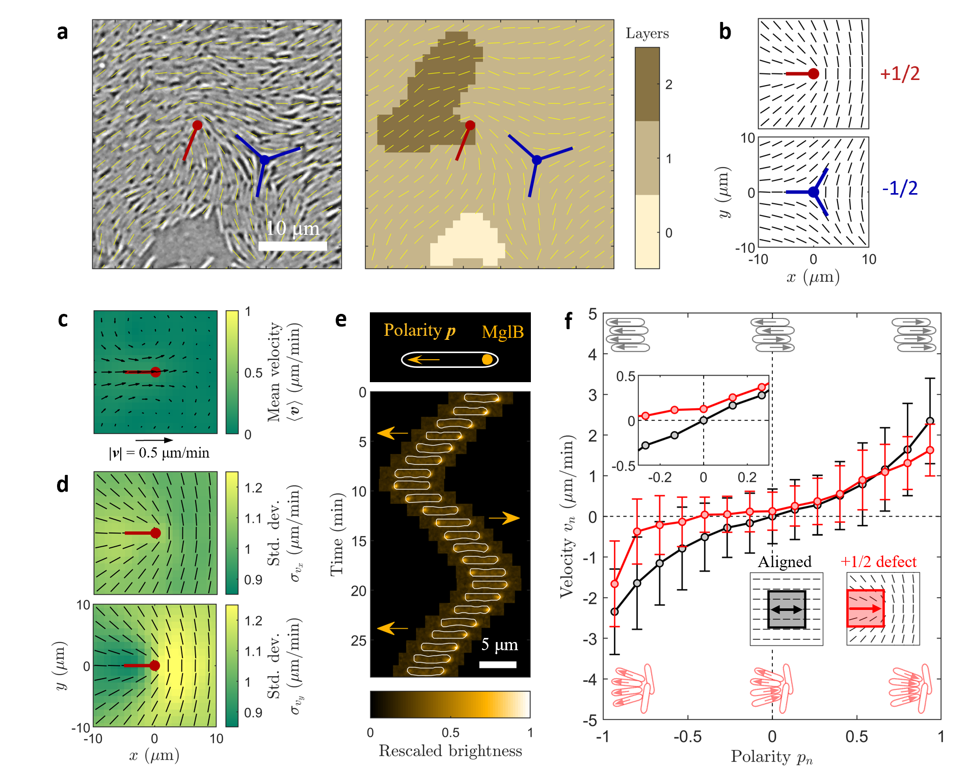
Here, we show that polarity fluctuations trigger the formation of new cell layers, which enables the starvation-induced development from monolayers to droplet-like fruiting bodies in the social bacterium Myxococcus xanthus [30]. Layers of M. xanthus cells contain both comet-like defects of topological charge and triangular defects of charge (Fig. 1a,b), indicating that the system is nematic. Previous work showed that the average cell flow around these defects is well-explained by an active nematic model [15] (see Fig. LABEL:Efig:defect_mean_velocity). Fig. 1c shows the mean cell velocity near a defect, which originates from the balance between the active nematic force density,
| (1) |
and an anisotropic cell-substrate friction, where is the nematic order parameter tensor, is the director of the nematic order, is the scalar order parameter, and is the activity coefficient [23, 17, 15]. The velocity-based flux through a circular boundary around a defect, , is negative, indicating a net influx of cells toward the defect core. Consequently, defects promote cell accumulation and layer formation (Fig. 1a). Similarly, defects produce a net outflux of cells and favor the formation of holes within the monolayer [15]. Thus, the nematic order is connected to colony morphology.

While the active nematic theory predicts a steady flow around defects, the nucleation of new layers is rare, sudden, and stochastic. To study these stochastic events, we measured the velocity fluctuations. Strikingly, the standard deviations of and , and , are both several times larger than the mean speed (Fig. 1d). We hypothesized that these velocity fluctuations are driven by fluctuations in local polar order around defects. To test this hypothesis, we experimentally measured cell polarity and traction forces with cellular-scale resolution simultaneously with the cell velocity, nematic order, and thickness fields near defects.

Instantaneous cell polarity is the main driver of cell flow
We measured cell polarity, , using the mglB::mVenus strain of M. xanthus. This strain expresses a fluorescent fusion to the MglB protein, which is localized to the rear pole of the cell (Fig. 1e) [31].
By simultaneously imaging the cells and the localization of MglB, we defined the polarity of each cell within the population (see SI Sec. LABEL:sec:experiment).
We calculated the average local polarity and velocity in square regions with a side m, the length of two cells, where the subscript denotes the component projected along the director.
To obtain and , we reoriented the regions either with aligned cells or near a defect as shown in Fig. 1f, and averaged the horizontal components of p and v, respectively inside the boxes (see SI Fig. LABEL:Efig:polarity_velocity).
For the defect, “right” was defined as the positive direction, while due to the left-right symmetry of the aligned region, and are equivalent.
In the areas with aligned cells (), does not drive any consistent cell flow, and the cell flow is driven by instantaneous polarity.
In these regions, is a monotonic function of (Fig. 1f, black), and the slope of versus increases as , indicating that the increase of local polar order leads to higher cell speed.
In contrast, near a defect, both the nematic and polar order drive cell motion.
Yet, instantaneous local polar order is still the major driver of the velocity field as seen in the monotonic - relationship (Fig. 1f, red).
However, compared to the aligned areas, this specific arrangement of cells around defects limits the local cell speed, leading to lower as .
Furthermore, at zero polarity , the cell velocity is non-zero (Fig. 1f inset, red); this is the flow driven by the net active nematic force (Fig. 1c).
Our results now show that this average velocity due to nematic forces is small compared to the velocities produced by polarity fluctuations (Fig. 1f).
Polarity-driven cell influx drives layer formation
The cell number was approximately constant as the cells did not grow or divide under our experimental conditions (see Methods).
The formation of a second layer on top of a monolayer thus requires a local influx of cells via motility.
The change in volume within a region is given by the velocity-based flux, , where is the thickness of the cell colony (see SI Fig. LABEL:Efig:volume).
As polarity fluctuations are one of the main drivers of cell flows around defects, they have a major contribution to the cell flux, and hence to layer formation.
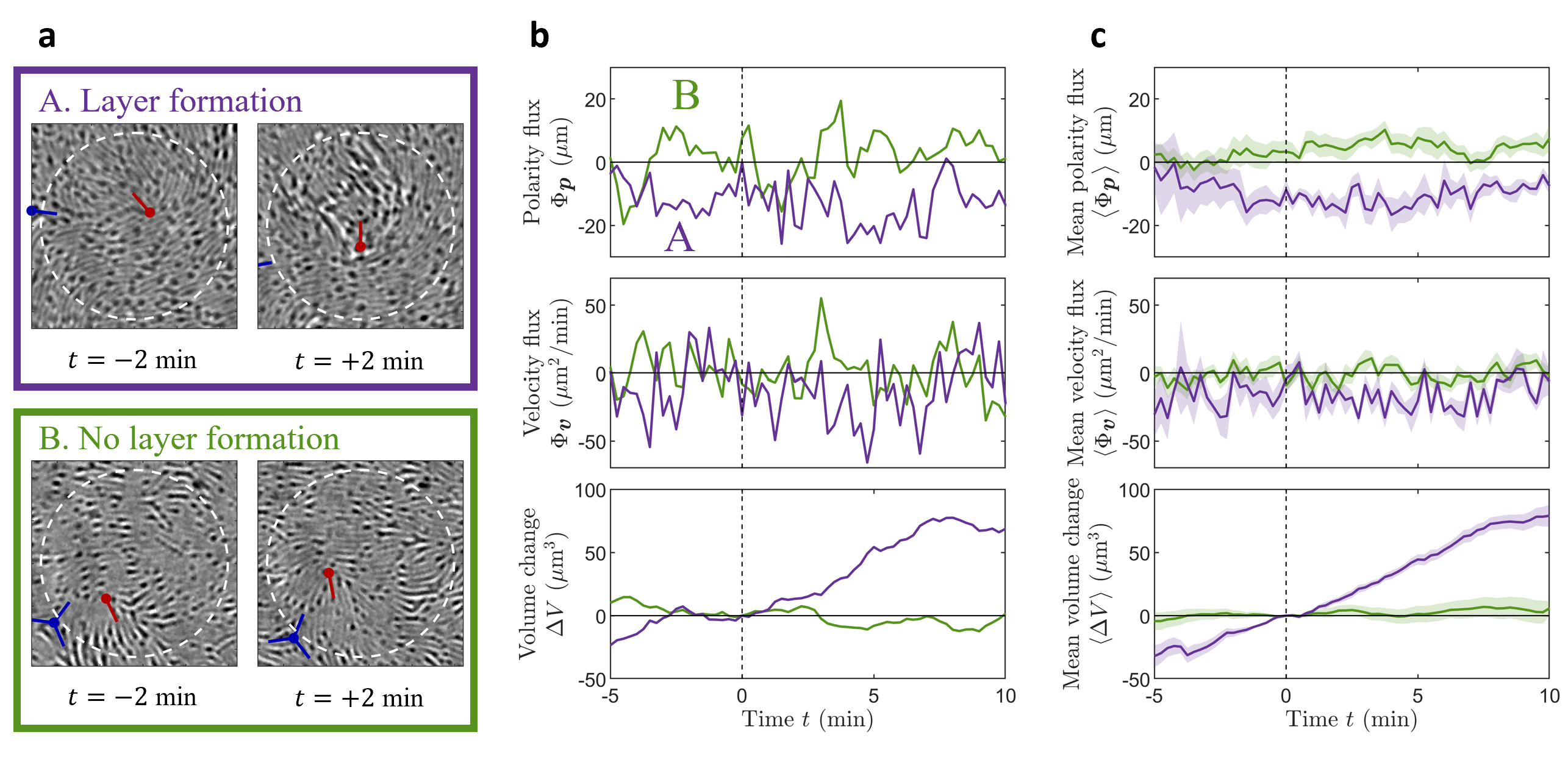
Fig. 2a shows two circular regions with radius , each surrounding a defect. A visible second layer appeared at min in region , while region remained as a monolayer. Fig. 2b shows the polarity-based flux across the boundaries of these two regions . For , was consistently negative starting from several minutes before the out-of-plane cell motion, while for region fluctuated around . The resulting velocity-based flux showed the same trends, with a net influx for the region but not for . We identified multiple regions around defects with and without layer formation, and show the mean polarity-based flux , velocity-based flux , and volume change in Fig. 2c. Their trends are consistent with the exemplary individual events in Fig. 2b, where and are directly related to layer formation.

Besides the average instantaneous flux , we calculated the flux based on the mean velocity near defects (Fig. 1c), and obtained . In , was averaged over all the defects, with or without second layer formation, while in reporting , we selected two separate subsets: those with second layer formation and those without. Note that is significantly weaker than the value of leading to layer formation (Fig. 2c, purple). This shows that most of the influx at defects is due to the strong velocity fluctuation induced by the local polar order, which explains why out-of-plane cell motion is not deterministic in our system. Instead of accumulating cells at a steady rate, when a second layer forms, it forms fast. Moreover, and do not always have a strong positive correlation, because the velocity of a cell in the colony is not directly determined by its polarity but also depends on its mechanical interactions with neighboring cells. This involves the active propelling forces generated by each individual cell, local cell-substrate friction, and cell-cell contact interactions, which introduce further stochasticity to the system.
Cellular tractions and layer formation
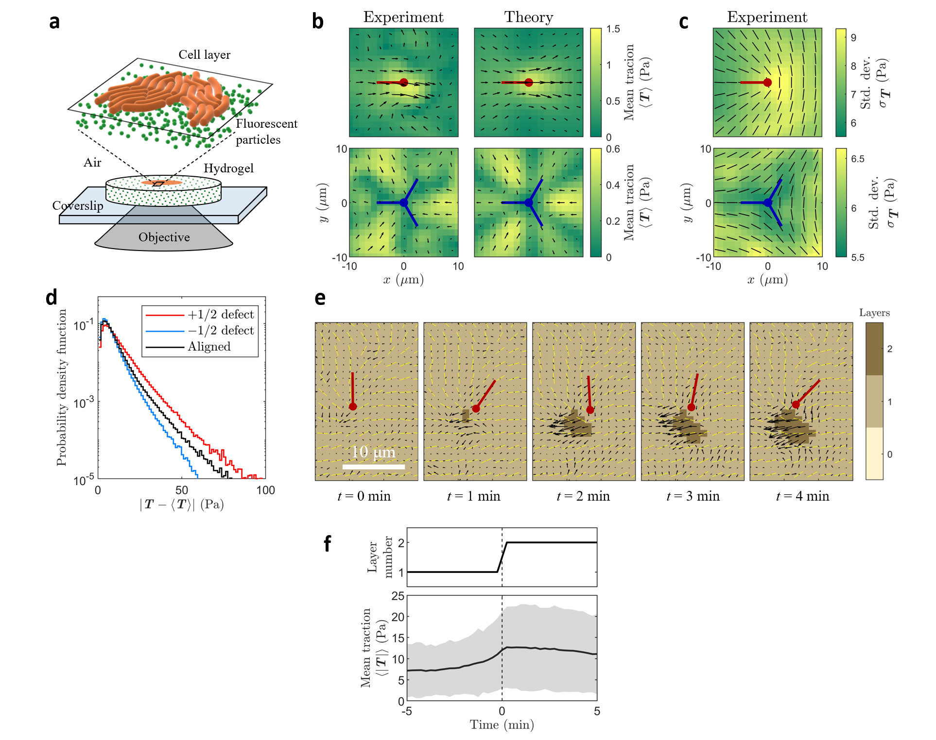
To probe cellular forces in M. xanthus colonies, we used traction force microscopy (TFM) [32, 33, 34] to measure the shear stress exerted on the solid substrate (the - plane) by the cells (Fig. 3a).
By tracking the embedded fluorescent particles, we measured the displacements of the substrate tangential to its surface, and in turn, reconstructed the traction (see SI Sec. LABEL:sec:TFM_method).
According to Eq. 1, the active nematic force is due to bending and splay in the nematic order, given by .
Consequently, should be large near the defects, where exhibits strong distortions (Fig. 1b).
This is confirmed by our measurements of the average traction field around defects (Fig. 3b).
We then set out to explain these measurements theoretically.
Since the cells glide on both the substrate and on neighboring cells, the total active force density has a cell-substrate component and a cell-cell component .
The mean traction reflects the cell-cell interactions: , where is the pressure within the cell layer (see SI Sec. LABEL:sec:theory).
Our measurements agree with the theoretical predictions (Fig. 3b), assuming the cell layer is an active nematic system and the average cell-cell active force is given by in Eq. 1 with , which corresponds to extensile active stresses.
Our experiments showed that, similar to , the instantaneous traction had a standard deviation about an order of magnitude larger than the mean (Fig. 3c). Unlike , the fluctuations were not controlled by . In regions where the cells were aligned (), there were still consistent, strong force fluctuations (Fig. 3d). Moreover, traction fluctuations were stronger at defects and weaker at defects (Fig. 3c,d), although both types of defects had large near their cores (see SI Fig. LABEL:Efig:divQ). Polarity can contribute to these stress fluctuations via , where represents the polar active force a cell generates while gliding on the substrate.
The fluctuations in polar active forces (in or ) will produce fluctuations in the pressure field in the cell colony. The local build-up of pressure can then trigger the formation of new cell layers. Fig. 3e shows an example of a second layer emerging near a defect, simultaneously with a strong increase in . We identified multiple such monolayer to double-layer transitions and found that increases at the reference time , when the second layer forms (Fig. 3f). The increase in upon second layer formation was about 5 Pa, which significantly exceeds Pa around defects (Fig. 3b). The traction did not relax immediately after second layer formation. Similar to , evolved slowly, on the time scale of minutes.

Cell reversals control local polar order and layer formation
Our results so far show that cell polarity produces strong fluctuations in traction and cell flux, which triggers layer formation.
How do cells control local polar order in the system?
During locomotion, M. xanthus cells can reverse their direction of motion on the minute time scale [35].
Cells control this reversal frequency in response to starvation to induce layer formation.
In a nutrient-rich environment, the average revresal time is min [36, 37] and the cells spread into a thin layer on a solid surface (Fig. 1a).
As nutrients become scarce, the cells increase , which leads to layer formation and ultimately to three-dimensional droplets called fruiting bodies [30, 38, 39].
To understand the effect of cellular reversal on surface traction, we probed the non-reversing mutant frzE while keeping the nutrient-rich environment invariant.
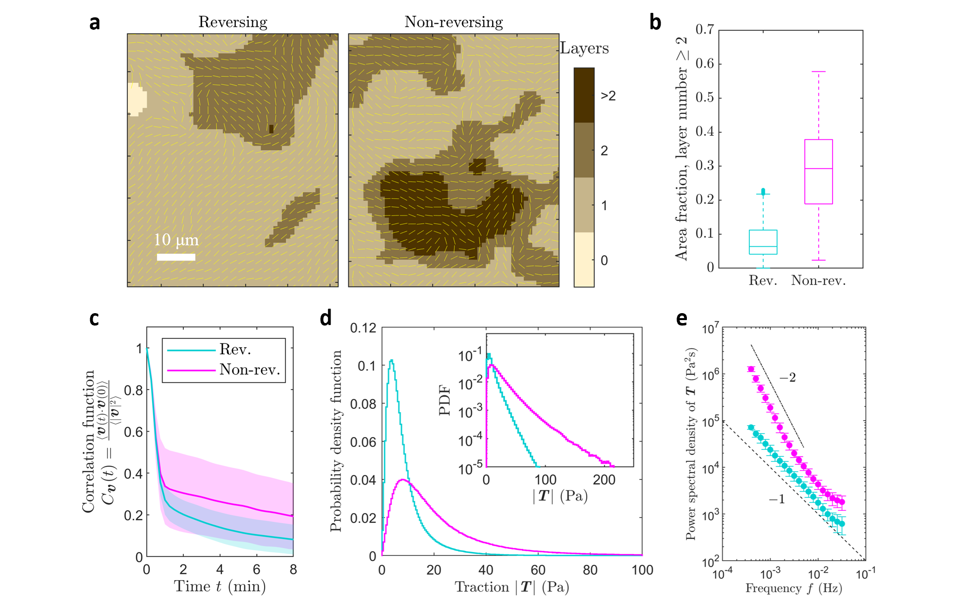
Fig. 4a shows exemplary maps of layer thickness for reversing and non-reversing cells.
Across multiple measurements, the non-reversing mutant generated more multi-layer regions than the reversing ones (Fig. 4b).
Longer reversal time enhances the local polar order in the system. With the polarity assay (see SI Sec. LABEL:sec:experiment), we measured the temporal autocorrelation functions of and , and , respectively, and showed that the correlation times of polarity and velocity were approximately equivalent. More importantly, they both increased along with the local polarity (see SI Fig. LABEL:Efig:polarity_correlation). Similarly, we measured for reversing and non-reversing strains (Fig. 4c), and found that the correlation time increased from min with reversals to min without, implying stronger local polar order in the non-reversing cell colonies. As a result, although the speeds of reversing and non-reversing cells were similar, the non-reversing populations generated more persistent flows, leading to enhanced aggregation.
As expected, the increased local polar order in the non-reversing cells leads to stronger stress fluctuations, as shown by the longer tail in the traction distribution (Fig. 4d). This increase in traction fluctuations happens predominantly at low frequencies (Fig. 4e). The power spectral densities (PSDs) show two different power laws: traction generated by reversing cells follows a power law across almost two decades in frequency , while the PSD for non-reversing cells approaches at low frequencies (Fig. 4e). Understanding the origin of these power laws requires further theoretical investigations.
In summary, our experiments reveal stochastic local polar order in thin M. xanthus colonies, which not only leads to strong fluctuations in cell velocity and mechanical stress but also triggers layer formation. Although active nematic theory explains the average flows generated around topological defects and the cell accumulation that promotes layer formation [15], it needs to be extended to capture the large stress fluctuations we measure or the stochasticity in layer formation. Our results show that the polarity fluctuations generated by collectives of self-propelled rod-shaped cells produce stronger forces and flows than the active nematic stresses around topological defects. We further show that M. xanthus colonies have found a simple knob —the cell reversal time —that they can use to control internal mechanical stress and colony morphology via tuning the local polar order. This control mechanism enables the colony to spread on a surface when nutrients are abundant and then initiate aggregation when food is scarce.
Acknowledgments: We thank the Confocal Imaging Facility, a Nikon Center of Excellence, in the Department of Molecular Biology at Princeton University for instrument use and technical advice. We thank Roy Welch and Lotte Sogaard-Andersen for providing M. xanthus strains. We thank Matthew Black, Aaron Bourque, Benjamin Bratton, Zemer Gitai, Guannan Liu, Howard Stone, Nan Xue, Cassidy Yang, and Rui Zhang for their assistance in the research and many useful discussions. This work was supported by the NSF through awards PHY-1806501 and PHY-2210346, and the Center for the Physics of Biological Function (PHY-1734030). NSW acknowledges National Institutes of Health Grant R01 GM082938.
Author contributions: E.H. and J.W.S. conceived the research. E.H. performed the experiments and data analysis. C.F. and R.A. derived the theoretical model. K.C. and M.D.K. provided key data processing and experimental techniques, respectively. N.S.W. and J.W.S. supervised the project. All authors designed the research and interpreted the results. E.H., C.F., R.A., N.S.W., and J.W.S. wrote the paper with input from the other authors.
References
- [1] Hans Gruler, Manfred Schienbein, Kurt Franke, and Anne de Boisfleury-chevance. Migrating Cells: Living Liquid Crystals. Molecular Crystals and Liquid Crystals Science and Technology. Section A. Molecular Crystals and Liquid Crystals, 260(1):565–574, February 1995.
- [2] T. B. Saw, A. Doostmohammadi, V. Nier, L. Kocgozlu, S. Thampi, Y. Toyama, P. Marcq, C. T. Lim, J. M. Yeomans, and B. Ladoux. Topological defects in epithelia govern cell death and extrusion. Nature, 544(7649):212–216, 2017.
- [3] Lakshmi Balasubramaniam, Amin Doostmohammadi, Thuan Beng Saw, Gautham Hari Narayana Sankara Narayana, Romain Mueller, Tien Dang, Minnah Thomas, Shafali Gupta, Surabhi Sonam, Alpha S. Yap, Yusuke Toyama, René-Marc Mège, Julia M. Yeomans, and Benoît Ladoux. Investigating the nature of active forces in tissues reveals how contractile cells can form extensile monolayers. Nature Materials, 20(8):1156–1166, August 2021.
- [4] Jordi Comelles, Soumya Ss, Linjie Lu, Emilie Le Maout, S Anvitha, Guillaume Salbreux, Frank Jülicher, Mandar M Inamdar, and Daniel Riveline. Epithelial colonies in vitro elongate through collective effects. eLife, 10:e57730, January 2021.
- [5] Josep-Maria Armengol-Collado, Livio Nicola Carenza, Julia Eckert, Dimitrios Krommydas, and Luca Giomi. Epithelia are multiscale active liquid crystals, 2022.
- [6] Guillaume Duclos, Christoph Erlenkämper, Jean-François Joanny, and Pascal Silberzan. Topological defects in confined populations of spindle-shaped cells. Nature Physics, 13(1):58–62, January 2017.
- [7] C. Blanch-Mercader, V. Yashunsky, S. Garcia, G. Duclos, L. Giomi, and P. Silberzan. Turbulent Dynamics of Epithelial Cell Cultures. Physical Review Letters, 120(20):208101, May 2018.
- [8] K. Kawaguchi, R. Kageyama, and M. Sano. Topological defects control collective dynamics in neural progenitor cell cultures. Nature, 545(7654):327–331, 2017.
- [9] Dmitri Volfson, Scott Cookson, Jeff Hasty, and Lev S. Tsimring. Biomechanical ordering of dense cell populations. Proceedings of the National Academy of Sciences, 105(40):15346–15351, October 2008.
- [10] D. Dell’Arciprete, M. L. Blow, A. T. Brown, F. D. C. Farrell, J. S. Lintuvuori, A. F. McVey, D. Marenduzzo, and W. C. K. Poon. A growing bacterial colony in two dimensions as an active nematic. Nat Commun, 9(1):4190, 2018.
- [11] Y. I. Yaman, E. Demir, R. Vetter, and A. Kocabas. Emergence of active nematics in chaining bacterial biofilms. Nat Commun, 10(1):2285, 2019.
- [12] M. M. Genkin, A. Sokolov, O. D. Lavrentovich, and I. S. Aranson. Topological defects in a living nematic ensnare swimming bacteria. Physical Review X, 7(1), 2017.
- [13] H. Li, X. Shi, M. Huang, X. Chen, M. Xiao, C. Liu, H. Chaté, and H. P. Zhang. Data-driven quantitative modeling of bacterial active nematics. Proceedings of the National Academy of Sciences, 116(3):777–785, 2019.
- [14] O. J. Meacock, A. Doostmohammadi, K. R. Foster, J. M. Yeomans, and W. M. Durham. Bacteria solve the problem of crowding by moving slowly. Nature Physics, 17(2):205–210, 2020.
- [15] K. Copenhagen, R. Alert, N. S. Wingreen, and J. W. Shaevitz. Topological defects promote layer formation in myxococcus xanthus colonies. Nature Physics, 17(2):211–215, 2021.
- [16] T. Sanchez, D. T. Chen, S. J. DeCamp, M. Heymann, and Z. Dogic. Spontaneous motion in hierarchically assembled active matter. Nature, 491(7424):431–4, 2012.
- [17] A. Doostmohammadi, J. Ignes-Mullol, J. M. Yeomans, and F. Sagues. Active nematics. Nat Commun, 9(1):3246, 2018.
- [18] G. Duclos, R. Adkins, D. Banerjee, M. S. E. Peterson, M. Varghese, I. Kolvin, A. Baskaran, R. A. Pelcovits, T. R. Powers, A. Baskaran, F. Toschi, M. F. Hagan, S. J. Streichan, V. Vitelli, D. A. Beller, and Z. Dogic. Topological structure and dynamics of three-dimensional active nematics. Science, 367(6482):1120–1124, 2020.
- [19] R. Zhang, A. Mozaffari, and J. J. de Pablo. Autonomous materials systems from active liquid crystals. Nature Reviews Materials, 6(5):437–453, 2021.
- [20] Pau Guillamat, Carles Blanch-Mercader, Guillaume Pernollet, Karsten Kruse, and Aurélien Roux. Integer topological defects organize stresses driving tissue morphogenesis. Nature Materials, 21(5):588–597, May 2022.
- [21] Y. Maroudas-Sacks, L. Garion, L. Shani-Zerbib, A. Livshits, E. Braun, and K. Keren. Topological defects in the nematic order of actin fibres as organization centres of hydra morphogenesis. Nature Physics, 17(2):251–259, 2020.
- [22] Carlos Pérez-González, Ricard Alert, Carles Blanch-Mercader, Manuel Gómez-González, Tomasz Kolodziej, Elsa Bazellieres, Jaume Casademunt, and Xavier Trepat. Active wetting of epithelial tissues. Nature Physics, 15(1):79–88, January 2019.
- [23] R. Aditi Simha and S. Ramaswamy. Hydrodynamic fluctuations and instabilities in ordered suspensions of self-propelled particles. Phys Rev Lett, 89(5):058101, 2002.
- [24] M. C. Marchetti, J. F. Joanny, S. Ramaswamy, T. B. Liverpool, J. Prost, Madan Rao, and R. Aditi Simha. Hydrodynamics of soft active matter. Reviews of Modern Physics, 85(3):1143–1189, 2013.
- [25] M. Bär, R. Großmann, S. Heidenreich, and F. Peruani. Self-propelled rods: Insights and perspectives for active matter. Annual Review of Condensed Matter Physics, 11(1):441–466, 2020.
- [26] F. Ginelli, F. Peruani, M. Bar, and H. Chate. Large-scale collective properties of self-propelled rods. Phys Rev Lett, 104(18):184502, 2010.
- [27] A. Baskaran and M. C. Marchetti. Hydrodynamics of self-propelled hard rods. Phys Rev E Stat Nonlin Soft Matter Phys, 77(1 Pt 1):011920, 2008.
- [28] L. Huber, R. Suzuki, T. Krüger, E. Frey, and A. R. Bausch. Emergence of coexisting ordered states in active matter systems. Science, 361(6399):255–258, 2018.
- [29] J. Denk and E. Frey. Pattern-induced local symmetry breaking in active-matter systems. Proc Natl Acad Sci U S A, 117(50):31623–31630, 2020.
- [30] D. Kaiser. Coupling cell movement to multicellular development in myxobacteria. Nat Rev Microbiol, 1(1):45–54, 2003.
- [31] D. Szadkowski, L. A. M. Carreira, and Lotte Søgaard-Andersen. A bipartite, low-affinity roadblock domain-containing gap complex regulates bacterial front-rear polarity. bioRxiv, page 2022.03.17.484758, 2022.
- [32] S. V. Plotnikov, B. Sabass, U. S. Schwarz, and C. M. Waterman. High-resolution traction force microscopy. Methods Cell Biol, 123:367–94, 2014.
- [33] B. Sabass, M. L. Gardel, C. M. Waterman, and U. S. Schwarz. High resolution traction force microscopy based on experimental and computational advances. Biophys J, 94(1):207–20, 2008.
- [34] B. Sabass, M. D. Koch, G. Liu, H. A. Stone, and J. W. Shaevitz. Force generation by groups of migrating bacteria. Proc Natl Acad Sci U S A, 114(28):7266–7271, 2017.
- [35] D.R. Zusman, Y.F. Inclan, and T. Mignot. Chapter 7, The Frz Chemosensory System of Myxococcus xanthus. In Myxobacteria - Multicellularity and Differentiation, D.E. Whitworth (Ed.). American Society for Microbiology (ASM), 2008.
- [36] W Shi, F K Ngok, and D R Zusman. Cell density regulates cellular reversal frequency in myxococcus xanthus. Proceedings of the National Academy of Sciences, 93(9):4142–4146, 1996.
- [37] S. Thutupalli, M. Sun, F. Bunyak, K. Palaniappan, and J. W. Shaevitz. Directional reversals enable myxococcus xanthus cells to produce collective one-dimensional streams during fruiting-body formation. J R Soc Interface, 12(109):20150049, 2015.
- [38] J. Munoz-Dorado, F. J. Marcos-Torres, E. Garcia-Bravo, A. Moraleda-Munoz, and J. Perez. Myxobacteria: Moving, killing, feeding, and surviving together. Front Microbiol, 7:781, 2016.
- [39] G. Liu, A. Patch, F. Bahar, D. Yllanes, R. D. Welch, M. C. Marchetti, S. Thutupalli, and J. W. Shaevitz. Self-driven phase transitions drive myxococcus xanthus fruiting body formation. Phys Rev Lett, 122(24):248102, 2019.
- [40] M. E. Black and J. W. Shaevitz. Rheological dynamics of active myxococcus xanthus populations during development, 2021.
- [41] J. Zhu, L. Chen, J. Shen, and V. Tikare. Coarsening kinetics from a variable-mobility cahn-hilliard equation: Application of a semi-implicit fourier spectral method. Physical Review E, 60(4):3564, 1999.
- [42] B. Qin, C. Fei, A. A. Bridges, A. A. Mashruwala, H. A. Stone, N. S. Wingreen, and B. L. Bassler. Cell position fates and collective fountain flow in bacterial biofilms revealed by light-sheet microscopy. Science, 369(6499):71–77, 2020.
- [43] Daniel Wall and Dale Kaiser. Type iv pili and cell motility. Molecular Microbiology, 32(1):01–10, 1999.
- [44] E. M. Mauriello, T. Mignot, Z. Yang, and D. R. Zusman. Gliding motility revisited: how do the myxobacteria move without flagella? Microbiol Mol Biol Rev, 74(2):229–49, 2010.
- [45] C. Kaimer and D. R. Zusman. Regulation of cell reversal frequency in myxococcus xanthus requires the balanced activity of chey-like domains in frze and frzz. Mol Microbiol, 100(2):379–95, 2016.
- [46] W. G. Herrick, T. V. Nguyen, M. Sleiman, S. McRae, T. S. Emrick, and S. R. Peyton. Peg-phosphorylcholine hydrogels as tunable and versatile platforms for mechanobiology. Biomacromolecules, 14(7):2294–304, 2013.
- [47] H. Li, X. Q. Shi, M. Huang, X. Chen, M. Xiao, C. Liu, H. Chate, and H. P. Zhang. Data-driven quantitative modeling of bacterial active nematics. Proc Natl Acad Sci U S A, 116(3):777–785, 2019.
- [48] A. J. Vromans and L. Giomi. Orientational properties of nematic disclinations. Soft Matter, 12(30):6490–5, 2016.
- [49] D. Blair and Dufresne E. The matlab particle tracking code repository. Particle-tracking code available at site.physics.georgetown.edu/matlab/, 2018.
- [50] N. G. Deen, P. Willems, M. van Sint Annaland, J. A. M. Kuipers, R. G. H. Lammertink, A. J. B. Kemperman, M. Wessling, and W. G. J. van der Meer. On image pre-processing for piv of single- and two-phase flows over reflecting objects. Experiments in Fluids, 49(2):525–530, 2010.
- [51] U. S. Schwarz and J. R. Soine. Traction force microscopy on soft elastic substrates: A guide to recent computational advances. Biochim Biophys Acta, 1853(11 Pt B):3095–104, 2015.
- [52] L.D. Landau and E.M. Lifshitz. Theory of Elasticity (Second Edition). Pergamon Press, 1970.
Supplemental Information Spontaneous polar order controls morphology of Myxococcus xanthus colonies