2Johns Hopkins University
3Shanghai Jiao Tong University
11email: yshen92@jhu.edu
TransNuSeg: A Lightweight Multi-Task Transformer for Nuclei Segmentation
Abstract
Nuclei appear small in size, yet, in real clinical practice, the global spatial information and correlation of the color or brightness contrast between nuclei and background, have been considered a crucial component for accurate nuclei segmentation. However, the field of automatic nuclei segmentation is dominated by Convolutional Neural Networks (CNNs), meanwhile, the potential of the recently prevalent Transformers has not been fully explored, which is powerful in capturing local-global correlations. To this end, we make the first attempt at a pure Transformer framework for nuclei segmentation, called TransNuSeg. Different from prior work, we decouple the challenging nuclei segmentation task into an intrinsic multi-task learning task, where a tri-decoder structure is employed for nuclei instance, nuclei edge, and clustered edge segmentation respectively. To eliminate the divergent predictions from different branches in previous work, a novel self distillation loss is introduced to explicitly impose consistency regulation between branches. Moreover, to formulate the high correlation between branches and also reduce the number of parameters, an efficient attention sharing scheme is proposed by partially sharing the self-attention heads amongst the tri-decoders. Finally, a token MLP bottleneck replaces the over-parameterized Transformer bottleneck for a further reduction in model complexity. Experiments on two datasets of different modalities, including MoNuSeg have shown that our methods can outperform state-of-the-art counterparts such as CA2.5-Net by 2-3% Dice with 30% fewer parameters. In conclusion, TransNuSeg confirms the strength of Transformer in the context of nuclei segmentation, which thus can serve as an efficient solution for real clinical practice. Code is available at https://github.com/zhenqi-he/transnuseg.
Keywords:
Lightweight Multi-Task Framework Shared Attention Heads Nuclei, Edge and Clustered Edge Segmentation.1 Introduction
Accurate cancer diagnosis, grading, and treatment decisions from medical images heavily rely on the analysis of underlying complex nuclei structures [method_for_nuclei]. Yet, due to the numerous nuclei contained in a digitized whole-slide image (WSI), or even in an image patch of deep learning input, dense annotation of nuclei contouring is extremely time-consuming and labor-expensive [a_review_and_comparison_of_breast_tumor]. Consequently, automated nuclei segmentation approaches have emerged to satisfy a broad range of computer-aided diagnostic systems, where the deep learning methods, particularly the convolutional neural networks [U-Net, Unet++, NucleiSegNet, HistoSeg, maskGANet] have received notable attention due to their simplicity and generalization ability.
In the literature work, the sole-decoder design in these UNet variants (Fig. 1 (a)) is susceptible to failures in splitting densely clustered nuclei when precise edge information is absent. Hence, deep contour-aware neural network (DCAN) [DCAN] with bi-decoder structure achieves improved instance segmentation performance by adopting multi-task learning, in which one decoder learns to segment the nuclei and the other recognizes edges as described in Fig. 1 (b). Similarly, CIA-Net [CIA-net] extends DCAN with an extra information aggregator to fuse the features from two decoders for more precise segmentation. Much recently, CA2.5-Net [CA2.5net] shows identifying the clustered edges in a multiple-task learning manner can achieve higher performance, and thereby proposes an extra output path to learn the segmentation of clustered edges explicitly. A significant drawback of the aforementioned multi-decoder networks is the ignorance of the prediction consistency between branches, resulting in sub-optimal performance and missing correlations between the learned branches. Specifically, a prediction mismatch between the nuclei and edge branches is observed in previous work [ClusterSeg], implying a direction for performance improvement. To narrow this gap, we propose a consistency distillation between the branches, as shown by the dashed line in Fig. 1 (c). Furthermore, to resolve the cost of involving more decoders, we propose an attention sharing scheme, along with an efficient token MLP bottleneck [token-MLP], which can both reduce the number of parameters.
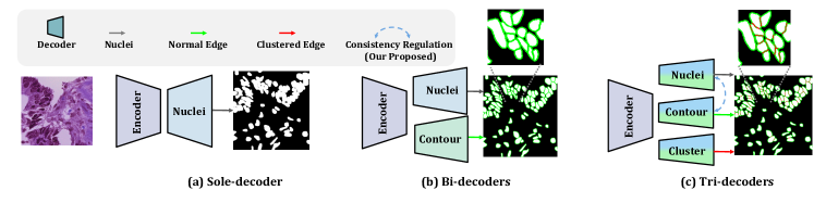
Additionally, existing methods are CNN-based, and their intrinsic convolution operation fails to capture global spatial information or the correlation amongst nuclei [DA-Net], which domain experts rely heavily on for accurate nuclei allocation. It suggests the presence of long-range correlation in practical nuclei segmentation tasks. Inspired by the capability in long-range global context capturing by Transformers [Attention_is_all_u_need], we make the first attempt to construct a tri-decoder based Transformer model to segment nuclei. In short, our major contributions are three-fold: (1) We propose a novel multi-task framework for nuclei segmentation, namely TransNuSeg, as the first attempt at a fully Swin-Transformer driven architecture for nuclei segmentation. (2) To alleviate the prediction inconsistency between branches, we propose a novel self distillation loss that regulates the consistency between the nuclei decoder and normal edge decoder. (3) We propose an innovative attention sharing scheme that shares attention heads amongst all decoders. By leveraging the high correlation between tasks, it can communicate the learned features efficiently across decoders and sharply reduce the number of parameters. Furthermore, the incorporation of a light-weighted MLP bottleneck leads to a sharp reduction of parameters at no cost of performance decline.
2 Methodology
2.0.1 Network Architecture Overview.
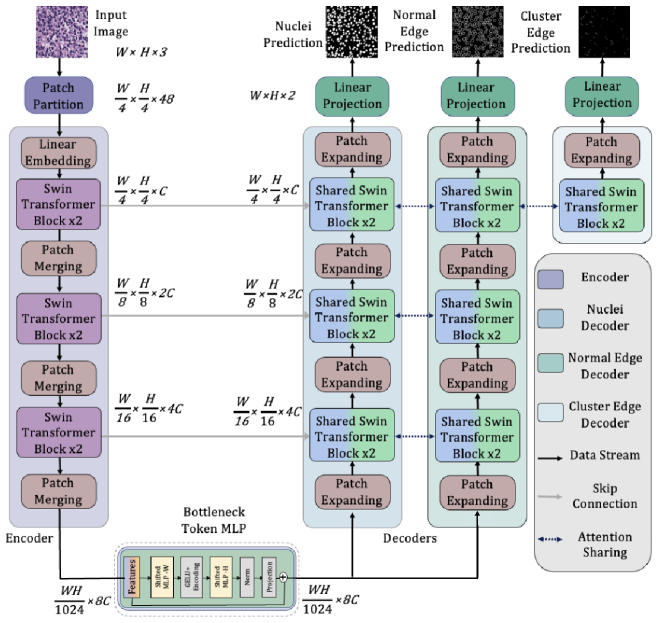
Fig. 2 illustrates the overall architecture of the proposed multi-task tri-decoder Transformer network, named TransNuSeg. Both the encoder and decoders utilize the Swin Transformer [Swin-Transformer] as the building blocks to capture the long-range feature correlations in the nuclei segmentation context. Our network consists of three individual output decoder paths for nuclei segmentation, normal edges segmentation, and clustered edges segmentation. Given the high dependency between edge and clustered edge, we are inspired to propose a novel attention sharing scheme, which can communicate the information and share learned features across decoders while also reducing the number of parameters. Additionally, a token MLP bottleneck is incorporated to further increase the model efficiency.
2.0.2 Attention Sharing Scheme.
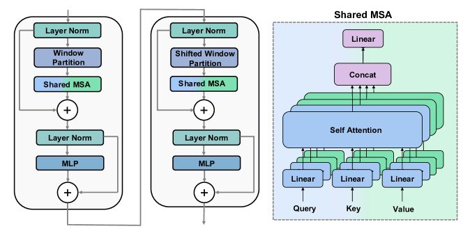
To capture the strong correlation between nuclei segmentation and contour segmentation between multiple decoders [shen2022federated], we introduce a novel attention sharing scheme that is designed as an enhancement to the multi-headed self-attention (MSA) module in the plain Transformer [Attention_is_all_u_need]. Based on the attention sharing scheme, we design a shared MSA module, which is similar in structure to vanilla MSA. Specifically, it consists of a LayerNorm layer [ba2016layer], residual connection, and feed-forward layer. Innovatively, it differs from the vanilla MSA by sharing a proportion of globally-shared self-attention (SA) heads amongst all the parallel Transformer blocks in decoders, while keeping the remaining SA heads unshared i. e. learn the weights separately. A schematic illustration of the shared MSA module in the Swin Transformer block is demonstrated in Fig. 3, as is formally formulated as follows:
| (1) |
writes for the concatenation, denotes the self-attention head whose output dimension is , and is a learnable matrix. The superscript and refer to the globally-shared and unshared weights across all decoders, respectively.
2.0.3 Token MLP Bottleneck.
To reduce the complexity of the model, we leverage a token MLP bottleneck as a light-weight alternative for the Swin Transformer bottleneck. Specifically, this approach involves shifting the latent features extracted by the encoder via two MLP blocks across the width and height channels, respectively [token-MLP]. The objective of this process is to attend to specific areas, which mimics the shifted window attention mechanism in Swin Transformer [Swin-Transformer]. The shifted features are then projected by a learnable MLP and normalized through a LayerNorm [ba2016layer] before being fed to a reprojection MLP layer.
2.0.4 Consistency Self Distillation.
To alleviate the inconsistency between the contour generated from the nuclei segmentation prediction and the predicted edge, we propose a novel consistency self distillation loss, denoted as . Formally, this regularization is defined as the dice loss between the contour generated from the nuclei branch prediction () using the Sobel operation () and the predicted edges from the normal edge decoder. Specifically, the self distillation loss is formulated by .
2.0.5 Multi-Task Learning Objective.
We employ a multi-task learning paradigm to train the tri-decoder network, aiming to improve model performance by leveraging the additional supervision signal from edges. Particularly, the nuclei semantic segmentation is considered the primary task, while the normal edge and clustered edge semantic segmentation are viewed as auxiliary tasks. All decoder branches follow a uniform scheme that combines the cross-entropy loss and the dice loss, with the balancing coefficients set to and respectively, as previous work [CA2.5net]. Subsequently, the overall loss is calculated as a weighted summation of semantic nuclei mask loss (), normal edge loss (), and clustered edge loss (), and the self distillation loss () i. e.
, where coefficients , and are set to , , respectively, and is initially set to with a decrease for every 10 epochs until it reaches .
| Dataset | Methods | DSC (%) | F1 (%) | Acc (%) | IoU (%) | ErCnt (%) |
|---|---|---|---|---|---|---|
| Microscopy | UNet | |||||
| UNet++ | ||||||
| TransUNet | ||||||
| SwinUNet | ||||||
| CA2.5-Net | ||||||
| Ours | ||||||
| Histology | UNet | |||||
| UNet++ | ||||||
| TransUNet | ||||||
| SwinUNet | ||||||
| CA2.5-Net | ||||||
| Ours |
| Methods | #Params () | FLOPs () | Training (s) |
|---|---|---|---|
| UNet [U-Net] | / | ||
| UNet++ [Unet++] | / | ||
| TransUNet [Trans-Unet] | / | ||
| SwinUNet [Swin-Unet] | / | ||
| CA2.5-Net [CA2.5net] | / | ||
| Ours (w/o MLP & w/o AS) | / | ||
| Ours (w/o MLP) | / | ||
| Ours (w/o AS) | / | ||
| Ours (full settings) | / |
3 Experiments
3.0.1 Dataset.
We evaluated the applicability of our approach across multiple modalities by conducting evaluations on microscopy and histology datasets. (1) Fluorescence Microscopy Image Dataset: This set combines three different data sources to simulate the heterogeneous nature of medical images [dataset_CA]. It consists of 524 fluorescence images, each with a resolution of pixels. (2) Histology Image Dataset: This set is the combination of the open dataset MoNuSeg [MICCAI2018] and another private histology dataset [ClusterSeg] of images. We crop each image in the MoNuSeg dataset into four partially overlapping images. The private dataset contains images sized at tessellated from 50 WSIs scanned at , and meticulously labeled by five pathologists according to the labeling guidelines of the MoNuSeg [MICCAI2018]. For both datasets, we randomly split of the samples on the patient level as the training set and the remaining as the test set.
3.0.2 Implementations.
All experiments are performed on one NVIDIA RTX GPU with GB memory. We use Adam optimizer with an initial learning rate of . We compare TransNuSeg with UNet [U-Net], UNet++ [Unet++], TransUNet [Trans-Unet], SwinUNet [Swin-Unet], and CA2.5-Net [CA2.5net]. We evaluate the results by using Dice Score (DSC), Intersection over Union (IoU), pixel-level accuracy (Acc), and F1-score(F1) as metrics, and ErCnt [ClusterSeg]. To ensure statistical significance, we run all methods five times with different fixed seeds and report the results as mean standard deviation.
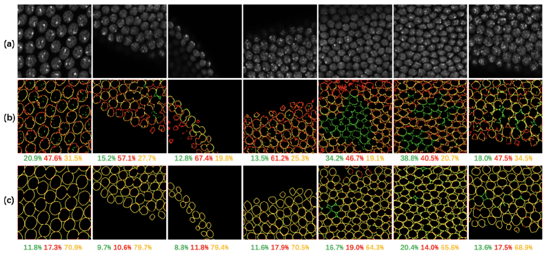
3.0.3 Results.
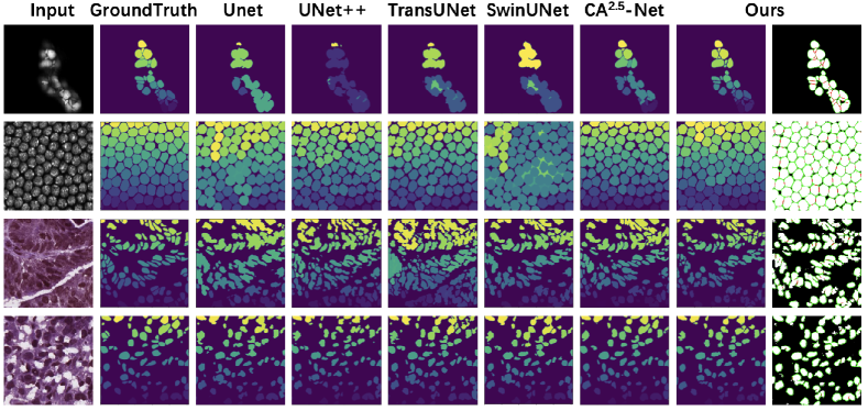
Table 1 shows the quantitative comparisons for the nuclei segmentation. The large margin between the SwinUNet and the other CNN-based or hybrid networks also confirms the superiority of the Transformer in fine-grained nuclei segmentation. More importantly, our method can outperform SwinUNet and the previous methods on both datasets. For example, in the histology image dataset, TransNuSeg improves the dice score, F1 score, accuracy, and IoU by , , , and respectively, over the second-best models. Similarly, in the fluorescence microscopy image dataset, our proposed model improves DSC by , while also leading to , and increment in F1 score, accuracy, and IoU to the second-best performance. For better visualization, representative samples and their segmentation results using different methods are demonstrated in Fig. 4. Furthermore, Table 2 compares the model complexity in terms of the number of parameters, floating point operations per second (FLOPs), and the training computational cost, where our approach can significantly reduce around of the training time compared to the state-of-the-art CNN multi-task method CA2.5-Net, while also boosting performance.
| MLP | AS | SD | Microscopy | Histology | ||||||
|---|---|---|---|---|---|---|---|---|---|---|
| DSC (%) | F1 (%) | Acc (%) | IoU (%) | DSC (%) | F1 (%) | Acc (%) | IoU (%) | |||
3.0.4 Ablation.
Our ablation study yields that token MLP bottleneck and attention sharing schemes can complementarily reduce the training cost while increasing efficiency, as shown in Table 2 (the last 4 rows). To further show the effectiveness of these schemes, as well as consistency self distillation, we conduct a comprehensive ablation study on both datasets. As described in Table 3, each component proportionally contributes to the improvement to reach the overall performance boost. Moreover, self distillation can enhance the intrinsic consistency between two branches, as visualized in Fig. 5.
4 Conclusion
In this paper, we make the first attempt at an efficient but effective multi-task Transformer framework for modality-agnostic nuclei segmentation. Specifically, our tri-decoder framework TransNuSeg leverages an innovative self distillation regularization to impose consistency between the different branches. Experimental results on two datasets demonstrate the excellence of our TransNuSeg against state-of-the-art counterparts for potential real-world clinical deployment. Additionally, our work opens a new architecture to perform nuclei segmentation tasks with Swin Transformer, where further investigations can be performed to explore the generalizability to the top of our methods with different modalities.
References
- [1] Jimmy Lei Ba, Jamie Ryan Kiros, and Geoffrey E Hinton. Layer normalization. arXiv preprint arXiv:1607.06450, 2016.
- [2] Hu Cao, Yueyue Wang, Joy Chen, et al. Swin-unet: Unet-like pure transformer for medical image segmentation, 2021.
- [3] Hao Chen, Xiaojuan Qi, Lequan Yu, and Pheng-Ann Heng. DCAN: deep contour-aware networks for accurate gland segmentation. CoRR, abs/1604.02677, 2016.
- [4] Jieneng Chen, Yongyi Lu, Qihang Yu, Xiangde Luo, Ehsan Adeli, Yan Wang, Le Lu, Alan L. Yuille, and Yuyin Zhou. Transunet: Transformers make strong encoders for medical image segmentation. CoRR, abs/2102.04306, 2021.
- [5] Ruoyu Guo, Maurice Pagnucco, and Yang Song. Learning with noise: Mask-guided attention model for weakly supervised nuclei segmentation. In Medical Image Computing and Computer Assisted Intervention – MICCAI 2021, pages 461–470, Cham, 2021. Springer International Publishing.
- [6] Jinghan Huang, Yiqing Shen, Dinggang Shen, and Jing Ke. Ca2.5-net nuclei segmentation framework with a microscopy cell benchmark collection. In Marleen de Bruijne, Philippe C. Cattin, Stéphane Cotin, Nicolas Padoy, Stefanie Speidel, Yefeng Zheng, and Caroline Essert, editors, Medical Image Computing and Computer Assisted Intervention – MICCAI 2021, pages 445–454, Cham, 2021. Springer International Publishing.
- [7] Humayun Irshad, Antoine Veillard, Ludovic Roux, and Daniel Racoceanu. Methods for nuclei detection, segmentation, and classification in digital histopathology: A review—current status and future potential. IEEE Reviews in Biomedical Engineering, 7:97–114, 2014.
- [8] Jing Ke, Yizhou Lu, Yiqing Shen, Junchao Zhu, Yijin Zhou, Jinghan Huang, Jieteng Yao, Xiaoyao Liang, Yi Guo, Zhonghua Wei, Sheng Liu, Qin Huang, Fusong Jiang, and Dinggang Shen. Clusterseg: A crowd cluster pinpointed nucleus segmentation framework with cross-modality datasets. Medical Image Analysis, page 102758, 2023.
- [9] Florian Kromp, Eva Bozsaky, Fikret Rifatbegovic, Lukas Fischer, Magdalena Ambros, Maria Berneder, Tamara Weiss, Daria Lazic, Wolfgang Dörr, Allan Hanbury, and et al. An annotated fluorescence image dataset for training nuclear segmentation methods. Scientific Data, 7(1), 2020.
- [10] Neeraj Kumar, Ruchika Verma, Deepak Anand, et al. A multi-organ nucleus segmentation challenge. IEEE Transactions on Medical Imaging, 39(5):1380–1391, 2020.
- [11] Andrew Lagree, Majidreza Mohebpour, Nicholas Meti, Khadijeh Saednia, Fang-I. Lu, Elzbieta Slodkowska, Sonal Gandhi, Eileen Rakovitch, Alex Shenfield, Ali Sadeghi-Naini, and et al. A review and comparison of breast tumor cell nuclei segmentation performances using deep convolutional neural networks. Scientific Reports, 11(1), 2021.
- [12] Shyam Lal, Devikalyan Das, Kumar Alabhya, Anirudh Kanfade, Aman Kumar, and Jyoti Kini. Nucleisegnet: Robust deep learning architecture for the nuclei segmentation of liver cancer histopathology images. Computers in biology and medicine, 128, 01 2021.
- [13] Ze Liu, Yutong Lin, Yue Cao, Han Hu, Yixuan Wei, Zheng Zhang, Stephen Lin, and Baining Guo. Swin transformer: Hierarchical vision transformer using shifted windows. CoRR, abs/2103.14030, 2021.
- [14] Olaf Ronneberger, Philipp Fischer, and Thomas Brox. U-net: Convolutional networks for biomedical image segmentation. CoRR, abs/1505.04597, 2015.
- [15] Yiqing Shen, Baiyun Liu, Ruize Yu, Yudong Wang, Shaokang Wang, Jiangfen Wu, and Weidao Chen. Federated learning for chronic obstructive pulmonary disease classification with partial personalized attention mechanism. arXiv preprint arXiv:2210.16142, 2022.
- [16] Jeya Maria Valanarasu and Vishal M. Patel. Unext: Mlp-based rapid medical image segmentation network. Lecture Notes in Computer Science, page 23–33, 2022.
- [17] Ashish Vaswani, Noam Shazeer, Niki Parmar, Jakob Uszkoreit, Llion Jones, Aidan N Gomez, Ł ukasz Kaiser, and Illia Polosukhin. Attention is all you need. In I. Guyon, U. Von Luxburg, S. Bengio, H. Wallach, R. Fergus, S. Vishwanathan, and R. Garnett, editors, Advances in Neural Information Processing Systems, volume 30. Curran Associates, Inc., 2017.
- [18] Changwei Wang, Rongtao Xu, Shibiao Xu, Weiliang Meng, and Xiaopeng Zhang. Da-net: Dual branch transformer and adaptive strip upsampling for retinal vessels segmentation. In Linwei Wang, Qi Dou, P. Thomas Fletcher, Stefanie Speidel, and Shuo Li, editors, Medical Image Computing and Computer Assisted Intervention – MICCAI 2022, pages 528–538. Springer Nature Switzerland, 2022.
- [19] Saad Wazir and Muhammad Moazam Fraz. HistoSeg: Quick attention with multi-loss function for multi-structure segmentation in digital histology images. In 2022 12th International Conference on Pattern Recognition Systems (ICPRS). IEEE, jun 2022.
- [20] Yanning Zhou, Omer Fahri Onder, Qi Dou, Efstratios Tsougenis, Hao Chen, and Pheng-Ann Heng. Cia-net: Robust nuclei instance segmentation with contour-aware information aggregation. CoRR, abs/1903.05358, 2019.
- [21] Zongwei Zhou, Md Mahfuzur Rahman Siddiquee, Nima Tajbakhsh, and Jianming Liang. Unet++: A nested u-net architecture for medical image segmentation. In Deep Learning in Medical Image Analysis and Multimodal Learning for Clinical Decision Support, pages 3–11, Cham, 2018. Springer International Publishing.