3D Shape-Based Myocardial Infarction Prediction Using Point Cloud Classification Networks
Abstract
Myocardial infarction (MI) is one of the most prevalent cardiovascular diseases with associated clinical decision-making typically based on single-valued imaging biomarkers. However, such metrics only approximate the complex 3D structure and physiology of the heart and hence hinder a better understanding and prediction of MI outcomes. In this work, we investigate the utility of complete 3D cardiac shapes in the form of point clouds for an improved detection of MI events. To this end, we propose a fully automatic multi-step pipeline consisting of a 3D cardiac surface reconstruction step followed by a point cloud classification network. Our method utilizes recent advances in geometric deep learning on point clouds to enable direct and efficient multi-scale learning on high-resolution surface models of the cardiac anatomy. We evaluate our approach on 1068 UK Biobank subjects for the tasks of prevalent MI detection and incident MI prediction and find improvements of 13% and 5% respectively over clinical benchmarks. Furthermore, we analyze the role of each ventricle and cardiac phase for 3D shape-based MI detection and conduct a visual analysis of the morphological and physiological patterns typically associated with MI outcomes.
Clinical relevance— The presented approach enables the fast and fully automatic pathology-specific analysis of full 3D cardiac shapes. It can thus be employed as a real-time diagnostic tool in clinical practice to discover and visualize more intricate biomarkers than currently used single-valued metrics and improve predictive accuracy of myocardial infarction.
Index Terms:
Myocardial Infarction, Point Cloud Networks, Cine MRI, 3D Cardiac Shape Analysis, Ejection Fraction, Geometric Deep Learning.I INTRODUCTION
Myocardial infarction (MI) is a common manifestation of coronary artery disease, the deadliest pathology in the world [1]. In current clinical practice, its diagnosis and treatment typically involve the acquisition of cardiac cine magnetic resonance (MR) images as the gold standard imaging modality for cardiac anatomy and function assessments [2]. However, current clinical decision-making is often guided by single-valued biomarkers, such as ejection fraction, which are directly calculated from 2D MR imaging (MRI) slices to evaluate cardiac anatomy and physiology on a purely global level [2]. Consequently, considerable research efforts have focused on developing methods that can take more comprehensive image information into account [3, 4, 5, 6, 7]. However, all these approaches still only approximate the true 3D structure of the heart based on 2D images or image-derived features and therefore neglect more complex and localized changes in 3D cardiac shapes, which play a crucial role in improving the understanding, prediction, and management of MI outcomes [8, 9, 10, 11, 12].
In this work, we investigate the utility of full 3D cardiac shape representations in the form of point clouds for the detection and prediction of MI events. To this end, we propose a novel fully-automatic MI detection pipeline, which first reconstructs 3D cardiac anatomy point clouds from raw cine MR images and then employs targeted point cloud networks for the MI classification task. The network architectures of its individual components are based on recent advances in point cloud-based deep learning to enable efficient multi-scale feature learning directly on anatomical surface data. Deep learning approaches for point cloud data have recently been increasingly used in the field of cardiac image analysis for a variety of applications, such as 3D surface reconstruction [13, 14, 15, 16, 17], image segmentation [14, 18], pathology classification [18], or 3D anatomy and function modeling [19, 20, 21, 22, 23]. In this work, we specifically study 3D anatomical representations of the left and right ventricles at both ends of the cardiac cycle and their effect on prior and future MI. To the best of our knowledge, this is the first MRI-based point cloud deep learning approach to focus on MI prediction directly from 3D cardiac shapes.
II METHODS
II-A Dataset
Our dataset consists of 1068 subjects of the UK Biobank study for which cine MR images were acquired using a balanced steady-state free precession (b-SSFP) protocol [24]. An MI event after the imaging date (incident MI) was recorded for 235 subjects, while 294 subjects suffered an MI event prior to imaging (prevalent MI). The remaining 539 subjects were selected to be free of any diseases associated with the cardiovascular system and are used as normal control cases for our analysis. We follow the disease definition as proposed in previous work [25] and use the UK Biobank field ID 42,000 to identify both incident and prevalent MI subjects.
II-B Infarction Detection Pipeline
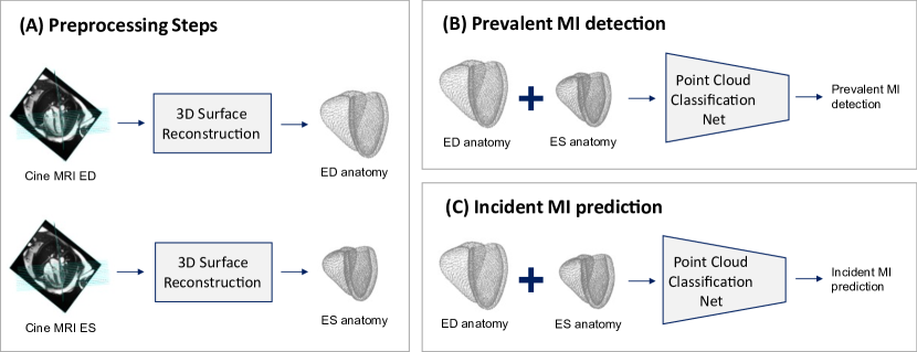
Our proposed 3D anatomy-based MI detection pipeline consists of multiple fully automatic steps as illustrated in Fig. 1. It receives the cine MRI acquisitions at both the end-diastolic (ED) and end-systolic (ES) phases of the cardiac cycle as inputs. Based on these inputs, we reconstruct the corresponding 3D biventricular anatomy models at both phases using a multi-step cardiac surface reconstruction approach [15] (Fig. 1-A). It first segments the left ventricular (LV) endocardium, LV epicardium, and right ventricular (RV) endocardium in the short-axis and four-chamber long-axis (LAX) slices of the MRI acquisition with separate pre-trained fully convolutional neural networks [26] and in the two-chamber LAX images using a U-Net with adversarial training. The resulting segmentation contours of all image slices are then placed into 3D space as sparse point clouds [27] before a point cloud completion network is employed to correct the motion-induced slice misalignment and output dense point cloud representations of the 3D cardiac anatomy.
These 3D cardiac anatomies are then used as inputs to point cloud classification networks for the tasks of prevalent MI classification (Fig. 1-B) and incident MI prediction (Fig. 1-C). For each task, we study both ES only and combined ED and ES anatomy inputs as implicit and explicit representations of 3D shape-based cardiac function. In the latter case, we concatenate the ED and ES point clouds before feeding them into the point cloud classification network, giving it direct access to all available anatomical information at both phases. In addition, we investigate the utility of the RV as part of a biventricular representation of 3D cardiac shape for MI, by using first only LV anatomies, and then combined LV and RV anatomies as network inputs. We analyze the effect of these two different shape inputs for both MI classification tasks and for each of the two temporal input types, resulting in a total of 8 different experiments.
II-C Point Cloud Classification Network
We choose PointNet [28] as the architectural basis of our point cloud classification network and adjust it for the task of binary MI classification of 3D point cloud representations of cardiac anatomy and function. To this end, we first use a sigmoid activation layer at the end of PointNet’s classification branch to obtain binary prediction probabilities as the network’s output. We then tune the drop-out probabilities in the last multi-layer perceptron part of the network based on a grid search procedure. We train our network using the binary cross entropy loss and the Adam optimizer with a mini-batch size of 20 and a learning rate of 1E-6 for 200 epochs on an RTX 2060S GPU with 8 GB memory.
III EXPERIMENTS AND RESULTS
III-A Prevalent Infarction Detection
In our first experiment, we assess whether the high-resolution 3D point cloud representations of the cardiac anatomy contain more information about prevalent MI events than corresponding global clinical benchmarks and whether a point cloud-based deep learning network is able to successfully extract them without any manual intervention. Furthermore, we analyze the importance of different cardiac substructures and cardiac phases for this task. To this end, we train four separate point cloud classification networks using the ES LV anatomy, the combined ED and ES LV anatomies, the ES biventricular anatomy, and the combined ED and ES biventricular anatomies as inputs to the respective networks. We then select widely used clinical metrics (ES volume, ejection fraction) for the LV and RV as our comparative benchmarks and input them as independent variables in four separate logistic regression models, each trained on the same dataset and task as the corresponding point cloud networks. We conduct a four-fold cross validation experiment in each case and report the results in terms of area under the receiver operating characteristic (AUROC) scores in Table I.
| Anatomy | Input | Method | AUROC |
|---|---|---|---|
| LV | ES Volume | Regression | 0.654 |
| ES 3D Shape | Proposed | 0.705 | |
| Ejection Fraction | Regression | 0.670 | |
| ED+ES 3D Shape | Proposed | 0.725 | |
| LV+RV | ES Volume | Regression | 0.641 |
| ES 3D Shape | Proposed | 0.699 | |
| Ejection Fraction | Regression | 0.671 | |
| ED+ES 3D Shape | Proposed | 0.758 |
We find that 3D shape-based point cloud classification networks outperform the respective clinical benchmarks for all cardiac phases and anatomical substructures with an average relative difference of 10% in terms of AUROC. As expected, the combined biventricular input at ED and ES achieves the highest score and a 13% outperformance of its respective clinical benchmark.
Following this quantitative evaluation, we further investigate which 3D anatomical shape features are typically associated with prevalent MI cases by the network. To this end, we select two representative cases corresponding to good and poor network predictions on the test dataset for both MI and normal cases and visualize them in Fig. 2.
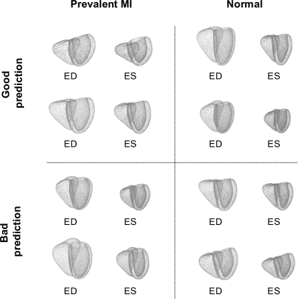
We observe that good network predictions for prevalent MI subjects are more likely to occur in cases of reduced myocardial thickening and smaller overall volume changes between the ED and ES phases, and vice versa for normal cases. Bad predictions more commonly happen when these associations are weakened or reversed.
III-B Incident Infarction Prediction
In addition to detecting prevalent MI events, we investigate whether 3D anatomy-based patterns learned by point cloud networks are also beneficial for the prediction of incident MI events. We follow a similar experimental setup as for prevalent MI classification in Sec. III-A and train four separate networks and their corresponding clinical regression benchmarks for the binary prediction of incident MI events using four-fold cross validation and the AUROC evaluation metric (Table II). We again use the full 3D shapes (LV ES, LV ED+ES, LV+RV ES, LV+RV ED+ES) as neural network inputs and the respective clinical metrics (LV ES volume, LV ejection fraction, LV+RV ES volume, LV+RV ejection fraction) as independent regression variables.
| Anatomy | Input | Method | AUROC |
|---|---|---|---|
| LV | ES Volume | Regression | 0.632 |
| ES 3D Shape | Proposed | 0.660 | |
| Ejection Fraction | Regression | 0.635 | |
| ED+ES 3D Shape | Proposed | 0.654 | |
| LV+RV | ES Volume | Regression | 0.620 |
| ES 3D Shape | Proposed | 0.651 | |
| Ejection Fraction | Regression | 0.618 | |
| ED+ES 3D Shape | Proposed | 0.646 |
We find that the 3D shape-based point cloud network is able to outperform the respective clinical benchmark for both cardiac phases and ventricles by 4% on average. The best score is achieved by the combined ventricular anatomy at ES with a 5% improvement. When visually examining the results, we observe similar patterns as in our prevalent MI detection experiments (Sec. III-A) with a generally higher probability of accurate MI prediction for smaller changes in myocardial thickness between ED and ES phases.
IV DISCUSSION AND CONCLUSION
We have presented a novel end-to-end point cloud-based deep learning pipeline for the detection of both prior and future MI events based on 3D cardiac shapes. In our experiments, the method has been able to outperform corresponding clinical benchmarks for both classification tasks using a variety of different inputs. On the one hand, this indicates that full 3D cardiac shapes contain more infarction-related information than current single-valued clinical biomarkers, which is in line with previous works [8, 9] and promises to improve both patient risk stratification and the implementation of preventive measures. On the other hand, it shows that the architectural design of our pipeline is adequately chosen to successfully extract relevant biomarkers directly from the 3D anatomical shapes. Hereby, the selected point cloud representation of cardiac surface data considerably reduces the memory requirements compared to previous voxelgrid-based approaches. Combined with the fully automatic pipeline design, this allows for faster execution speeds, wider applicability, and easy scaling to both higher resolutions and large numbers of patients in real time.
In our experiments, we also find better predictive performance for prevalent compared to incident MI cases. We hypothesize that this is primarily caused by the more easily visible morphological changes of post-MI cardiac remodeling, which the network is able to capture. While the addition of RV information achieved mixed results, the inclusion of anatomies at both ED and ES phases generally improved predictive accuracy, which corroborates previous findings on the importance of 3D LV contraction information for MI detection [8, 9]. While we focused on the role of 3D shapes in this study, we believe that the pipeline can be easily extended to include other patient-specific information with a potential to further improve the understanding of MI events.
ACKNOWLEDGMENT
This research has been conducted using the UK Biobank Resource under Application Number ‘40161’. The authors express no conflict of interest. The work of M. Beetz was supported by the Stiftung der Deutschen Wirtschaft (Foundation of German Business). A. Banerjee is a Royal Society University Research Fellow and is supported by the Royal Society Grant No. URF\R1\221314. The work of A. Banerjee was partially supported by the British Heart Foundation (BHF) Project under Grant PG/20/21/35082. The works of V. Grau and L. Li were supported by the CompBioMed 2 Centre of Excellence in Computational Biomedicine (European Commission Horizon 2020 research and innovation programme, grant agreement No. 823712). L. Li was partially supported by the SJTU 2021 Outstanding Doctoral Graduate Development Scholarship.
References
- [1] M. A. Khan et al., “Global epidemiology of ischemic heart disease: results from the global burden of disease study,” Cureus, vol. 12, no. 7, 2020.
- [2] M. Reindl et al., “Role of cardiac magnetic resonance to improve risk prediction following acute ST-elevation myocardial infarction,” Journal of Clinical Medicine, vol. 9, no. 4, p. 1041, 2020.
- [3] O. Bernard et al., “Deep learning techniques for automatic MRI cardiac multi-structures segmentation and diagnosis: is the problem solved?” IEEE Transactions on Medical Imaging, vol. 37, no. 11, pp. 2514–2525, 2018.
- [4] F. Isensee et al., “Automatic cardiac disease assessment on cine-MRI via time-series segmentation and domain specific features,” in ACDC Challenge, International Workshop on Statistical Atlases and Computational Models of the Heart, 2018, pp. 120–129.
- [5] J. M. Wolterink et al., “Automatic segmentation and disease classification using cardiac cine MR images,” in ACDC Challenge, International Workshop on Statistical Atlases and Computational Models of the Heart, 2018, pp. 101–110.
- [6] N. Zhang et al., “Deep learning for diagnosis of chronic myocardial infarction on nonenhanced cardiac cine MRI,” Radiology, vol. 291, no. 3, pp. 606–617, 2019.
- [7] L. Li et al., “MyoPS: A benchmark of myocardial pathology segmentation combining three-sequence cardiac magnetic resonance images,” Medical Image Analysis, p. 102808, 2023.
- [8] A. Suinesiaputra et al., “Statistical shape modeling of the left ventricle: myocardial infarct classification challenge,” IEEE Journal of Biomedical and Health Informatics, vol. 22, no. 2, pp. 503–515, 2017.
- [9] J. Corral Acero et al., “Understanding and improving risk assessment after myocardial infarction using automated left ventricular shape analysis,” JACC: Cardiovascular Imaging, 2022.
- [10] M. Beetz et al., “Interpretable cardiac anatomy modeling using variational mesh autoencoders,” Frontiers in Cardiovascular Medicine, p. 3258, 2022.
- [11] M. Beetz et al., “Post-infarction risk prediction with mesh classification networks,” in International Workshop on Statistical Atlases and Computational Models of the Heart, 2023, pp. 291–301.
- [12] M. Beetz et al., “Mesh U-Nets for 3D cardiac deformation modeling,” in International Workshop on Statistical Atlases and Computational Models of the Heart, 2023, pp. 245–257.
- [13] X.-Y. Zhou et al., “One-stage shape instantiation from a single 2D image to 3D point cloud,” in International Conference on Medical Image Computing and Computer Assisted Intervention, 2019, pp. 30–38.
- [14] M. Ye et al., “PC-U net: Learning to jointly reconstruct and segment the cardiac walls in 3D from CT data,” in International Workshop on Statistical Atlases and Computational Models of the Heart, 2020, pp. 117–126.
- [15] M. Beetz et al., “Biventricular surface reconstruction from cine MRI contours using point completion networks,” in 2021 IEEE 18th International Symposium on Biomedical Imaging (ISBI). IEEE, 2021, pp. 105–109.
- [16] X. Chen et al., “Shape registration with learned deformations for 3D shape reconstruction from sparse and incomplete point clouds,” Medical Image Analysis, vol. 74, p. 102228, 2021.
- [17] M. Beetz et al., “Point2Mesh-Net: Combining point cloud and mesh-based deep learning for cardiac shape reconstruction,” in International Workshop on Statistical Atlases and Computational Models of the Heart, 2023, pp. 280–290.
- [18] Y. Chang and C. Jung, “Automatic cardiac MRI segmentation and permutation-invariant pathology classification using deep neural networks and point clouds,” Neurocomputing, vol. 418, pp. 270–279, 2020.
- [19] M. Beetz et al., “Generating subpopulation-specific biventricular anatomy models using conditional point cloud variational autoencoders,” in International Workshop on Statistical Atlases and Computational Models of the Heart, 2021, pp. 75–83.
- [20] M. Beetz et al., “Predicting 3D cardiac deformations with point cloud autoencoders,” in International Workshop on Statistical Atlases and Computational Models of the Heart, 2021, pp. 219–228.
- [21] M. Beetz et al., “Combined generation of electrocardiogram and cardiac anatomy models using multi-modal variational autoencoders,” in 2022 IEEE 19th International Symposium on Biomedical Imaging (ISBI), 2022, pp. 1–4.
- [22] M. Beetz et al., “Multi-domain variational autoencoders for combined modeling of MRI-based biventricular anatomy and ECG-based cardiac electrophysiology,” Frontiers in Physiology, p. 991, 2022.
- [23] L. Li et al., “Deep computational model for the inference of ventricular activation properties,” in International Workshop on Statistical Atlases and Computational Models of the Heart, 2023, pp. 369–380.
- [24] S. E. Petersen et al., “UK Biobank’s cardiovascular magnetic resonance protocol,” Journal of Cardiovascular Magnetic Resonance, vol. 18, no. 8, pp. 1–7, 2015.
- [25] W. Bai et al., “A population-based phenome-wide association study of cardiac and aortic structure and function,” Nature Medicine, vol. 26, no. 10, pp. 1654–1662, 2020.
- [26] W. Bai et al., “Automated cardiovascular magnetic resonance image analysis with fully convolutional networks,” Journal of Cardiovascular Magnetic Resonance, vol. 20, no. 65, pp. 1–12, 2018.
- [27] A. Banerjee et al., “A completely automated pipeline for 3D reconstruction of human heart from 2D cine magnetic resonance slices,” Philosophical Transactions of the Royal Society A: Mathematical, Physical and Engineering Sciences, vol. 379, no. 2212, p. 20200257, 2021.
- [28] C. R. Qi et al., “Pointnet: Deep learning on point sets for 3D classification and segmentation,” in Proceedings of the IEEE Conference on Computer Vision and Pattern Recognition, 2017, pp. 652–660.