TriFormer: A Multi-modal Transformer Framework For Mild Cognitive Impairment Conversion Prediction
Abstract
The prediction of mild cognitive impairment (MCI) conversion to Alzheimer’s disease (AD) is important for early treatment to prevent or slow the progression of AD. To accurately predict the MCI conversion to stable MCI or progressive MCI, we propose TriFormer, a novel transformer-based framework with three specialized transformers to incorporate multi-modal data. TriFormer uses I) an image transformer to extract multi-view image features from medical scans, II) a clinical transformer to embed and correlate multi-modal clinical data, and III) a modality fusion transformer that produces an accurate prediction based on fusing the outputs from the image and clinical transformers. Triformer is evaluated on the Alzheimer’s Disease Neuroimaging Initiative (ADNI) 1 and ADNI2 datasets and outperforms previous state-of-the-art single and multi-modal methods.
Index Terms— Alzheimer’s disease, Transformer, MRI, Multi-modality
1 INTRODUCTION
Mild cognitive impairment (MCI) patients exhibit a memory impairment earlier than the expected age. It is a transitional stage from Congnitively normal (CN) to Alzheimer’s disease (AD) where around 10% to 15% MCI patients end up progressing to AD every year [1]. Patients having MCI can either progress to AD within several years defined as progressive MCI (pMCI) or stay at the same MCI stage defined as stable MCI (sMCI). Previous studies have shown that early nonpharmacological therapy and treatment can delay the progression from MCI to AD. However, the prerequisite to early intervention is accurately predicting the likelihood of MCI conversion to AD for MCI patients at the early stage. Utilizing multi-modal clinical data such as cognitive test results, genetic information, and imaging data including T1w or T2w Magnetic resonance imaging (MRI) and Positron emission tomography (PET) could help more accurately predict MCI conversion [2].
Convolutional neural networks have been widely applied to AD classification and prediction from imaging data. Valliani et al. [3] fine-tuned a pretrained ResNet-50 to classify AD and CN based on 2D axial slices. Wen et al. [4] leveraged 3D spatial information by using a 3D CNN and outperformed previous 2D-based methods in AD classification and MCI conversion prediction. However, both 2D and 3D CNNs have a strong inductive bias towards local receptive fields, which could limit the performance on high dimensional data [5]. Recently, transformers have been shown to be effective in capturing global long-range dependency within imaging [6] and sequential data [7]. They also have no indictive bias compared with CNNs. Despite the performance advantages, few studies have attempted transformers for predicting the progression of MCI to pMCI/sMCI or classifying AD. This is mainly due to their prohibitive computational costs on 3D medical imaging data. A novel tractable transformer-based network is needed to enhance the performance of AD classification and MCI conversion predictions. Furthermore, several studies have shown that using multi-modal data can significantly improve the accuracy of MCI conversion prediction and AD classification. Qiu et al. [8] used multi-modal data including age, gender, Mini-Mental State Exam (MMSE) and MRI to diagnose AD using patch-based 3D CNN. Pan et al. [9] proposed a spatially-constrained Fisher representation network for AD diagnosis using MRI and PET imaging data. Guan et al. [2] proposed a knowledge distilling network using multi-modal data for MCI conversion prediction. These works typically used a simple multi-layer perceptual (MLP) network to weight different clinical data. To the best of our knowledge, there is no existing multi-modal work adopting a transformer to explicitly leverage the correlation between clinical features for more accurate MCI predictions.
In this paper, we propose a novel transformer-based network, TriFormer, which is made up of three transformers to exploit multi-model data including T1 weighted MRI and clinical data to predict MCI conversion. Our contributions are summarized as follows: 1. We propose a 2.5D Vision Transformer (ViT) to efficiently extract multi-view image features. 2. With our proposed clinical transformer, TriFormer becomes the first transformer to embed and correlate between different clinical features for MCI conversion prediction. 3. We further propose a modality fusion transformer to aggregate the multi-modal features from the clinical and image transformers. The three transformers are put together to construct Triformer, which improves the performance of MCI conversion prediction yielding state-of-the-art results.
2 METHODS
As shown in Figure 1, the proposed TriFormer contains three transformer modules: (1) an image feature extractor containing a 2.5D ViT to extract global image features from coronal, sagittal, and axial views, (2) a second transformer to distill information from clinical data, (3) a third modality fusion transformer which aggregates multi-modal representations and provide final predictions.
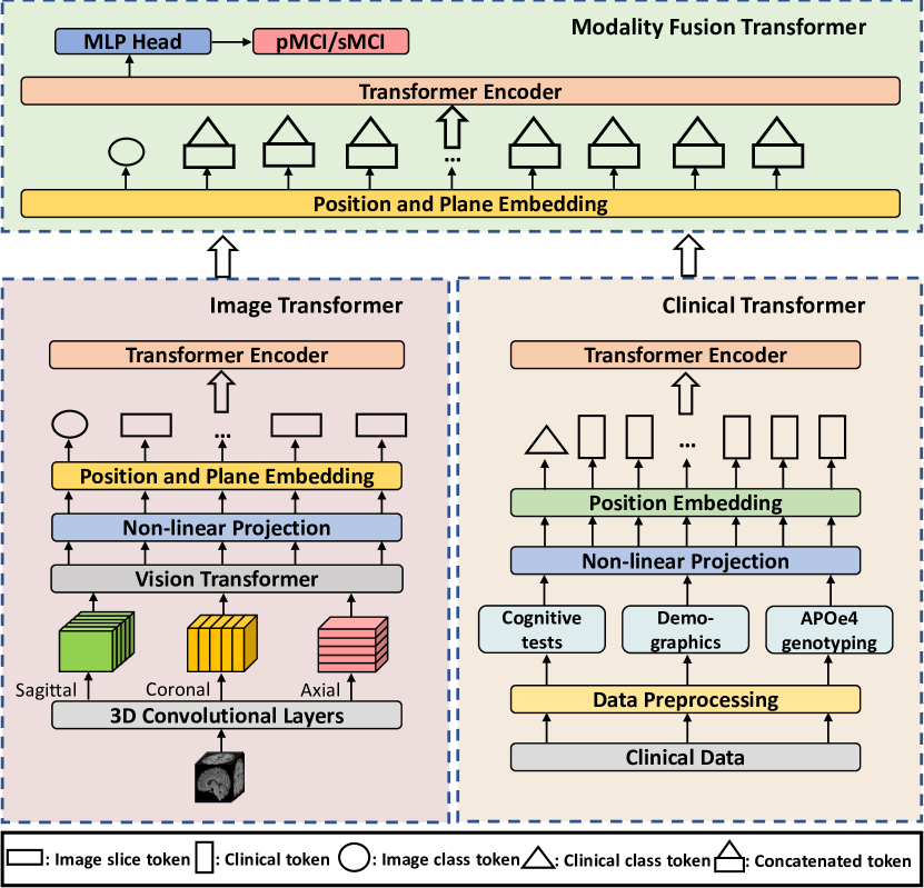
2.1 Image Feature Extractor
Given a MRI image with dimension , the feature embedding layer which contains two 3D convolutional layers project input into a C-dimensional latent space. Then the extracted volumes are sliced into 2D along the three spatial dimensions For instance, we obtain slices of features with the dimension of from the axial view. As a result, concatenate them to slices of image features. Compared to directly feeding the raw input image slices into the module, feature embedding helps preserve more spatial information from neighbouring slices. We then adopt ViT [6] as our feature extraction module to explore the global semantic information from the embedded features and encode them into compact feature vectors. Specifically, image features are fed into a shared-parameter ViT slice by slice predicting feature vectors. Following the image feature extraction stage, we apply another transformer encoder to explore the correlation between feature vectors from different planes and slices. In this stage, we introduce a novel positional embedding layer to inject the plane information. Instead of using sinusoidal positional encoding, we provide a unique identifier for each plane to make the transformer location-aware. A plane separation token is further used for the model better to differentiate the different views. The final input to the transformer encoder is formulated as:
| (1) | |||
where is the image feature, is the image class token, is the plane separation token, , , are the image slice features from all three views.
| (2) |
where is the position embedding of each token.
| (3) |
where is the plane feature with the view information of where each image slice token is from.
| (4) |
All the features and embeddings are added together before feeding into the image transformer encoder.
2.2 Clinical Feature Extractor
Each clinical modality is first normalized between 0 and 1 and passed through a non-linear projection layer containing an MLP layer followed by a ReLU activation function. Then all clinical modalities are stacked together and passed through a learnable positional embedding layer with a clinical class token prepended before feeding into the transformer encoder. Instead of separately inputting each clinical feature to the transformer encoder of the modality fusion transformer, the clinical class token is used to inject into all the clinical features at once using a single vector.
2.3 Modality Fusion Transformer
The clinical class token is first duplicated times and each concatenated with an image slice token from the image feature extractor. These aggregated features are then fed into a modality fusion transformer which has the same positional and plane embedding layers as the image transformer. Lastly, the class token is passed through MLP layers to perform a binary classification between pMCI and sMCI.
3 RESULTS AND DISCUSSION
3.1 Dataset
We conducted our experiments on two public datasets, the Alzheimer’s Disease Neuroimaging Initiative (ADNI)1 and ADNI2. ADNI3 was not included due to the lack of MCI patients with sufficient follow-up timepoints. All the T1-weighted images were first reoriented to the standard space and cropped automatically using FSL. Non-linear registration to the MNI152 template, skull stripping and bias field correction were applied for data normalization. Finally, all images were resampled to 128x128x128 with a voxel size of using ANTs. Before feeding into TriFormer, all image intensity values were normalized between 0 and 1. Only baseline images were included in this study and the data split was based on the patient level to avoid data leakage. We included 12 types of clinical data: age, gender, education years, APOe4 genotyping, Clinical Dementia Rating Sum of Boxes (CDRSB), Alzheimer’s disease assessment scale (ADAS) (ADAS11, ADAS13), MMSE score and Rey Auditory Verbal Learning Test (RAVLT) (immediate, learning, forgetting, percent forgetting). The subjects were separated into three groups: AD, CN and MCI. The MCI group was subdivided into two groups: sMCI and pMCI. Patients were diagnosed as MCI at all available timepoints with at least 24 months records were defined as sMCI while patients were diagnosed as MCI at baseline but converted to AD within 36 months without changing back to CN or MCI at all available timepoints were defined as pMCI. Details of the demographics of patients are shown in Table 1.
| Dataset | Label | No. of Subjects | Age Range | Male/Female |
|---|---|---|---|---|
| ADNI1 | pMCI | 137 | 55.2-88.3 | 81/56 |
| sMCI | 100 | 57.8-87.9 | 60/40 | |
| AD | 169 | 55.1-90.1 | 87/82 | |
| CN | 206 | 59.9-89.6 | 103/103 | |
| ADNI2 | pMCI | 57 | 55.0-84.6 | 30/27 |
| sMCI | 117 | 55.9-91.3 | 65/52 | |
| AD | 102 | 55.9-88.3 | 58/44 | |
| CN | 137 | 56.2-85.6 | 67/70 |
3.2 Implementation
TriFormer is implemented in Pytorch and trained on an NVIDIA Tesla V100 GPU. Data augmentation methods including Random corona-view flipping and gaussian noising are applied during the training. We also utilize AD and CN patients to augment training data considering MCI group is small as previous studies have shown learning from AD and CN can improve the MCI prediction performance [2]. TriFomer is trained for 50 epochs using the Adam optimizer with a batch size of 2 and the cross-entropy loss. All three transformers contain 6 transformer layers with 8 heads. Image dimensions are 128 each and are 32 and 512, respectively. All the embedding layers’ dimensions are 256. We first train TriFormer on the ADNI1 dataset and evaluate it on the ADNI2 dataset, then we swap the two datasets to train on ADNI2 and evaluate on the ADNI1 dataset. An 80%/20% train-validation is applied to each experiment. The weights with the highest AUC value on the validation set is selected as the final weights. We report the average results across 5 repeats to reduce variance in the experimental results.
3.3 Comparisons with state-of-the-art methods
Table 2 compares TriFormer’s area under the curve (AUC) and accuracy (ACC) metrics with other state-of-the-art (SOTA) methods. The results indicate TriFormer can effectively utilize multi-modal data to outperform single-modality works such as [5]. Distinct improvements can also be observed when comparing TriFormer to the previous CNN-based multi-modal works [2, 8, 9, 10]. Finally, the TriFormer also outperforms another transformer [11] that fuses cortical features from MRI in predicting MCI conversion. The proposed TriFormer network outperforms previous SOTA methods on both ADNI1 and ADNI2.
3.4 Ablation studies
We further investigate the effectiveness of the three constituent transformers in TriFormer on the ADNI1 dataset. First, we compare the 2.5D ViT with Wen et al. [4] which uses a CNN as a feature extractor. It improves the ACC of the previous method by 3.45%, which demonstrates the advantage of using a transformer to extract multi-view features. Then we show that our clinical transformer can better capture the correlation between multi-modal clinical data compared to the MLP classifier. Further, we apply both imaging data and clinical data as multi-modal inputs associated with a MLP classifier to demonstrate the usage of multi-modal data outperforms single-modality inputs. We finally demonstrate that the modality fusion transformer with the multi-plane information and extracted multi-modal clinical information from the clinical transformer can further boost the performance compared to the normal MLP classifier in both accuracy and AUC metrics. The full ablation study is shown in Table 3.
| Models | Metrics | |||
|---|---|---|---|---|
| Image | Clinical Data | Fusion | AUC | ACC |
| 3D CNN [4] | ✗ | ✗ | 65.98 | 70.69 |
| 2.5D ViT | ✗ | ✗ | 71.88 | 74.14 |
| ✗ | MLP | ✗ | 90.62 | 81.61 |
| ✗ | Transformer | ✗ | 91.07 | 82.37 |
| 2.5D ViT | Transformer | MLP | 91.21 | 82.57 |
| 2.5D ViT | Transformer | Transformer | 91.47 | 84.10 |
4 CONCLUSION
In this work, we propose a novel transformer-based network, TriFormer, which predicts the conversion of MCI using multi-modal data including 3D MRI and multi-modal clinical data. The TriFormer effectively extracts multi-view image features and also captures the correlation between different clinical modalities using two of the three transformers. Then, with a third modality fusion transformer with plane positional information, the framework as a whole can better utilize both imaging and clinical features and a normal MLP classifier. Since medical image analysis makes constant use of multi-modal data, this work could benefit other medical research fields that require multi-modal data.
5 Acknowledgments
This research was supported by the Medical Research Future Fund (MRF1201961) and the Motor Accident Insurance Commission (MAIC), The Queensland Government, Australia (grant number: 2014000857). We would like to thank the Alzheimer’s Disease Neuroimaging Initiative (ADNI) for data collection and sharing.
References
- [1] Ross A Dunne et al., “Mild cognitive impairment: the manchester consensus,” Age and ageing, vol. 50, pp. 72–80, 2021.
- [2] Hao Guan, Chaoyue Wang, and Dacheng Tao, “Mri-based alzheimer’s disease prediction via distilling the knowledge in multi-modal data,” NeuroImage, vol. 244, pp. 118586, 2021.
- [3] Aly Valliani and Ameet Soni, “Deep residual nets for improved alzheimer’s diagnosis,” in Proceedings of the 8th ACM International Conference on Bioinformatics, Computational Biology, and Health Informatics, 2017, pp. 615–615.
- [4] Junhao Wen et al., “Convolutional neural networks for classification of alzheimer’s disease: Overview and reproducible evaluation,” Medical image analysis, vol. 63, pp. 101694, 2020.
- [5] Jinseong Jang and Dosik Hwang, “M3t: Three-dimensional medical image classifier using multi-plane and multi-slice transformer,” in Proceedings of the IEEE/CVF Conference on Computer Vision and Pattern Recognition (CVPR), June 2022, pp. 20718–20729.
- [6] Alexey Dosovitskiy et al., “An image is worth 16x16 words: Transformers for image recognition at scale,” arXiv preprint arXiv:2010.11929, 2020.
- [7] Jacob Devlin, Ming-Wei Chang, Kenton Lee, and Kristina Toutanova, “Bert: Pre-training of deep bidirectional transformers for language understanding,” arXiv preprint arXiv:1810.04805, 2018.
- [8] Shangran Qiu et al., “Development and validation of an interpretable deep learning framework for alzheimer’s disease classification,” Brain, vol. 143, pp. 1920, 2020.
- [9] Yongsheng Pan et al., “Spatially-constrained fisher representation for brain disease identification with incomplete multi-modal neuroimages,” IEEE transactions on medical imaging, vol. 39, no. 9, pp. 2965–2975, 2020.
- [10] Weiming Lin et al., “Convolutional neural networks-based mri image analysis for the alzheimer’s disease prediction from mild cognitive impairment,” Frontiers in neuroscience, vol. 12, pp. 777, 2018.
- [11] Guowei Zheng et al., “A transformer-based multi-features fusion model for prediction of conversion in mild cognitive impairment,” Methods, vol. 204, pp. 241–248, 2022.