MIDLMedical Imaging with Deep Learning
\jmlrpages
\jmlryear2023
\jmlrworkshopShort Paper – MIDL 2023 submission
\jmlrvolume– Under Review
\midlauthor\NameRuining Deng\midljointauthortextJoint first author: contributed equally\nametag1 \Emailr.deng@vanderbilt.edu
\NameCan Cui\midlotherjointauthor\nametag1 \Emailcan.cui.1@vanderbilt.edu
\NameQuan Liu\midlotherjointauthor\nametag1 \Emailquan.liu@vanderbilt.edu
\NameTianyuan Yao\nametag1 \Emailtianyuan.yao@vanderbilt.edu
\NameLucas W. Remedios\nametag1
\Emaillucas.w.remedios@vanderbilt.edu
\NameShunxing Bao\nametag1
\Emailshunxing.bao@vanderbilt.edu
\NameBennett A. Landman\nametag1 \Emailbennett.landman@vanderbilt.edu
\NameLee E. Wheless\nametag2,3
\Emaillee.e.wheless@vumc.org
\NameLori A. Coburn\nametag2,3
\Emaillori.coburn@vumc.org
\NameKeith T. Wilson\nametag2,3 \Emailkeith.wilson@vumc.org
\NameYaohong Wang\nametag2 \Emailyaohong.wang@vumc.org
\NameShilin Zhao\nametag2 \Emailshilin.zhao.1@vumc.org
\NameAgnes B. Fogo\nametag2 \Emailagnes.fogo@vumc.org
\NameHaichun Yang\nametag2 \Emailhaichun.yang@vumc.org
\NameYucheng Tang\nametag4 \Emailyuchengt@nvidia.com
\NameYuankai Huo\midljointauthortextCorresponding author\nametag1 \EmailYuankai.huo@vanderbilt.edu
\addr1 Vanderbilt University, Nashville, TN, USA
\addr2 Vanderbilt University Medical Center, Nashville, TN, USA
\addr3 Veterans Affairs Tennessee Valley Healthcare System, Nashville, TN, USA
\addr4 NVIDIA Cooperation, Redmond, WA, USA
Segment Anything Model (SAM) for Digital Pathology:
Assess Zero-shot Segmentation on Whole Slide Imaging
Abstract
The segment anything model (SAM) was released as a foundation model for image segmentation. The promptable segmentation model was trained by over 1 billion masks on 11M licensed and privacy-respecting images. The model supports zero-shot image segmentation with various segmentation prompts (e.g., points, boxes, masks). It makes the SAM attractive for medical image analysis, especially for digital pathology where the training data are rare. In this study, we evaluate the zero-shot segmentation performance of SAM model on representative segmentation tasks on whole slide imaging (WSI), including (1) tumor segmentation, (2) non-tumor tissue segmentation, (3) cell nuclei segmentation. Core Results: The results suggest that the zero-shot SAM model achieves remarkable segmentation performance for large connected objects. However, it does not consistently achieve satisfying performance for dense instance object segmentation, even with 20 prompts (clicks/boxes) on each image. We also summarized the identified limitations for digital pathology: (1) image resolution, (2) multiple scales, (3) prompt selection, and (4) model fine-tuning. In the future, the few-shot fine-tuning with images from downstream pathological segmentation tasks might help the model to achieve better performance in dense object segmentation.
keywords:
segment anything, SAM model, digital pathology, medical image analysis.1 Introduction
Large language models (e.g., ChatGPT [Brown et al.(2020)Brown, Mann, Ryder, Subbiah, Kaplan, Dhariwal, Neelakantan, Shyam, Sastry, Askell, et al.] and GPT-4 [OpenAI(2023)]), are leading a paradigm shift in natural language processing with strong zero-shot and few-shot generalization capabilities. This development has encouraged researchers to develop large-scale vision foundation models. While the first successful ”foundation models” [Bommasani et al.(2021)Bommasani, Hudson, Adeli, Altman, Arora, von Arx, Bernstein, Bohg, Bosselut, Brunskill, et al.] in computer vision have focused on pre-training approaches (e.g., CLIP [Radford et al.(2021)Radford, Kim, Hallacy, Ramesh, Goh, Agarwal, Sastry, Askell, Mishkin, Clark, et al.] and ALIGN [Jia et al.(2021)Jia, Yang, Xia, Chen, Parekh, Pham, Le, Sung, Li, and Duerig]) and generative AI applications (e.g., DALL·E [Ramesh et al.(2021)Ramesh, Pavlov, Goh, Gray, Voss, Radford, Chen, and Sutskever]), they have not been specifically designed for image segmentation tasks [Kirillov et al.(2023)Kirillov, Mintun, Ravi, Mao, Rolland, Gustafson, Xiao, Whitehead, Berg, Lo, et al.]. Segmenting objects (e.g., tumor, tissue, cell nuclei) for whole slide imaging (WSI) data is an essential task for digital pathology, deep learning models typically necessitate well-delineated training data. Obtaining these gold-standard data from clinical experts can be challenging due to privacy regulations, intensive manual efforts, insufficient reproducibility, and complicated annotation processes [Huo et al.(2021)Huo, Deng, Liu, Fogo, and Yang]. Hence, zero-shot image segmentation [Wang et al.(2019)Wang, Zheng, Yu, and Miao] is desired, where the model can accurately segment pathological images without prior exposure to the domain data during training.
Recently, the ”Segment Anything Model” (SAM) [Kirillov et al.(2023)Kirillov, Mintun, Ravi, Mao, Rolland, Gustafson, Xiao, Whitehead, Berg, Lo, et al.] was proposed as a foundation model for image segmentation. The model has been trained on over 1 billion masks on 11 million licensed and privacy-respecting images. Furthermore, the model supports zero-shot image segmentation with various segmentation prompts (e.g., points, boxes, and masks). This feature makes it particularly attractive for pathological image analysis where the labeled training data are rare and expensive.
In this study, we assess the zero-shot segmentation performance of the SAM model on representative segmentation tasks, including (1) tumor segmentation [Liu et al.(2021)Liu, Louis, Lu, Jha, Zhao, Deng, Yao, Roland, Yang, Zhao, et al.], (2) tissue segmentation [Deng et al.(2023)Deng, Liu, Cui, Yao, Long, Asad, Womick, Zhu, Fogo, Zhao, et al.], and (3) cell nuclei segmentation [Li et al.(2021)Li, Yang, He, Jha, Fogo, Wheless, Zhao, and Huo]. Our study reveals that the SAM model has some limitations and performance gaps compared to state-of-the-art (SOTA) domain-specific models.
2 Experiments and Performance
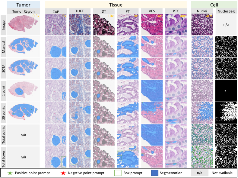
We obtained the source code and the trained model from https://segment-anything.com. To ensure scalable assessments, all experiments were performed directly using Python, rather than relying on the Demo website. The results are presented in Figure 1 and Table 1.
| Method | Prompts | Tumor | Tissue | Cell | |||||
| 0.5 | 5 | 10 | 40 | 40 | |||||
| Tumor | CAP | TUFT | DT | PT | VES | PTC | Nuclei | ||
| SOTA | no prompt | 71.98 | 96.50 | 96.59 | 81.01 | 89.80 | 85.05 | 77.23 | 81.77 |
| SAM | 1 point | 58.71 | 78.08 | 80.11 | 58.93 | 49.72 | 65.26 | 67.03 | 1.95 |
| SAM | 20 points | 74.98 | 80.12 | 79.92 | 60.35 | 66.57 | 68.51 | 64.63 | 41.65 |
| SAM | total points | n/a | 88.10 | 89.65 | 70.21 | 73.19 | 67.04 | 67.61 | 69.50 |
| SAM | total boxes | n/a | 95.23 | 96.49 | 89.97 | 86.77 | 87.44 | 87.18 | 88.30 |
total points/boxes: we place points/boxes on every single instance object (based on the known ground truth) as a theoretical upper bound of SAM. Note that it is impractical in real applications.
Tumor Segmentation.The whole-slide images (WSIs) of skin cancer patients were obtained from the Cancer Genome Atlas (TCGA) datasets (TCGA Research Network: https://www.cancer.gov/tcga). We employed SimTriplet [Liu et al.(2021)Liu, Louis, Lu, Jha, Zhao, Deng, Yao, Roland, Yang, Zhao, et al.] approach as the SOTA method, with the same testing cohort to make a fair comparison. In order to be compatible with the SAM segmentation model, the WSI inputs were scaled down 80 times from a resolution of 40, resulting in an average size of 8601279 pixels. We evaluated two different scenarios: (1) SAM with a single positive point prompt, and (2) SAM with 20 point prompts (10 positive and 10 negative points). The prompts were randomly selected from manual annotations, with positive prompt points being selected from the tumor region and negative prompt points being selected from the non-tumor region.
Tissue Segmentation. A total of 1,751 regions of interest (ROIs) images were obtained from 459 WSIs belonging to 125 patients diagnosed with Minimal Change Diseases. These images were manually segmented to identify six structurally normal pathological primitives [Jayapandian et al.(2021)Jayapandian, Chen, Janowczyk, Palmer, Cassol, Sekulic, Hodgin, Zee, Hewitt, O’Toole, et al.], using digital renal biopsies from the NEPTUNE study [Barisoni et al.(2013)Barisoni, Nast, Jennette, Hodgin, Herzenberg, Lemley, Conway, Kopp, Kretzler, Lienczewski, et al.]. To form a test cohort for multi-tissue segmentation, we captured 8,359 patches measuring 256256 pixels. For comparison, We employed Omni-Seg [Deng et al.(2023)Deng, Liu, Cui, Yao, Long, Asad, Womick, Zhu, Fogo, Zhao, et al.] approach as the SOTA method. The tissue types consist of the glomerular unit (CAP), glomerular tuft (TUFT), distal tubular (DT), proximal tubular (PT), arteries (VES), and peritubular capillaries (PTC). For the SAM method, we evaluated four different scenarios: (1) SAM with a single positive point prompt, (2) SAM with 20 point prompts (10 positive and 10 negative points), and (3)/(4) SAM with all points/boxes on every single instance object, which served as a theoretical upper bound for SAM. We randomly selected point prompts from the manual annotations and eroded each connected component with a 10x10 filter to generate at most one random point. For the box prompts, we used the bounding box of each connected component.
Cell nuclei Segmentation. The dataset for nuclei segmentation was obtained from the MoNuSeg challenge [Kumar et al.(2019)Kumar, Verma, Anand, Zhou, Onder, Tsougenis, Chen, Heng, Li, Hu, et al.]. It contains H&E stained images at 40 magnification with 10001000 pixels from the TCGA dataset, along with corresponding annotations of nuclear boundaries. The MoNuSeg dataset includes 30 images for training and 14 for testing. We evaluated the performance of SAM models against the BEDs model [Li et al.(2021)Li, Yang, He, Jha, Fogo, Wheless, Zhao, and Huo], a competitive nuclei segmentation model trained on the MoNuSeg training data. The prompt method and evaluation are as described in Tissue Segmentation.
3 Limitations on Digital Pathology
The SAM models achieve remarkable performance under zero-shot learning scenarios. However, we identified several limitations during our assessment.
Image resolution. The average training image resolution of SAM is 33004950 pixels [Kirillov et al.(2023)Kirillov, Mintun, Ravi, Mao, Rolland, Gustafson, Xiao, Whitehead, Berg, Lo, et al.], which is significantly smaller than Giga-pixel WSI data ( pixels). Moreover, analyzing WSI data at the patch level may result in an impractical number of interactions, even if only a small number of points or bounding boxes are marked for each patch.
Multiple scales. Multi-scale is a significant feature in digital pathology. Different tissue types have their optimal image resolution (as shown in Table 1). For instance, at the optimal resolution for CAP segmentation (5 scale), it is difficult to achieve good segmentation for PTC. However, zooming in (40 scale) would result in nearly 100 times more patches.
Prompt selection. Firstly, to achieve decent segmentation performance in zero-shot learning scenarios, a considerable number of prompts are still necessary. Secondly, the segmentation performance heavily depends on the quality of prompt selection. Another concern related to segmentation performance is inter-rater and intra-rater reproducibility of prompt-based segmentation.
Model fune-tuning. Currently, tedious manual prompt placements are still necessary for segmentation tasks with significant domain heterogeneities. A reasonable online/offline fine-tuning strategy is necessary to propagate the knowledge obtained from manual prompts to larger-scale automatic segmentation on Giga-pixel WSI data.
4 Conclusion
The zero-shot setting of SAM enables domain users to segment heterogeneous objects in digital pathology without undergoing a heavy training process. The results suggest that the zero-shot SAM model achieves remarkable segmentation performance for large connected objects. However, it does not consistently achieve satisfying performance for dense instance object segmentation, even with 20 prompts (clicks/boxes) on each image. Nonetheless, several limitations still exist and require further investigation for digital pathology.
This research was supported by NIH R01DK135597 (Huo), The Leona M. and Harry B. Helmsley Charitable Trust grant G-1903-03793 and G-2103-05128, NSF CAREER 1452485, NSF 2040462, NCRR Grant UL1 RR024975-01 (now at NCATS Grant 2 UL1 TR000445-06), NIH NIDDK DK56942 (ABF), DoD HT94252310003 (Yang), the VA grants I01BX004366 and I01CX002171, VUMC Digestive Disease Research Center supported by NIH grant P30DK058404, NVIDIA hardware grant, resources of ACCRE at Vanderbilt University.
References
- [Barisoni et al.(2013)Barisoni, Nast, Jennette, Hodgin, Herzenberg, Lemley, Conway, Kopp, Kretzler, Lienczewski, et al.] Laura Barisoni, Cynthia C Nast, J Charles Jennette, Jeffrey B Hodgin, Andrew M Herzenberg, Kevin V Lemley, Catherine M Conway, Jeffrey B Kopp, Matthias Kretzler, Christa Lienczewski, et al. Digital pathology evaluation in the multicenter nephrotic syndrome study network (neptune). Clinical Journal of the American Society of Nephrology, 8(8):1449–1459, 2013.
- [Bommasani et al.(2021)Bommasani, Hudson, Adeli, Altman, Arora, von Arx, Bernstein, Bohg, Bosselut, Brunskill, et al.] Rishi Bommasani, Drew A Hudson, Ehsan Adeli, Russ Altman, Simran Arora, Sydney von Arx, Michael S Bernstein, Jeannette Bohg, Antoine Bosselut, Emma Brunskill, et al. On the opportunities and risks of foundation models. arXiv preprint arXiv:2108.07258, 2021.
- [Brown et al.(2020)Brown, Mann, Ryder, Subbiah, Kaplan, Dhariwal, Neelakantan, Shyam, Sastry, Askell, et al.] Tom Brown, Benjamin Mann, Nick Ryder, Melanie Subbiah, Jared D Kaplan, Prafulla Dhariwal, Arvind Neelakantan, Pranav Shyam, Girish Sastry, Amanda Askell, et al. Language models are few-shot learners. Advances in neural information processing systems, 33:1877–1901, 2020.
- [Deng et al.(2023)Deng, Liu, Cui, Yao, Long, Asad, Womick, Zhu, Fogo, Zhao, et al.] Ruining Deng, Quan Liu, Can Cui, Tianyuan Yao, Jun Long, Zuhayr Asad, R Michael Womick, Zheyu Zhu, Agnes B Fogo, Shilin Zhao, et al. Omni-seg: A scale-aware dynamic network for renal pathological image segmentation. IEEE Transactions on Biomedical Engineering, 2023.
- [Huo et al.(2021)Huo, Deng, Liu, Fogo, and Yang] Yuankai Huo, Ruining Deng, Quan Liu, Agnes B Fogo, and Haichun Yang. Ai applications in renal pathology. Kidney international, 99(6):1309–1320, 2021.
- [Jayapandian et al.(2021)Jayapandian, Chen, Janowczyk, Palmer, Cassol, Sekulic, Hodgin, Zee, Hewitt, O’Toole, et al.] Catherine P Jayapandian, Yijiang Chen, Andrew R Janowczyk, Matthew B Palmer, Clarissa A Cassol, Miroslav Sekulic, Jeffrey B Hodgin, Jarcy Zee, Stephen M Hewitt, John O’Toole, et al. Development and evaluation of deep learning–based segmentation of histologic structures in the kidney cortex with multiple histologic stains. Kidney international, 99(1):86–101, 2021.
- [Jia et al.(2021)Jia, Yang, Xia, Chen, Parekh, Pham, Le, Sung, Li, and Duerig] Chao Jia, Yinfei Yang, Ye Xia, Yi-Ting Chen, Zarana Parekh, Hieu Pham, Quoc Le, Yun-Hsuan Sung, Zhen Li, and Tom Duerig. Scaling up visual and vision-language representation learning with noisy text supervision. In International Conference on Machine Learning, pages 4904–4916. PMLR, 2021.
- [Kirillov et al.(2023)Kirillov, Mintun, Ravi, Mao, Rolland, Gustafson, Xiao, Whitehead, Berg, Lo, et al.] Alexander Kirillov, Eric Mintun, Nikhila Ravi, Hanzi Mao, Chloe Rolland, Laura Gustafson, Tete Xiao, Spencer Whitehead, Alexander C Berg, Wan-Yen Lo, et al. Segment anything. arXiv preprint arXiv:2304.02643, 2023.
- [Kumar et al.(2019)Kumar, Verma, Anand, Zhou, Onder, Tsougenis, Chen, Heng, Li, Hu, et al.] Neeraj Kumar, Ruchika Verma, Deepak Anand, Yanning Zhou, Omer Fahri Onder, Efstratios Tsougenis, Hao Chen, Pheng-Ann Heng, Jiahui Li, Zhiqiang Hu, et al. A multi-organ nucleus segmentation challenge. IEEE transactions on medical imaging, 39(5):1380–1391, 2019.
- [Li et al.(2021)Li, Yang, He, Jha, Fogo, Wheless, Zhao, and Huo] Xing Li, Haichun Yang, Jiaxin He, Aadarsh Jha, Agnes B Fogo, Lee E Wheless, Shilin Zhao, and Yuankai Huo. Beds: Bagging ensemble deep segmentation for nucleus segmentation with testing stage stain augmentation. In 2021 IEEE 18th International Symposium on Biomedical Imaging (ISBI), pages 659–662. IEEE, 2021.
- [Liu et al.(2021)Liu, Louis, Lu, Jha, Zhao, Deng, Yao, Roland, Yang, Zhao, et al.] Quan Liu, Peter C Louis, Yuzhe Lu, Aadarsh Jha, Mengyang Zhao, Ruining Deng, Tianyuan Yao, Joseph T Roland, Haichun Yang, Shilin Zhao, et al. Simtriplet: Simple triplet representation learning with a single gpu. In Medical Image Computing and Computer Assisted Intervention–MICCAI 2021: 24th International Conference, Strasbourg, France, September 27–October 1, 2021, Proceedings, Part II 24, pages 102–112. Springer, 2021.
- [OpenAI(2023)] OpenAI. Gpt-4 technical report, 2023.
- [Radford et al.(2021)Radford, Kim, Hallacy, Ramesh, Goh, Agarwal, Sastry, Askell, Mishkin, Clark, et al.] Alec Radford, Jong Wook Kim, Chris Hallacy, Aditya Ramesh, Gabriel Goh, Sandhini Agarwal, Girish Sastry, Amanda Askell, Pamela Mishkin, Jack Clark, et al. Learning transferable visual models from natural language supervision. In International conference on machine learning, pages 8748–8763. PMLR, 2021.
- [Ramesh et al.(2021)Ramesh, Pavlov, Goh, Gray, Voss, Radford, Chen, and Sutskever] Aditya Ramesh, Mikhail Pavlov, Gabriel Goh, Scott Gray, Chelsea Voss, Alec Radford, Mark Chen, and Ilya Sutskever. Zero-shot text-to-image generation. In International Conference on Machine Learning, pages 8821–8831. PMLR, 2021.
- [Wang et al.(2019)Wang, Zheng, Yu, and Miao] Wei Wang, Vincent W Zheng, Han Yu, and Chunyan Miao. A survey of zero-shot learning: Settings, methods, and applications. ACM Transactions on Intelligent Systems and Technology (TIST), 10(2):1–37, 2019.