2023
\jmlrworkshopFull Paper – MIDL 2023 submission
\midlauthor\NameMd Mostafijur Rahman \Emailmostafijur.rahman@utexas.edu and
\NameRadu Marculescu \Emailradum@utexas.edu
\addrThe University of Texas at Austin
Multi-scale Hierarchical Vision Transformer with Cascaded Attention Decoding for Medical Image Segmentation
Abstract
Transformers have shown great success in medical image segmentation. However, transformers may exhibit a limited generalization ability due to the underlying single-scale self-attention (SA) mechanism. In this paper, we address this issue by introducing a Multi-scale hiERarchical vIsion Transformer (MERIT) backbone network, which improves the generalizability of the model by computing SA at multiple scales. We also incorporate an attention-based decoder, namely Cascaded Attention Decoding (CASCADE), for further refinement of multi-stage features generated by MERIT. Finally, we introduce an effective multi-stage feature mixing loss aggregation (MUTATION) method for better model training via implicit ensembling. Our experiments on two widely used medical image segmentation benchmarks (i.e., Synapse Multi-organ, ACDC) demonstrate the superior performance of MERIT over state-of-the-art methods. Our MERIT architecture and MUTATION loss aggregation can be used with downstream medical image and semantic segmentation tasks.
keywords:
Medical image segmentation, Vision transformer, Multi-scale transformer, Feature-mixing augmentation, Self-attention.1 Introduction
Automatic medical image segmentation has become an important step in disease diagnosis nowadays. Since the emergence of UNet [Ronneberger et al.(2015)Ronneberger, Fischer, and Brox], U-shaped convolutional neural networks (CNNs) [Oktay et al.(2018)Oktay, Schlemper, Folgoc, Lee, Heinrich, Misawa, Mori, McDonagh, Hammerla, Kainz, et al., Huang et al.(2020)Huang, Lin, Tong, Hu, Zhang, Iwamoto, Han, Chen, and Wu, Zhou et al.(2018)Zhou, Rahman Siddiquee, Tajbakhsh, and Liang, Fan et al.(2020)Fan, Ji, Zhou, Chen, Fu, Shen, and Shao] have become de facto methods for medical image segmentation. By producing high-resolution segmentation maps through aggregating multi-stage features via skip connections, UNet variants, such as UNet++ [Zhou et al.(2018)Zhou, Rahman Siddiquee, Tajbakhsh, and Liang] and UNet3Plus [Huang et al.(2020)Huang, Lin, Tong, Hu, Zhang, Iwamoto, Han, Chen, and Wu], have shown good performance in medical image segmentation. However, the spatial context of the convolution operation limits the CNN-based methods ability to learn the long-range relations among pixels [Cao et al.(2021)Cao, Wang, Chen, Jiang, Zhang, Tian, and Wang]. Some works [Chen et al.(2018)Chen, Tan, Wang, and Hu, Oktay et al.(2018)Oktay, Schlemper, Folgoc, Lee, Heinrich, Misawa, Mori, McDonagh, Hammerla, Kainz, et al., Fan et al.(2020)Fan, Ji, Zhou, Chen, Fu, Shen, and Shao] try to address this issue by embedding attention mechanisms in the encoder or decoder. Despite the significant efforts made in this direction, the CNN-based methods still have insufficient ability to capture long-range dependencies.
With the emergence of Vision transformers [Dosovitskiy et al.(2020)Dosovitskiy, Beyer, Kolesnikov, Weissenborn, Zhai, Unterthiner, Dehghani, Minderer, Heigold, Gelly, et al.], many works [Cao et al.(2021)Cao, Wang, Chen, Jiang, Zhang, Tian, and Wang, Chen et al.(2021)Chen, Lu, Yu, Luo, Adeli, Wang, Lu, Yuille, and Zhou, Dong et al.(2021)Dong, Wang, Fan, Li, Fu, and Shao, Wang et al.(2022b)Wang, Huang, Tang, Meng, Su, and Song] try to address the above problem using a transformer encoder, specifically for medical image segmentation. Transformers capture long-range dependencies by learning correlations among all the input patches using self-attention (SA). Recently, hierarchical vision transformers, such as pyramid vision transformer (PVT) [Wang et al.(2021)Wang, Xie, Li, Fan, Song, Liang, Lu, Luo, and Shao] with spatial reduction attention, Swin transformer [Liu et al.(2021)Liu, Lin, Cao, Hu, Wei, Zhang, Lin, and Guo] with window-based attention, and MaxViT [Tu et al.(2022)Tu, Talebi, Zhang, Yang, Milanfar, Bovik, and Li] with multi-axis attention have been introduced to improve performance. Indeed, these hierarchical vision transformers are very effective for medical image segmentation tasks [Cao et al.(2021)Cao, Wang, Chen, Jiang, Zhang, Tian, and Wang, Dong et al.(2021)Dong, Wang, Fan, Li, Fu, and Shao, Wang et al.(2022b)Wang, Huang, Tang, Meng, Su, and Song]. However, these transformer-based architectures have two limitations: 1) self-attention is performed with a single attention window (scale) which has limited feature processing ability, and 2) the self-attention modules used in transformers have limited ability to learn spatial relations among pixels [Chu et al.(2021)Chu, Tian, Zhang, Wang, Wei, Xia, and Shen].
More recently, PVTv2 [Wang et al.(2022c)Wang, Xie, Li, Fan, Song, Liang, Lu, Luo, and Shao] embeds convolution layers in transformer encoders, while CASCADE [Rahman et al.(2023)Rahman, Marculescu, and et al.] introduces an attention-based decoder to address the limitation of learning spatial relations among pixels. Although these methods enable learning, the local (spatial) relations among pixels, they still have limited ability to capture features of multi-scale (e.g., small, large) organs/lesions/objects due to computing self-attention in a single-scale attention window. To address this limitation, we introduce a novel multi-scale hierarchical vision transformer (MERIT) backbone which computes self-attention across multiple attention windows to improve the generalizability of the model. We also incorporate multiple CASCADE decoders to produce better high-resolution segmentation maps by effectively aggregating and enhancing multi-scale hierarchical features. Finally, we introduce a novel effective multi-stage (hierarchical) feature-mixing loss aggregation (MUTATION) strategy for implicit ensembling/augmentation which produces new synthetic predictions by mixing hierarchical prediction maps from the decoder. The aggregated loss from these synthetic predictions improves the performance of medical image segmentation. Our contributions are as follows:
-
•
Novel Network Architecture: We propose a novel multi-scale hierarchical vision transformer (MERIT) for 2D medical image segmentation which captures both multi-scale and multi-resolution features. Besides, we incorporate a cascaded attention-based decoder for better hierarchical multi-scale feature aggregation and refinement.
-
•
Multi-stage Feature-mixing Loss Aggregation: We propose a new simple, yet effective way, namely MUTATION, to create synthetic predictions by mixing features during loss calculation; this improves the medical image segmentation performance.
-
•
New State-of-the-art Results: We perform rigorous experiments and ablation studies on two medical image segmentation benchmarks, namely Synapse multi-organ and ACDC cardiac diagnosis. Our implementation of MERIT using two instances (with different windows for SA) of MaxViT [Tu et al.(2022)Tu, Talebi, Zhang, Yang, Milanfar, Bovik, and Li] backbone with CASCADE decoder and MUTATION loss aggregation strategy produces new state-of-the-art (SOTA) results on Synapse multi-organ and ACDC segmentation benchmarks.
2 Related Work
2.1 Vision transformers
Dosovitskiy et al. [Dosovitskiy et al.(2020)Dosovitskiy, Beyer, Kolesnikov, Weissenborn, Zhai, Unterthiner, Dehghani, Minderer, Heigold, Gelly, et al.] build the first vision transformer (ViT), which can learn long-range (global) relations among the pixels through SA. Recent works focus on improving ViT in different ways, such as designing new SA blocks [Liu et al.(2021)Liu, Lin, Cao, Hu, Wei, Zhang, Lin, and Guo, Tu et al.(2022)Tu, Talebi, Zhang, Yang, Milanfar, Bovik, and Li], incorporating CNNs [Wang et al.(2022c)Wang, Xie, Li, Fan, Song, Liang, Lu, Luo, and Shao, Tu et al.(2022)Tu, Talebi, Zhang, Yang, Milanfar, Bovik, and Li], or introducing new architectural designs [Wang et al.(2021)Wang, Xie, Li, Fan, Song, Liang, Lu, Luo, and Shao, Xie et al.(2021)Xie, Wang, Yu, Anandkumar, Alvarez, and Luo]. Liu et al. [Liu et al.(2021)Liu, Lin, Cao, Hu, Wei, Zhang, Lin, and Guo] introduce a sliding window attention mechanism in the hierarchical Swin transformer. In DeiT [Touvron et al.(2021)Touvron, Cord, Douze, Massa, Sablayrolles, and Jégou], authors explore data-efficient training strategies to minimize the computational cost for ViT. SegFormer [Xie et al.(2021)Xie, Wang, Yu, Anandkumar, Alvarez, and Luo] proposes a positional-encoding-free hierarchical transformer using Mix-FFN blocks. In PVT, authors [Wang et al.(2021)Wang, Xie, Li, Fan, Song, Liang, Lu, Luo, and Shao] develop a pyramid vision transformer using a spatial reduction attention mechanism. The authors extend the PVT to PVTv2 [Wang et al.(2022c)Wang, Xie, Li, Fan, Song, Liang, Lu, Luo, and Shao] by embedding an overlapping patch embedding, a linear complexity attention layer, and a convolutional feed-forward network. Recently, in MaxViT [Tu et al.(2022)Tu, Talebi, Zhang, Yang, Milanfar, Bovik, and Li], authors propose a multi-axis self-attention mechanism to build a hierarchical hybrid CNN transformer.
Although vision transformers have shown excellent promise, they have limited spatial information processing ability; also, there is little effort in designing multi-scale transformer backbones [Lin et al.(2022)Lin, Chen, Xu, Zhang, Lu, and Zhang]. In this paper, we address these very limitations by introducing a multi-scale hierarchical vision transformer with attention-based decoding.
2.2 Medical image segmentation
Medical image segmentation can be formulated as a dense prediction task of classifying the pixels of lesions or organs in endoscopy, CT, MRI, etc. [Dong et al.(2021)Dong, Wang, Fan, Li, Fu, and Shao, Chen et al.(2021)Chen, Lu, Yu, Luo, Adeli, Wang, Lu, Yuille, and Zhou]. U-shaped architectures [Ronneberger et al.(2015)Ronneberger, Fischer, and Brox, Oktay et al.(2018)Oktay, Schlemper, Folgoc, Lee, Heinrich, Misawa, Mori, McDonagh, Hammerla, Kainz, et al., Zhou et al.(2018)Zhou, Rahman Siddiquee, Tajbakhsh, and Liang, Huang et al.(2020)Huang, Lin, Tong, Hu, Zhang, Iwamoto, Han, Chen, and Wu, Lou et al.(2021)Lou, Guan, and Loew] are commonly used in medical image segmentation because of their sophisticated encoder-decoder architecture. Ronneberger et al. [Ronneberger et al.(2015)Ronneberger, Fischer, and Brox] introduce UNet, an encoder-decoder architecture that aggregates features from multiple stages through skip connections. In UNet++ [Zhou et al.(2018)Zhou, Rahman Siddiquee, Tajbakhsh, and Liang], authors use nested encoder-decoder sub-networks that are linked using dense skip connections. Besides, UNet3Plus [Huang et al.(2020)Huang, Lin, Tong, Hu, Zhang, Iwamoto, Han, Chen, and Wu] explores the full-scale skip connections having intra-connections among the decoder blocks.
Transformers are nowadays widely used in medical image segmentation [Cao et al.(2021)Cao, Wang, Chen, Jiang, Zhang, Tian, and Wang, Chen et al.(2021)Chen, Lu, Yu, Luo, Adeli, Wang, Lu, Yuille, and Zhou, Dong et al.(2021)Dong, Wang, Fan, Li, Fu, and Shao]. In TransUNet [Chen et al.(2021)Chen, Lu, Yu, Luo, Adeli, Wang, Lu, Yuille, and Zhou], authors propose a hybrid CNN transformer architecture to learn both local and global relations among pixels. Swin-Unet [Cao et al.(2021)Cao, Wang, Chen, Jiang, Zhang, Tian, and Wang] introduces a pure U-shaped transformer using Swin transformer [Liu et al.(2021)Liu, Lin, Cao, Hu, Wei, Zhang, Lin, and Guo] blocks. Recently, in CASTFormer [You et al.(2022)You, Zhao, Liu, Dong, Chinchali, Staib, s Duncan, et al.], authors introduce a class-aware transformer with adversarial training.
Some studies explore attention mechanisms with CNN [Oktay et al.(2018)Oktay, Schlemper, Folgoc, Lee, Heinrich, Misawa, Mori, McDonagh, Hammerla, Kainz, et al., Fan et al.(2020)Fan, Ji, Zhou, Chen, Fu, Shen, and Shao] and transformer-based architectures [Dong et al.(2021)Dong, Wang, Fan, Li, Fu, and Shao] for medical image segmentation. In PraNet [Fan et al.(2020)Fan, Ji, Zhou, Chen, Fu, Shen, and Shao], authors utilize the reverse attention [Chen et al.(2018)Chen, Tan, Wang, and Hu]. PolypPVT [Dong et al.(2021)Dong, Wang, Fan, Li, Fu, and Shao] uses PVTv2 [Wang et al.(2022c)Wang, Xie, Li, Fan, Song, Liang, Lu, Luo, and Shao] as the encoder and adopts a CBAM [Woo et al.(2018)Woo, Park, Lee, and Kweon] attention block in the decoder with other modules. In CASCADE [Rahman et al.(2023)Rahman, Marculescu, and et al.], authors propose a cascaded decoder using attention modules for feature refinement. Due to its remarkable performance in medical image segmentation, we incorporate the CASCADE decoder with our architecture.
3 Method
In this section, we first introduce our proposed multi-scale hierarchical vision transformer (MERIT) backbone and decoder. We then describe an overall architecture combining our MERIT (i.e., MaxViT [Tu et al.(2022)Tu, Talebi, Zhang, Yang, Milanfar, Bovik, and Li]) with the decoder (i.e., CASCADE [Rahman et al.(2023)Rahman, Marculescu, and et al.]). Finally, we introduce a new hierarchical feature-mixing loss aggregation method.
3.1 Multi-scale hierarchical vision transformer (MERIT)
To improve the generalizability of the model across small and large objects in an image, we propose two designs based on the MERIT backbone network, i.e., Cascaded and Parallel.
fig:cascaded_architecture
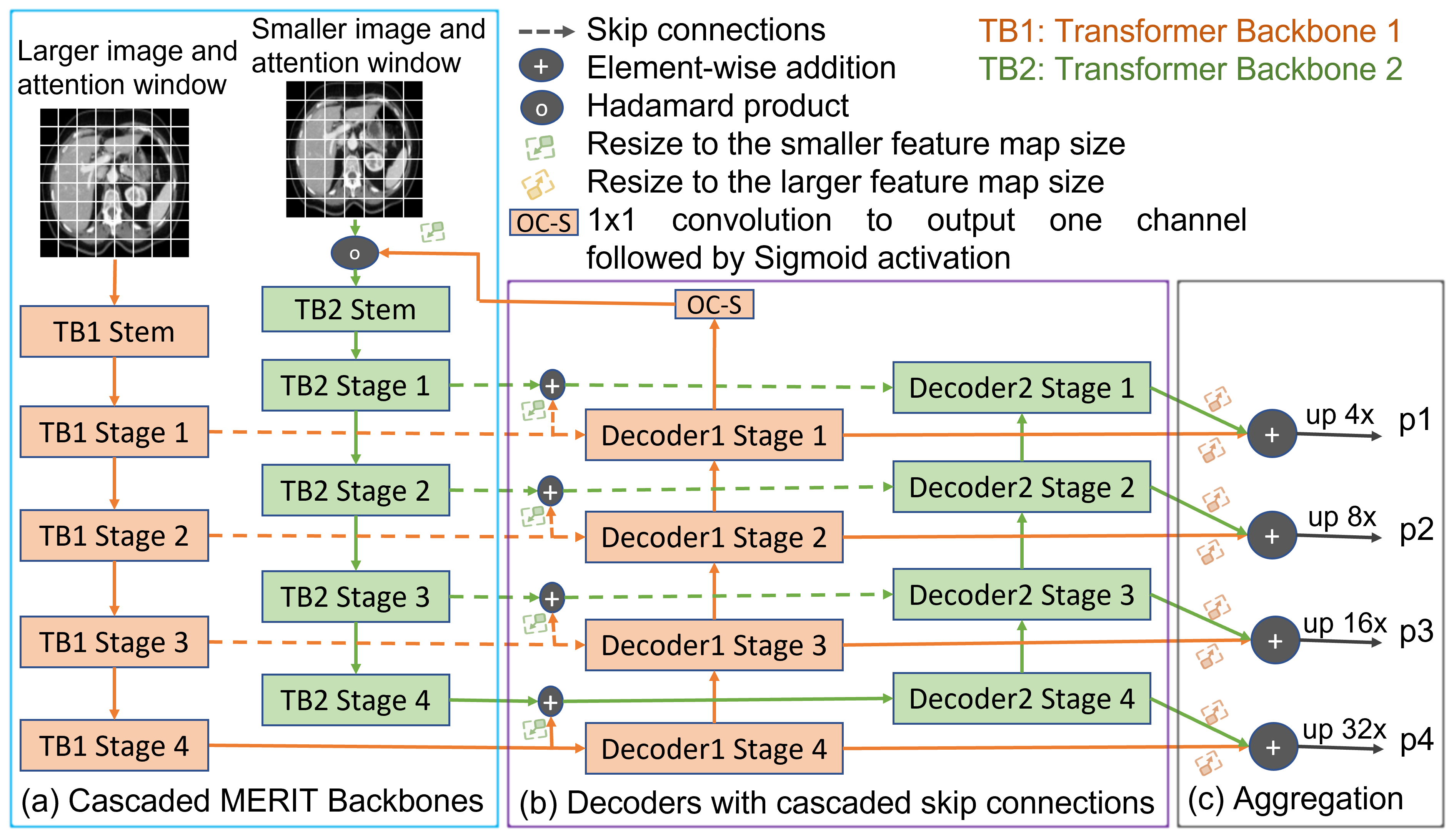
3.1.1 Cascaded MERIT
In the cascaded design of our MERIT, we add (i.e., cascade) feedback from a backbone to the next backbone. We extract the hierarchical features from four different stages of the backbone network. Then, we cascade these features with the features from the previous backbone and pass them to the skip connections and bottleneck modules of respective decoders, except the first decoder. We also pass feedback from the decoder of one backbone (except the last) to the next backbone. This design captures the multi-scale, as well as multi-resolution features due to using multiple attention windows and hierarchical features. It also refines the features well due to adding feedback from the decoder of a backbone to the next backbone and using cascaded skip connections. Fig. LABEL:fig:cascaded_architecture(a) presents the Cascaded MERIT architecture with two backbone networks. For each backbone network, the images with resolution (H, W) are first put into a Stem layer (TB1 Stem, TB2 Stem in Fig. LABEL:fig:cascaded_architecture(a)) which reduces the feature resolution to (H/4, W/4). Afterward, these features are passed through four stages of transformer backbones (this reduces feature resolution by 2 times at each stage except the fourth). The features from the last stage of the first decoder are combined with the input image to cascade it (feature) with the second backbone in Fig. LABEL:fig:cascaded_architecture(a). To do this, we reduce the number of channels to one and produce logits by applying a 1x1 convolution followed by Sigmoid activation. We also resize the feature map to the input resolution (i.e., in our implementation) of Backbone 2.
3.1.2 Parallel MERIT
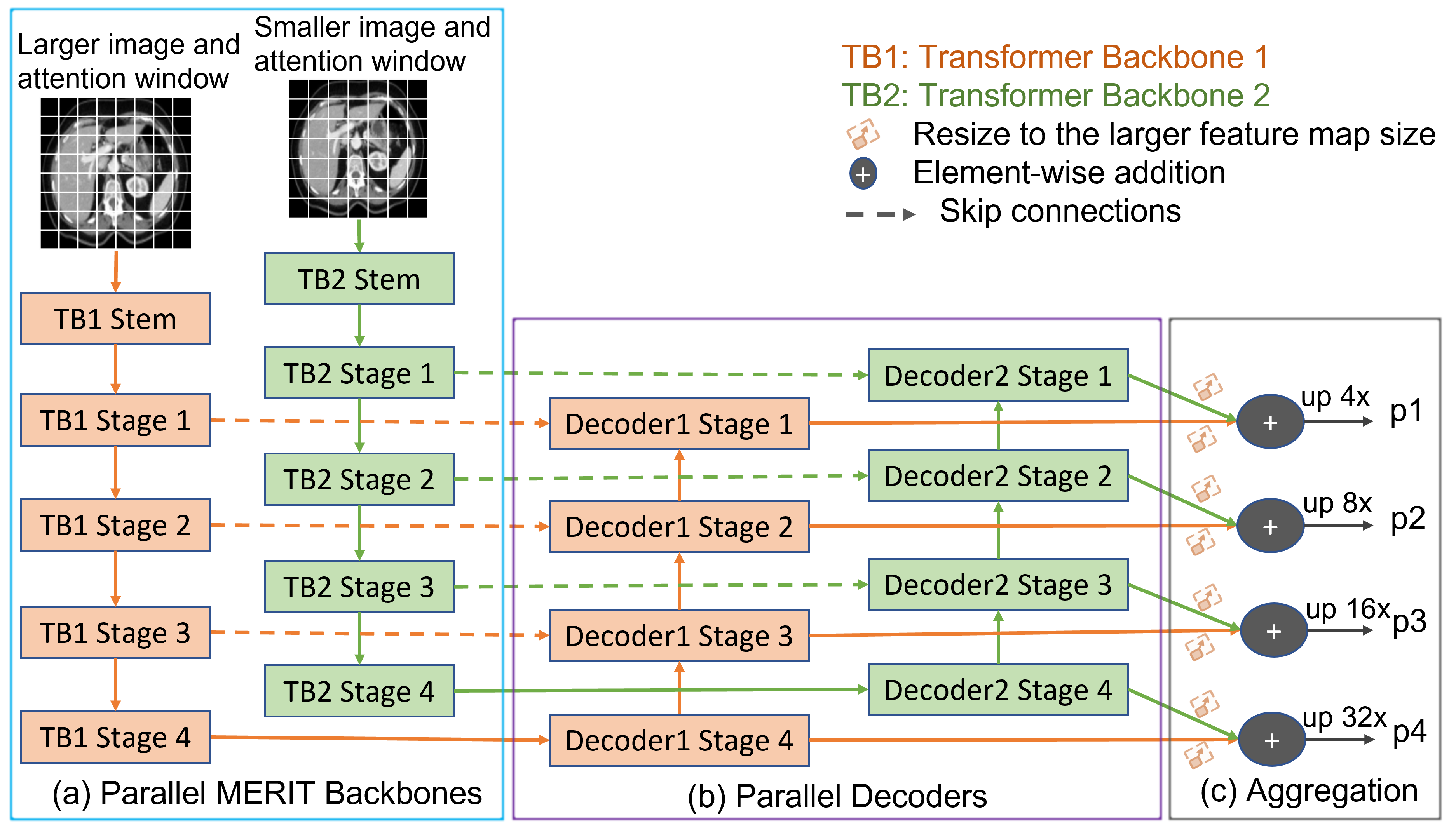
Unlike Cascaded MERIT, in the parallel design of our MERIT backbone, we pass input images of multiple resolutions/scales in parallel into separate hierarchical transformer backbone encoders with different attention windows. Similar to Cascaded MERIT, we extract the hierarchical features from four different stages of the backbone networks and pass those features to the respective parallel decoders. This design also captures multi-scale features due to using hierarchical backbones with multiple attention windows. Fig. LABEL:fig:parallel_architecture(a) in Appendix A presents a design for the Parallel MERIT with two backbone networks. The input images are passed through similar steps in the backbone networks as in Cascaded MERIT. However, the Parallel MERIT shares information among the backbone networks only at the very end during the feature aggregation step (Fig. LABEL:fig:parallel_architecture(c) in Appendix A).
3.2 Decoder
We propose using a separate decoder for each transformer backbone. As shown in Fig. LABEL:fig:cascaded_architecture(b), we use cascaded skip connections in the decoder of our cascaded MERIT architecture. Here, we add the skip connections from the first backbone to the skip connections of the second backbone network. In this case, we share information across backbones in three phases, i.e., during backbone cascading, skip connections cascading, and aggregating prediction maps. This sharing of information helps to capture richer information than the single-resolution backbone, as well as our Parallel MERIT.
Unlike Fig. LABEL:fig:cascaded_architecture(b), in Fig. LABEL:fig:parallel_architecture(b) in Appendix A, we have two parallel decoders for our parallel backbones. Each decoder has four stages that correspond to four stages of the transformer backbone. We only aggregate the multi-stage prediction maps produced by the decoders in Fig. LABEL:fig:parallel_architecture(b) at the aggregation step shown in Fig. LABEL:fig:parallel_architecture(c).
3.3 Overall Architecture
In our experiments, we use one of the most recent SOTA transformers, MaxViT [Tu et al.(2022)Tu, Talebi, Zhang, Yang, Milanfar, Bovik, and Li]. We use two instances of MaxViT-S (standard) backbone with and attention windows to create our MERIT backbone. Each MaxViT backbone has two Stem blocks followed by four stages that consist of multiple (i.e., 2, 2, 5, 2) MaxViT blocks. Each MaxViT block is built with a Mobile Convolution Block (MBConv), a Block Attention having Block Self-Attention (SA) followed by a Feed Forward Network (FFN), a Grid Attention having a Grid SA followed by an FFN. We note that although we use the MaxViT backbone in our experiments, other transformer backbones can easily be used with our MERIT.
Pure transformers have limited (spatial) contextual information processing ability among pixels. As a result, the transformer-based models face difficulties in locating discriminative local features. To address this issue, we adopt a recent attention-based cascaded decoder, CASCADE [Rahman et al.(2023)Rahman, Marculescu, and et al.], for multi-stage feature refinement and aggregation. CASCADE decoder uses the attention gate (AG) [Oktay et al.(2018)Oktay, Schlemper, Folgoc, Lee, Heinrich, Misawa, Mori, McDonagh, Hammerla, Kainz, et al.] for cascaded feature aggregation and the convolutional attention module (CAM) for robust feature map enhancement. CASCADE decoder has four CAM blocks for the four stages of hierarchical features from the transformer backbone and three AGs for three skip connections. CASCADE decoder aggregates the multi-resolution features by combining the upsampled features from the previous stage of the decoder with the features from the skip connections using AG. Then, CASCADE decoder processes the aggregated features using the CAM module (consists of channel attention [Hu et al.(2018)Hu, Shen, and Sun] followed by spatial attention [Chen et al.(2017)Chen, Zhang, Xiao, Nie, Shao, Liu, and Chua]) which groups pixels together and suppresses background information. Lastly, CASCADE decoder sends the output from the CAM block of each stage to a prediction head to produce prediction maps.
We produce four prediction maps from the four stages of the CASCADE decoder. As shown in Fig. LABEL:fig:cascaded_architecture(c) and Fig. LABEL:fig:parallel_architecture(c) in Appendix A, we aggregate (additive) the prediction maps for each stage of our two decoders. We generate the final prediction map, , using Equation 1:
| (1) |
where , , , and represent the prediction maps, and , , , and are the weights of each prediction heads. We use the value of 1.0 for , , , and . Finally, we apply Softmax activation on to get the multi-class segmentation output.
3.4 Multi-stage feature-mixing loss aggregation (MUTATION)
We now introduce a simple, yet effective multi-stage feature mixing loss aggregation strategy for image segmentation, which enables better model training. Our intention is to create new prediction maps combining the available prediction maps. So, we take all the prediction maps from different stages of a network as input and aggregate the losses of prediction maps generated using non-empty subsets of prediction maps. For example, if a network produces 4 prediction maps, our multi-stage feature-mixing loss aggregation produces a total of prediction maps including 4 original maps. This mixing strategy is simple, it does not require additional parameters to calculate, and it does not introduce inference overheads. Due to its potential benefits, this strategy can be used with any multi-stage image segmentation or dense prediction networks. Algorithm 3.4 presents the steps to produce new prediction maps and loss aggregation.
Multi-stage Feature-Mixing Loss Aggregation \LinesNumbered\KwIn the ground truth mask A list ; , where each element is a prediction map \KwOut; the aggregated loss find all non-empty subsets of prediction map indices, ; \tcp is the set of non-empty subsets of \ForEach
; \tcp is a new prediction map \ForEach ; \tcp is any loss function (e.g., CrossEntropy, DICE)
tab:multi_organ_results
Architectures Average Aorta GBb KLb KRb Liver PCb SPb SMb DICE HD95a UNet [Ronneberger et al.(2015)Ronneberger, Fischer, and Brox] 70.11 44.69 84.00 56.70 72.41 62.64 86.98 48.73 81.48 67.96 AttnUNet [Oktay et al.(2018)Oktay, Schlemper, Folgoc, Lee, Heinrich, Misawa, Mori, McDonagh, Hammerla, Kainz, et al.] 71.70 34.47 82.61 61.94 76.07 70.42 87.54 46.70 80.67 67.66 R50+UNet [Chen et al.(2021)Chen, Lu, Yu, Luo, Adeli, Wang, Lu, Yuille, and Zhou] 74.68 36.87 84.18 62.84 79.19 71.29 93.35 48.23 84.41 73.92 R50+AttnUNet [Chen et al.(2021)Chen, Lu, Yu, Luo, Adeli, Wang, Lu, Yuille, and Zhou] 75.57 36.97 55.92 63.91 79.20 72.71 93.56 49.37 87.19 74.95 SSFormerPVT [Wang et al.(2022b)Wang, Huang, Tang, Meng, Su, and Song] 78.01 25.72 82.78 63.74 80.72 78.11 93.53 61.53 87.07 76.61 PolypPVT [Dong et al.(2021)Dong, Wang, Fan, Li, Fu, and Shao] 78.08 25.61 82.34 66.14 81.21 73.78 94.37 59.34 88.05 79.4 TransUNet [Chen et al.(2021)Chen, Lu, Yu, Luo, Adeli, Wang, Lu, Yuille, and Zhou] 77.48 31.69 87.23 63.13 81.87 77.02 94.08 55.86 85.08 75.62 SwinUNet [Cao et al.(2021)Cao, Wang, Chen, Jiang, Zhang, Tian, and Wang] 79.13 21.55 85.47 66.53 83.28 79.61 94.29 56.58 90.66 76.60 MT-UNet [Wang et al.(2022a)Wang, Xie, Lin, Iwamoto, Han, Chen, and Tong] 78.59 26.59 87.92 64.99 81.47 77.29 93.06 59.46 87.75 76.81 MISSFormer [Huang et al.(2021)Huang, Deng, Li, and Yuan] 81.96 18.20 86.99 68.65 85.21 82.00 94.41 65.67 91.92 80.81 CASTformer [You et al.(2022)You, Zhao, Liu, Dong, Chinchali, Staib, s Duncan, et al.] 82.55 22.73 89.05 67.48 86.05 82.17 95.61 67.49 91.00 81.55 PVT-CASCADE [Rahman et al.(2023)Rahman, Marculescu, and et al.] 81.06 20.23 83.01 70.59 82.23 80.37 94.08 64.43 90.1 83.69 TransCASCADE [Rahman et al.(2023)Rahman, Marculescu, and et al.] 82.68 17.34 86.63 68.48 87.66 84.56 94.43 65.33 90.79 83.52 Parallel MERIT (Ours) 84.22 16.51 88.38 73.48 87.21 84.31 95.06 69.97 91.21 84.15 Cascaded MERIT (Ours) 84.90 13.22 87.71 74.40 87.79 84.85 95.26 71.81 92.01 85.38
4 Experiments
In this section, we demonstrate the superiority of our proposed MERIT architectures by comparing the results with SOTA methods. We introduce datasets, evaluation metrics, and implementation details in Appendix B. More experiments and ablation studies to answer questions related to our architectures are given in Appendix C.1-C.7.
4.1 Results on Synapse multi-organ segmentation
Table LABEL:tab:multi_organ_results presents the results of Synapse multi-organ segmentation; it can be seen that both variants of our MERIT significantly outperform all the SOTA CNN- and transformer-based 2D medical image segmentation methods. Among all the methods, our Cascaded MERIT achieves the best average DICE score (84.90%). Cascaded MERIT outperforms two popular methods on this dataset, such as TransUNet and SwinUNet by 7.42% and 5.57%, respectively, when compared to their original reported DICE scores. Cascaded MERIT achieves 2.22% better DICE than the existing best method, TransCASCADE (82.68% DICE), on this dataset. When we compare the HD95 distance of all the methods, we find that both variants of our MERIT achieve a lower HD95 distance. Cascaded MERIT has the lowest HD95 distance (13.22) which is 18.47 lower than TransUNet (HD95 of 31.69) and 4.12 lower than the best SOTA method, TransCASCADE (HD95 of 17.34).
If we look into the DICE score of individual organs, we observe that proposed MERIT variants significantly outperform SOTA methods on six out of eight organs. We also can conclude that Cascaded MERIT performs better both in large and small organs, though it exhibits greater improvement for small organs. We believe that both MERIT variants demonstrate better performance due to using the multi-scale hierarchical transformer encoder with cascaded attention-based decoding and MUTATION loss aggregation.
tab:acdc_results
Architectures
Avg DICE
RVa
Myoa
LVa
R50+UNet [Chen et al.(2021)Chen, Lu, Yu, Luo, Adeli, Wang, Lu, Yuille, and
Zhou]
87.55
87.10
80.63
94.92
R50+AttnUNet [Chen et al.(2021)Chen, Lu, Yu, Luo, Adeli, Wang, Lu, Yuille, and
Zhou]
86.75
87.58
79.20
93.47
ViT+CUP [Chen et al.(2021)Chen, Lu, Yu, Luo, Adeli, Wang, Lu, Yuille, and
Zhou]
81.45
81.46
70.71
92.18
R50+ViT+CUP [Chen et al.(2021)Chen, Lu, Yu, Luo, Adeli, Wang, Lu, Yuille, and
Zhou]
87.57
86.07
81.88
94.75
TransUNet [Chen et al.(2021)Chen, Lu, Yu, Luo, Adeli, Wang, Lu, Yuille, and
Zhou]
89.71
88.86
84.53
95.73
SwinUNet [Cao et al.(2021)Cao, Wang, Chen, Jiang, Zhang, Tian, and
Wang]
90.00
88.55
85.62
95.83
MT-UNet [Wang et al.(2022a)Wang, Xie, Lin, Iwamoto, Han, Chen, and
Tong]
90.43
86.64
89.04
95.62
MISSFormer [Huang et al.(2021)Huang, Deng, Li, and Yuan]
90.86
89.55
88.04
94.99
PVT-CASCADE [Rahman et al.(2023)Rahman, Marculescu, and et al.]
91.46
88.9
89.97
95.50
TransCASCADE [Rahman et al.(2023)Rahman, Marculescu, and et al.]
91.63
89.14
90.25
95.50
Parallel MERIT (Ours)
92.32
90.87
90.00
96.08
Cascaded MERIT (Ours)
91.85
90.23
89.53
95.80
a More details in Appendix B.1
4.2 Results on ACDC cardiac organ segmentation
Table LABEL:tab:acdc_results reports three cardiac organ segmentation results of different methods on the ACDC dataset for MRI data modality. Both our Parallel and Cascaded MERIT have better DICE scores than all other SOTA methods. Our Parallel MERIT achieves the best average DICE score (92.32%) which outperforms TransUNet and SwinUNet by 2.61% and 2.32%, respectively. Parallel MERIT also shows the best DICE scores in RV (90.87%) and LV (96.08%) segmentation. We can conclude from these results that our method performs the best across different medical imaging data modalities.
5 Conclusion
In this paper, we have introduced a novel multi-scale hierarchical transformer architecture (MERIT) that can capture both the multi-scale and multi-resolution features necessary for medical image segmentation. We have also incorporated an attention-based cascaded decoder to further refine features. Moreover, we have proposed a novel multi-stage feature mixing loss aggregation (MUTATION) strategy for implicit ensembling/augmentation which ensures better model training and boosts the performance without introducing additional hyper-parameters and inference overhead. Our experimental results on two well-known multi-class medical image segmentation benchmarks demonstrate the superiority of our proposed method over all SOTA approaches. Finally, we believe that our proposed MERIT architectures and MUTATION loss aggregation strategy will improve other downstream medical image segmentation and semantic segmentation tasks.
References
- [Cao et al.(2021)Cao, Wang, Chen, Jiang, Zhang, Tian, and Wang] Hu Cao, Yueyue Wang, Joy Chen, Dongsheng Jiang, Xiaopeng Zhang, Qi Tian, and Manning Wang. Swin-unet: Unet-like pure transformer for medical image segmentation. arXiv preprint arXiv:2105.05537, 2021.
- [Chen et al.(2021)Chen, Lu, Yu, Luo, Adeli, Wang, Lu, Yuille, and Zhou] Jieneng Chen, Yongyi Lu, Qihang Yu, Xiangde Luo, Ehsan Adeli, Yan Wang, Le Lu, Alan L Yuille, and Yuyin Zhou. Transunet: Transformers make strong encoders for medical image segmentation. arXiv preprint arXiv:2102.04306, 2021.
- [Chen et al.(2017)Chen, Zhang, Xiao, Nie, Shao, Liu, and Chua] Long Chen, Hanwang Zhang, Jun Xiao, Liqiang Nie, Jian Shao, Wei Liu, and Tat-Seng Chua. Sca-cnn: Spatial and channel-wise attention in convolutional networks for image captioning. In Proceedings of the IEEE conference on computer vision and pattern recognition, pages 5659–5667, 2017.
- [Chen et al.(2018)Chen, Tan, Wang, and Hu] Shuhan Chen, Xiuli Tan, Ben Wang, and Xuelong Hu. Reverse attention for salient object detection. In Proceedings of the European conference on computer vision (ECCV), pages 234–250, 2018.
- [Chu et al.(2021)Chu, Tian, Zhang, Wang, Wei, Xia, and Shen] Xiangxiang Chu, Zhi Tian, Bo Zhang, Xinlong Wang, Xiaolin Wei, Huaxia Xia, and Chunhua Shen. Conditional positional encodings for vision transformers. arXiv preprint arXiv:2102.10882, 2021.
- [Dong et al.(2021)Dong, Wang, Fan, Li, Fu, and Shao] Bo Dong, Wenhai Wang, Deng-Ping Fan, Jinpeng Li, Huazhu Fu, and Ling Shao. Polyp-pvt: Polyp segmentation with pyramid vision transformers. arXiv preprint arXiv:2108.06932, 2021.
- [Dosovitskiy et al.(2020)Dosovitskiy, Beyer, Kolesnikov, Weissenborn, Zhai, Unterthiner, Dehghani, Minderer, Heigold, Gelly, et al.] Alexey Dosovitskiy, Lucas Beyer, Alexander Kolesnikov, Dirk Weissenborn, Xiaohua Zhai, Thomas Unterthiner, Mostafa Dehghani, Matthias Minderer, Georg Heigold, Sylvain Gelly, et al. An image is worth 16x16 words: Transformers for image recognition at scale. arXiv preprint arXiv:2010.11929, 2020.
- [Fan et al.(2020)Fan, Ji, Zhou, Chen, Fu, Shen, and Shao] Deng-Ping Fan, Ge-Peng Ji, Tao Zhou, Geng Chen, Huazhu Fu, Jianbing Shen, and Ling Shao. Pranet: Parallel reverse attention network for polyp segmentation. In International conference on medical image computing and computer-assisted intervention, pages 263–273. Springer, 2020.
- [Hu et al.(2018)Hu, Shen, and Sun] Jie Hu, Li Shen, and Gang Sun. Squeeze-and-excitation networks. In Proceedings of the IEEE conference on computer vision and pattern recognition, pages 7132–7141, 2018.
- [Huang et al.(2020)Huang, Lin, Tong, Hu, Zhang, Iwamoto, Han, Chen, and Wu] Huimin Huang, Lanfen Lin, Ruofeng Tong, Hongjie Hu, Qiaowei Zhang, Yutaro Iwamoto, Xianhua Han, Yen-Wei Chen, and Jian Wu. Unet 3+: A full-scale connected unet for medical image segmentation. In ICASSP 2020-2020 IEEE International Conference on Acoustics, Speech and Signal Processing (ICASSP), pages 1055–1059. IEEE, 2020.
- [Huang et al.(2021)Huang, Deng, Li, and Yuan] Xiaohong Huang, Zhifang Deng, Dandan Li, and Xueguang Yuan. Missformer: An effective medical image segmentation transformer. arXiv preprint arXiv:2109.07162, 2021.
- [Lin et al.(2022)Lin, Chen, Xu, Zhang, Lu, and Zhang] Ailiang Lin, Bingzhi Chen, Jiayu Xu, Zheng Zhang, Guangming Lu, and David Zhang. Ds-transunet: Dual swin transformer u-net for medical image segmentation. IEEE Transactions on Instrumentation and Measurement, 2022.
- [Liu et al.(2021)Liu, Lin, Cao, Hu, Wei, Zhang, Lin, and Guo] Ze Liu, Yutong Lin, Yue Cao, Han Hu, Yixuan Wei, Zheng Zhang, Stephen Lin, and Baining Guo. Swin transformer: Hierarchical vision transformer using shifted windows. In Proceedings of the IEEE/CVF International Conference on Computer Vision, pages 10012–10022, 2021.
- [Loshchilov and Hutter(2017)] Ilya Loshchilov and Frank Hutter. Decoupled weight decay regularization. arXiv preprint arXiv:1711.05101, 2017.
- [Lou et al.(2021)Lou, Guan, and Loew] Ange Lou, Shuyue Guan, and Murray Loew. Dc-unet: rethinking the u-net architecture with dual channel efficient cnn for medical image segmentation. In Medical Imaging 2021: Image Processing, volume 11596, pages 758–768. SPIE, 2021.
- [Oktay et al.(2018)Oktay, Schlemper, Folgoc, Lee, Heinrich, Misawa, Mori, McDonagh, Hammerla, Kainz, et al.] Ozan Oktay, Jo Schlemper, Loic Le Folgoc, Matthew Lee, Mattias Heinrich, Kazunari Misawa, Kensaku Mori, Steven McDonagh, Nils Y Hammerla, Bernhard Kainz, et al. Attention u-net: Learning where to look for the pancreas. arXiv preprint arXiv:1804.03999, 2018.
- [Rahman et al.(2023)Rahman, Marculescu, and et al.] Md Mostafijur Rahman, Radu Marculescu, and et al. Medical image segmentation via cascaded attention decoding. In Proceedings of the IEEE/CVF Winter Conference on Applications of Computer Vision, pages 6222–6231, 2023.
- [Ronneberger et al.(2015)Ronneberger, Fischer, and Brox] Olaf Ronneberger, Philipp Fischer, and Thomas Brox. U-net: Convolutional networks for biomedical image segmentation. In International Conference on Medical image computing and computer-assisted intervention, pages 234–241. Springer, 2015.
- [Touvron et al.(2021)Touvron, Cord, Douze, Massa, Sablayrolles, and Jégou] Hugo Touvron, Matthieu Cord, Matthijs Douze, Francisco Massa, Alexandre Sablayrolles, and Hervé Jégou. Training data-efficient image transformers & distillation through attention. In International Conference on Machine Learning, pages 10347–10357. PMLR, 2021.
- [Tu et al.(2022)Tu, Talebi, Zhang, Yang, Milanfar, Bovik, and Li] Zhengzhong Tu, Hossein Talebi, Han Zhang, Feng Yang, Peyman Milanfar, Alan Bovik, and Yinxiao Li. Maxvit: Multi-axis vision transformer. ECCV, 2022.
- [Wang et al.(2022a)Wang, Xie, Lin, Iwamoto, Han, Chen, and Tong] Hongyi Wang, Shiao Xie, Lanfen Lin, Yutaro Iwamoto, Xian-Hua Han, Yen-Wei Chen, and Ruofeng Tong. Mixed transformer u-net for medical image segmentation. In ICASSP 2022-2022 IEEE International Conference on Acoustics, Speech and Signal Processing (ICASSP), pages 2390–2394. IEEE, 2022a.
- [Wang et al.(2022b)Wang, Huang, Tang, Meng, Su, and Song] Jinfeng Wang, Qiming Huang, Feilong Tang, Jia Meng, Jionglong Su, and Sifan Song. Stepwise feature fusion: Local guides global. arXiv preprint arXiv:2203.03635, 2022b.
- [Wang et al.(2021)Wang, Xie, Li, Fan, Song, Liang, Lu, Luo, and Shao] Wenhai Wang, Enze Xie, Xiang Li, Deng-Ping Fan, Kaitao Song, Ding Liang, Tong Lu, Ping Luo, and Ling Shao. Pyramid vision transformer: A versatile backbone for dense prediction without convolutions. In Proceedings of the IEEE/CVF International Conference on Computer Vision, pages 568–578, 2021.
- [Wang et al.(2022c)Wang, Xie, Li, Fan, Song, Liang, Lu, Luo, and Shao] Wenhai Wang, Enze Xie, Xiang Li, Deng-Ping Fan, Kaitao Song, Ding Liang, Tong Lu, Ping Luo, and Ling Shao. Pvt v2: Improved baselines with pyramid vision transformer. Computational Visual Media, 8(3):415–424, 2022c.
- [Wightman(2019)] Ross Wightman. Pytorch image models. https://github.com/rwightman/pytorch-image-models, 2019.
- [Woo et al.(2018)Woo, Park, Lee, and Kweon] Sanghyun Woo, Jongchan Park, Joon-Young Lee, and In So Kweon. Cbam: Convolutional block attention module. In Proceedings of the European conference on computer vision (ECCV), pages 3–19, 2018.
- [Xie et al.(2021)Xie, Wang, Yu, Anandkumar, Alvarez, and Luo] Enze Xie, Wenhai Wang, Zhiding Yu, Anima Anandkumar, Jose M Alvarez, and Ping Luo. Segformer: Simple and efficient design for semantic segmentation with transformers. Advances in Neural Information Processing Systems, 34:12077–12090, 2021.
- [You et al.(2022)You, Zhao, Liu, Dong, Chinchali, Staib, s Duncan, et al.] Chenyu You, Ruihan Zhao, Fenglin Liu, Siyuan Dong, Sandeep P Chinchali, Lawrence Hamilton Staib, James s Duncan, et al. Class-aware adversarial transformers for medical image segmentation. In Advances in Neural Information Processing Systems, 2022.
- [Zhou et al.(2018)Zhou, Rahman Siddiquee, Tajbakhsh, and Liang] Zongwei Zhou, Md Mahfuzur Rahman Siddiquee, Nima Tajbakhsh, and Jianming Liang. Unet++: A nested u-net architecture for medical image segmentation. In Deep learning in medical image analysis and multimodal learning for clinical decision support, pages 3–11. Springer, 2018.
Appendix A Parallel MERIT Architecture
Due to the page limitation, our parallel MERIT architecture is given in Fig. LABEL:fig:parallel_architecture. This architecture is described in Section 3.1.2 of the main text.
fig:parallel_architecture
Appendix B Experimental Setup
This section first describes datasets, then introduces evaluation metrics, and finally provides the implementation details of our proposed architecture and experiments.
B.1 Datasets
Synapse multi-organ dataset. There are 30 abdominal CT scans with 3779 axial contrast-enhanced abdominal CT images in the Synapse multi-organ dataset111\hrefhttps://www.synapse.org/#!Synapse:syn3193805/wiki/217789https://www.synapse.org/#!Synapse:syn3193805/wiki/217789 . Each CT scan has 85-198 slices of resolution pixels, having a voxel spatial resolution of ([0:54-0:54] [0:98-0:98][2:5-5:0]). We extract 2D slices from the CT scans and segment 8 abdominal organs, such as the aorta, gallbladder (GB), left kidney (KL), right kidney (KR), liver, pancreas (PC), spleen (SP), and stomach (SM). Following the experimental protocol of TransUNet [Chen et al.(2021)Chen, Lu, Yu, Luo, Adeli, Wang, Lu, Yuille, and Zhou], we split the dataset into 18 scans (2211 axial slices) for training, and 12 for validation.
ACDC dataset. The ACDC dataset222\hrefhttps://www.creatis.insa-lyon.fr/Challenge/acdc/https://www.creatis.insa-lyon.fr/Challenge/acdc/ contains 100 cardiac MRI scans collected from different patients. We extract 2D slices from each MRI scan and segment three organs, such as the right ventricle (RV), left ventricle (LV), and myocardium (Myo). Following MT-UNet [Wang et al.(2022a)Wang, Xie, Lin, Iwamoto, Han, Chen, and Tong], we split the dataset into 70 (1304 axial slices), 10 (182 axial slices), and 20 cases for training, validation, and testing, respectively.
B.2 Evaluation metrics.
In our experiments on the Synapse Multi-organ dataset, we use DICE and 95% Hausdorff Distance (95%HD) as the evaluation metrics. However, we use only DICE scores as an evaluation metric for the ACDC dataset. The DICE similarity scores and 95%HD distance (95th percentile of the distances between boundary points in and ) are calculated using Equations 2 and 3, respectively.
| (2) |
| (3) |
where and are the ground truth mask and predicted segmentation map, respectively.
B.3 Implementation details
We use PyTorch 1.12.0 with CUDA 11.6 in all of our experiments. Besides, we use a single NVIDIA RTX A6000 GPU with 48GB of memory to train all the models. We utilize the Pytorch pre-trained weights on ImageNet from timm library [Wightman(2019)] for MaxViT backbone networks. We use the input resolutions and attention windows of and , respectively, in our (dual-scale) MERIT. We augment data using only random rotation and flipping. We train our model using AdamW [Loshchilov and Hutter(2017)] optimizer with a weight decay and learning rate of 0.0001. We optimize the combined DICE and Cross-Entropy (CE) loss in Equation 4 with and (weights are selected empirically in Appendix C.7) in all our experiments:
| (4) |
where and are the weight for the DICE () and CE () losses, respectively.
We train each model a maximum of 300 epochs with a batch size of 24 for Synapse multi-organ segmentation. For ACDC cardiac organ segmentation, we use a batch size of 12 and train each model for a maximum of 400 epochs.
tab:compare_baseline_results
Architectures Input Resolutions Params (M)/ FLOPS (G) Inference Time (ms) Synapse Multi-organ ACDC MaxViT 65.25/10.43 19.79 77.11 90.56 MaxViT 65.25/14.19 20.58 78.53 90.98 MaxViT with CASCADE decoder 82.62/14.2 21.84 79.83 90.87 MaxViT with CASCADE decoder 82.62/19.11 23.07 80.20 91.15 Parallel MERIT (our) , 147.86/33.31 37.01 84.22 92.32 Cascaded MERIT (our) , 147.86/33.31 37.06 84.90 91.85
Appendix C Ablation Studies
In this section, we present a wide range of ablation studies to answer different intrinsic questions related to our proposed architectures, loss aggregation, and experiments; these are described in the following subsections.
C.1 Comparison with the baseline method
We compare our proposed methods with baseline hierarchical MaxViT architecture. In the case of MaxViT, we do the same multi-stage prediction for a fair comparison. We also use a similar experimental setting except using MUTATION with our architectures. Table LABEL:tab:compare_baseline_results presents the results of these experiments. We can see from Table LABEL:tab:compare_baseline_results that our proposed architectures with MUTATION loss (see ”our” entries in Table LABEL:tab:compare_baseline_results) improve the baseline hierarchical resolution MaxViT (see row entries in Table LABEL:tab:compare_baseline_results) by 6.37% and 1.34% DICE scores (with more FLOPS and longer inference time) in Synapse multi-organ and ACDC datasets, respectively. We can also see that our MERIT architecture has 147.86M parameters which is larger than resolution MaxViT with CASCADE decoder (see row entries in Table LABEL:tab:compare_baseline_results), but with a 4.7% better DICE score in Synapse multi-organ. We think that this increase of parameters/FLOPS/inference time worth it given the improvement in performance.
tab:multi_scale_results
Architectures Input Resolutions Attention Windows Params (M)/ FLOPS (G) Avg DICE (%) (single) MaxViT 82.62/14.2 79.83 (single) MaxViT 82.62/19.11 80.20 Parallel Double MaxViT , , 147.86/28.4 80.81 Parallel Double MaxViT , , 147.86/38.22 82.15 Cascaded Double MaxViT , , 147.86/28.4 81.06 Cascaded Double MaxViT , , 147.86/38.22 83.02 Parallel MERIT (our) , , 147.86/33.31 82.91 Cascaded MERIT (our) , , 147.86/33.31 83.35
tab:tiny_vs_small_results
Architectures Input Resolution Attention Windows Params (M)/ FLOPS (G) Avg DICE (%) MaxViT-Tiny 224224 77 36.86/6.57 77.84 MaxViT-Tiny 256256 88 36.86/8.61 78.43 Parallel MERIT-Tiny (ours) 256256, 224224 88, 77 65.41/15.18 81.34 Cascaded MERIT-Tiny (ours) 256256, 224224 88, 77 65.41/15.18 81.82 MaxViT-Small 224224 77 82.62/14.2 79.83 MaxViT-Small 256256 88 82.62/19.11 80.20
C.2 Effect of multi-scale backbone
We have conducted experiments on the Synapse multi-organ dataset to show the effect of our multi-scale backbone on medical image segmentation. In Table LABEL:tab:multi_scale_results, we present the results of all the methods with the CASCADE decoder (no MUTATION) to make a fair comparison of our proposed architecture. It can be seen from Table LABEL:tab:multi_scale_results that the input resolution has an impact on DICE score improvement. More precisely, resolution backbones have better DICE scores than the resolution backbones. As shown, our Cascaded MERIT achieves the best DICE score (83.35%) which improves the baseline resolution MaxViT (see row entries in Table LABEL:tab:multi_scale_results) by 3.15%. When comparing with the double backbone architectures with the same input scale (attention window), we can see that our multi-scale (attention window) double backbone architectures achieve better DICE scores due to their additive advantage of multi-scale feature extraction. We note that our Parallel/Cascaded MERIT (33.31G) has a significantly lower computational complexity/FLOPS than the resolution Parallel/Cascaded Double MaxViT (38.22G) due to using one and another resolution inputs. Despite that, our Parallel and Cascaded MERIT outperform the Double MaxViT by 0.76% and 0.33%, respectively. These improvements in the DICE score support the claim regarding the benefit of calculating SA in multiple scale attention windows.
We have conducted an additional set of experiments by implementing a tiny version of MERIT using the tiny MaxViT backbones, to clarify that performance improvement is due to the effect of multi-scale SA, not because of using a model with more parameters. As shown in Table LABEL:tab:tiny_vs_small_results, when comparing against the Small MaxViT backbone which has more model parameters, both of our Tiny MERIT backbones perform better. Our Tiny Cascaded MERIT backbone outperforms the Small MaxViT backbone (see row entries in Table LABEL:tab:tiny_vs_small_results) by up to 1.62% DICE score for a 256256 input resolution, while having smaller model parameters and fewer FLOPS. Therefore, again, we can conclude from the empirical evaluation that our multi-scale SA calculation improves the performance of medical image segmentation.
tab:cascade_mutation_effects_results Architectures CASCADE decoder MUTATION Avg DICE (%) Parallel MERIT No No 80.44 Parallel MERIT No Yes 81.06 Parallel MERIT Yes No 82.91 Parallel MERIT (our) Yes Yes 84.22 Cascaded MERIT No No 80.76 Cascaded MERIT No Yes 82.03 Cascaded MERIT Yes No 83.35 Cascaded MERIT (our) Yes Yes 84.90
fig:qualitative_results
C.3 Effect of CASCADE decoder and MUTATION loss aggregation in MERIT
We have conducted some experiments on Synapse multi-organ dataset to demonstrate the effect of CASCADE decoder and MUTATION loss aggregation strategy on our MERIT architectures. Table LABEL:tab:cascade_mutation_effects_results presents the results of our Parallel MERIT with or without CASCADE decoder and MUTATION. We can see from Table LABEL:tab:cascade_mutation_effects_results that Parallel and Cascaded MERIT without both CASCADE decoder and MUTATION have the lowest DICE scores. CASCADE decoder significantly increases the DICE scores (2.47-2.59%) due to capturing the spatial (contextual) relations among pixels (usually limited in vision transformer), while MUTATION alone marginally improves the DICE (0.62-1.27%). However, when MUTATION is used with the outputs from CASCADE decoder, it achieves the best DICE scores (84.22%, 84.90%) improving CASCADE decoder by 1.31-1.55%. We believe the reason behind this is that MUTATION works well with the refined features of the CASCADE decoder. Therefore, we can conclude that the synthesized prediction maps generated via combinatory aggregation (MUTATION) help us improve the performance of the model; this is why we prefer combinatory loss aggregation over linear aggregation. We believe that our combinatory loss aggregation (MUTATION) can be used as a beneficial ensembling/augmentation method in other downstream semantic and medical image segmentation tasks.
tab:cascade_aggregation_results Architectures Aggregation in CASCADE decoder Avg DICE (%) Parallel MERIT Concatenation 84.18 Parallel MERIT Concatenation 84.22 Cascaded MERIT Additive 84.88 Cascaded MERIT Additive 84.90
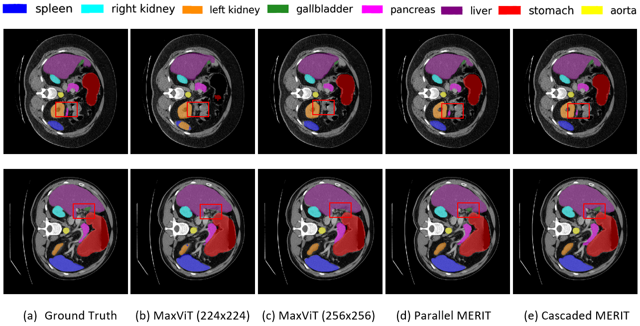
C.4 Qualitative results on Synapse Multi-organ Segmentation
Fig. LABEL:fig:qualitative_results shows the qualitative results of the baseline hierarchical MaxViT and our proposed MERIT architectures. As shown in the figure, our MERIT architecture can segment the small organs (see the red rectangular box) well. In contrast, the single scale MaxViT architecture with both and input resolutions fail to segment that small organ. Our MERIT architecture also segments the larger organ much better than the single scale MaxViT. We believe the reason behind this better segmentation of both small and large organs by our MERIT architectures is the use of multi-scale SA.
C.5 Effect of different aggregations in CASCADE decoder
Table LABEL:tab:cascade_aggregation_results presents the results of concatenation and additive aggregations in CASCADE decoder on Synapse multi-organ dataset. We can see from Table LABEL:tab:cascade_aggregation_results that our MERIT architectures with additive aggregation in CASCADE decoder are marginally better (0.04-0.02%) than the concatenation. Therefore, we can conclude from these results that the aggregation techniques do not have much impact on the CASCADE decoder of our architectures while using MUTATION. However, concatenation aggregation-based methods usually have additional computational overheads due to increasing the number of channels after the aggregation, while additive aggregation keeps the number of channels the same. Consequently, we recommend using additive aggregation in our MERIT architectures due to its computational benefits.
tab:interpolations_results Architectures Interpolations Avg DICE (%) Parallel MERIT nearest-exact 81.67 Parallel MERIT area 81.76 Parallel MERIT bicubic 83.58 Parallel MERIT bilinear 84.22 Cascaded MERIT nearest-exact 82.27 Cascaded MERIT area 82.38 Cascaded MERIT bicubic 84.05 Cascaded MERIT bilinear 84.90
fig:loss_curve

C.6 Effect of different interpolations in MERIT
We have conducted some experiments on Synapse multi-organ dataset to choose the best interpolations methods for our proposed MERIT architectures. Table LABEL:tab:interpolations_results presents the results of Parallel and Cascaded MERIT using nearest-exact (nearest neighbor), area, bicubic, and bilinear interpolation methods from Pytorch. The nearest-exact interpolation shows the lowest DICE scores while bilinear and bicubic interpolation achieve the best and second best DICE scores, respectively. Therefore, we recommend using bilinear interpolation in our proposed MERIT architectures to re-scale the features and prediction maps.
C.7 Choosing weight for DICE and CE losses
We optimize the combined DICE and CE loss during the training of our models. Here, we have conducted some experiments to choose the best weight pairs to combine these two losses. Fig. LABEL:fig:loss_curve presents the DICE scores for different weight pairs for losses. We can see in the graph that the model shows the worst DICE score when using only the CE loss. We get the best DICE score for the weights pair (, ) = (0.7, 0.3) which we have used in all of our experiments.
Appendix D Supplementary Materials
We make our source code publicly available at .