Fluctuation-induced dispersion forces on thin DNA films
Abstract
In this work, the calculation of Casimir forces across thin DNA films is carried out based on the Lifshitz theory. The variations of Casimir forces due to the DNA thicknesses, volume fractions of containing water, covering media and substrates are investigated. For a DNA film suspended in the air or water, the Casimir force is attractive, and its magnitude increases with decreasing the thickness of DNA films and the water volume fraction. For DNA films deposited on a dielectric(silica) substrate, the Casimir force is attractive for the air environment. However, the Casimir force shows unusual features in a water environment. Under specific conditions, switching signs of the Casimir force from attractive to repulsive can be achieved by increasing the DNA-film thickness. Finally, the Casimir force for DNA films deposited on a metallic substrate are investigated. The Casimir force is dominant by the repulsive interactions at a small DNA-film thickness for both the air and water environment. In a water environment, the Casimir force turns out to be attractive at a large DNA-film thickness, and a stable Casimir equilibrium can be found. In addition to the adhesion stability, our finding could be applicable to the problems of condensation and de-condensation of DNA, due to the fluctuation-induced dispersion forces.
I Introduction
The dispersion force is generated by the fluctuating dipoles, resulting from the zero-point vacuum fluctuation and thermal fluctuation Feinberg et al. (1989). When the consuming time for propagating waves between the fluctuation dipoles is larger or comparable with the lifetime of fluctuating dipoles, the retardation effect (or wave effect) can modified the separation-dependence decaying laws of dispersion force Rodriguez et al. (2011). Specifically, the dispersion force is known as the van der Waals force for closely spaced objects or interfaces Parsegian (2005), where the retardation is negligible. The retardation effect manifests when the separation distance is large, and the dispersion force is also named as retarded van der Waals forceDagastine et al. (2002) or the Casimir force Bordag et al. (2009); Klimchitskaya et al. (2009). In some configurations, the retardation effect can be apparent even at the separation of several nanometersLee and Sigmund (2001). The dispersion force and its free energy play an important role in various disciplines, ranging from nanomechanicsGong et al. (2021); Somers et al. (2018); Ge et al. (2020a); Munkhbat et al. (2021); Esteso et al. (2015), wetting phenomenaHough and White (1980); Boinovich and Emelyanenko (2011); Squarcini et al. (2022), to ice pre-melting and formation Li et al. (2022); Luengo-Márquez and MacDowell (2021); Esteso et al. (2020); Fiedler et al. (2020) etc. In addition, the dispersion force and its free energy across organic films were also investigated intensely Lu and Podgornik (2015); Blackwell et al. (2021); Baranov et al. (2019); Klimchitskaya et al. (2020, 2021, 2022). It was reported that the attractive dispersion force would make the organic films more stable, while the repulsive force has an opposite effectBaranov et al. (2019); Klimchitskaya et al. (2020, 2021, 2022).
Deoxyribonucleic acid (DNA) composed of two helical polynucleotide chains is one of the most important substances in biology. Along with its biological functions, the material properties of DNA are of great interest for the state-of-art of nanotechnology, motivated by the promising applications in a variety of fields, such as self-assembly of colloidal nanoparticles Nykypanchuk et al. (2008); Chou et al. (2014); Rogers et al. (2016); Cui et al. (2022), DNA-based nanomedicinesCampolongo et al. (2010); Hu et al. (2018); Weiden and Bastings (2021); Gu et al. (2021), organic nanophotonics Steckl (2007); Bui et al. (2019) etc. As one of the crucial elements, the DNA films have been widely applied in many bio-organic nanoscale devices Kawabe et al. (2000); Zhang et al. (2012); Khazaeinezhad et al. (2017); Jung et al. (2017). The DNA films are generally deposited on inorganic substrates using the spin coating process Jung et al. (2017). The structure stability of double-stranded DNA is determined by the hydrogen bonds between nucleotides and the base-stacking interactionsYakovchuk et al. (2006). However, the adhesion stability of DNA films placed on a substrate is dependent on the surface forces Boinovich and Emelyanenko (2011), such as ionic or electrostatic forces, intra-hydrogen bonds, dispersion forces etc. The dispersion force is an important ingredient at the surface forces, particularly, when the thickness of a bio-organic film is miniaturized to a sub-micro scaleBaranov et al. (2019). Moreover, the other surface forces (e.g., intra-hydrogen bonds) could be absent at the surface of some specific substrates. Then, it is expected that the contribution from the dispersion force becomes more prominent, and the quantitative calculations of this force are necessary.
In this work, we study the Casimir force of DNA films within the framework of Lifshitz theory. The influences of Casimir forces due to DNA-film thicknesses, water volume fractions, background media and substrates are investigated by numerical calculations. We find that the Casimir pressure is attractive for a DNA film suspended in the air or water, and its magnitude increases by decreasing the DNA-film thickness and water volume fraction. For a DNA film placed on a silica substrate in the air background, the Casimir pressure shows a similar trend as that in the suspended configuration. However, the Casimir pressure exhibits rich features when the setup is immersed in the water. Under specific water volume fraction, switching sign of the Casimir pressure across a wet DNA film is revealed by increasing the DNA-film thickness. Finally, the Casimir pressure for a DNA film placed on a metallic substrate is also calculated. It is found that the Casimir pressure is dominant by the repulsive interactions at a small DNA-film thickness for both the air and water environment. Interestingly, a stable Casimir equilibrium is found when the DNA film is immersed in the water. Our findings could be applicable to the problems of adhesive stability, condensation and de-condensation of DNA films, due to the fluctuation-induced dispersion forces.
II Theoretical models
We consider a DNA film with thickness sandwiched between a cladding medium and a substrate. The thicknesses of the cladding layer and substrate are assumed to be semi-infinite. In addition, the whole system is in thermal equilibrium at room temperature . The Casimir pressure of the DNA film is calculated based on the framework of the Lifshitz theory Klimchitskaya et al. (2009, 2020):
| (1) |
where the prime in summation denotes a prefactor 1/2 for the term , is the Boltzmann’s constant, is the vertical wavevector in the DNA film, is the parallel wavevector, is the speed of light in vacuum, is the permittivity of the dry DNA film, are the discrete Matsubara frequencies, is the reduced Planck constant, are the reflection coefficients for the DNA film, where the superscripts and correspond to the polarizations of transverse electric () and transverse magnetic () modes, respectively. The subscripts and denote the reflection coefficients at the top and bottom interfaces of DNA film, respectively.
The reflection coefficients for an electromagnetic wave incident from the DNA film to a medium (with permittivity ) is given as Klimchitskaya et al. (2020):
| (2) | |||||
| (3) |
where is the vertical wavevector in the medium 1. Here, the medium 1 can be the air, water, silica or gold.
The reflection coefficients are strongly dependent on the permittivity at different Matsubara frequencies. Here, the dielectric functions of used materials are fitted by a model of the modified harmonic oscillator, which is adopted from a recent literatureMoazzami Gudarzi and Aboutalebi (2021):
| (4) |
where corresponds to the oscillator strength for the -th resonance frequency , is a power exponent. In addition to the Kramers-Kronig relations, the influences of the electronic dielectric constant, optical bandgap, density, and chemical composition are taken into account in the Eq.(4), where the parameters for the dry DNA, water and silica are shown in Table 1.
| DNA: | (eV) | ||
| =1 | 1.766 | 0.0056 | 1.03 |
| =2 | 1.431 | 12.95 | 1.67 |
| Water: | |||
| =1 | 73.48 | 8.1 | 0.988 |
| =2 | 2.534 | 0.016 | 1.1 |
| =3 | 0.755 | 16.1 | 1.751 |
| Silica: | |||
| =1 | 1.843 | 0.0725 | 1.678 |
| =2 | 1.105 | 15.33 | 1.71 |
It is worth mentioning that the dielectric function of the dry DNA given by the parameters in Table 1 matches the measured data of DNA over a wide range of frequencies, from zero frequency to the far ultraviolet Moazzami Gudarzi and Aboutalebi (2021); Wittlin et al. (1986); Inagaki et al. (1974); Weidlich et al. (1987); Paulson et al. (2018). Based on the Clausius-Mossotti equation, the permittivity for a wet DNA (denoted by ) is given by the following form Baranov et al. (2019); Hough and White (1980):
| (5) |
where is the permittivity of water, and is the volume fraction of water in the DNA film.
The dielectric function of gold is given by summing up the Drude model and the modified harmonic oscillator, which is written as: Moazzami Gudarzi and Aboutalebi (2021)
| (6) |
where the parameters =6.5, =5.9 (eV), =1.42, =9.1 (eV), and =0.06 (eV). We find that the Casimir calculations based on the gold permittivity in the Eq.(6) are the same as those given by the generalized Drude-Lorentz modelGe et al. (2020b).
III Results and discussions
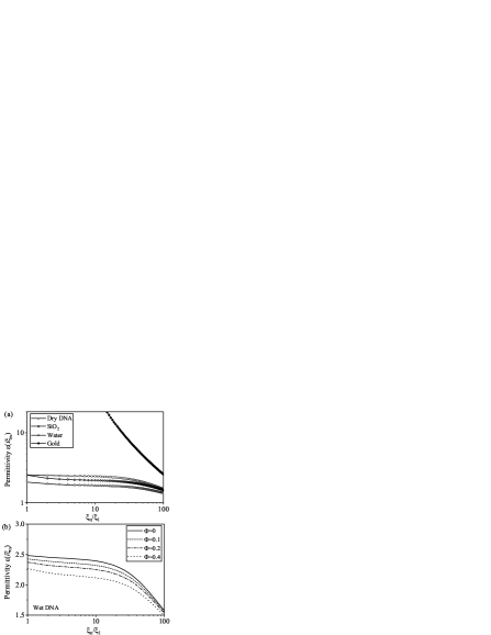
Figure 1(a) shows the permittivity of the applied materials evaluated in the imaginary frequency. The results show that the permittivity of a dry DNA is larger than those of the water and silica for the Matsubara term . The permittivity of water is the smallest over a wide range of frequencies. Figure 1(b) shows the permittivity of DNA under different water volume fractions. As expected, the permittivity of a wet DNA decreases by increasing the magnitude of , due to the elevated contribution from the low-refractive-index water.
To predict the sign of Casimir pressure, the permittivity at is significant since it plays a dominant role at a large thickness (or separation) as reported in Baranov et al. (2019); Esteso et al. (2016). The static permittivity for the silica, DNA and water are about 3.9, 4.2 and 81, respectively. However, the dielectric function of a wet DNA film at shows a different trend, compared with the high-frequency one. The static permittivity increases from 4.2 to 11.9, with increasing from 0 to 0.6.
III.1 The Casimir pressures for suspended DNA films
We first consider the Casimir force of a DNA film suspended in a homogeneous background medium. The Casimir force would be attractive as reported for suspended peptide films Klimchitskaya et al. (2020). The absolute Casimir pressure versus the thickness of suspended DNA film is shown in Fig. 2(a), where the solid and dash lines represent the background media to be the air and water, respectively. It is found that the magnitude of Casimir pressure decreases monotonously by increasing the DNA thickness. The Casimir pressure for the air is larger than the case of the water at a small thickness, while it is smaller at a larger thickness.
The sign and magnitude of the Casimir pressure are dependent on the dielectric responses of materials. Considering the DNA film is surrounded by medium 1 and medium 2, the Casimir pressure would be proportional to the permittivity contrasts of the media Dzyaloshinskii et al. (1961)
| (7) |
where and is the permittivity of the medium 1 and medium 2, respectively. We have when the DNA film is suspended in the air and water, respectively. The permittivity contrasts between DNA film and the air are larger than those of water for 0. It is known that the high frequency components are dominant for the calculation of Casimir force at a small separationYang et al. (2010). As a result, the Casimir pressure for the air is larger than the that of the water at a small DNA thickness. By contrast, the dielectric contrast between DNA film and the water is much larger than that of the air at =0, which is the leading term for a large DNA-film thickness. Thus, there is no surprise that the Casimir pressure in water environment is larger than the configuration of the air for a large thickness (e.g., 500 nm).
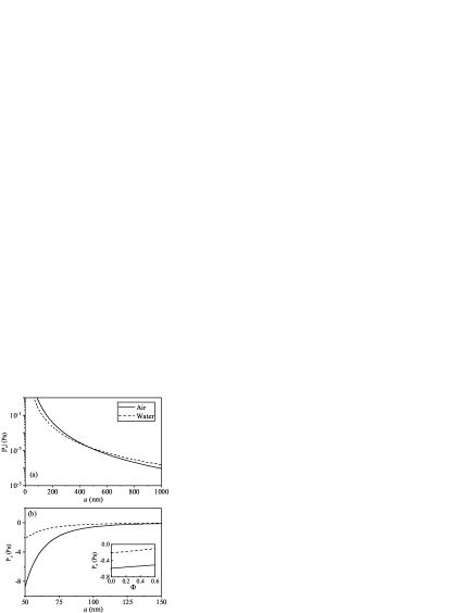
On the other hand, it would be interesting to consider the Casimir pressure across a wet DNA film. As an example, we set the thickness =100 nm, and the Casimir force versus the volume fraction of water in DNA film is shown in the inset of Fig. 2(b). The results show that the magnitude increases with decreasing the value of . At the limit =0, the attractive Casimir pressures at the air and water environment are about 0.2 and 0.6 Pa, respectively. The magnitude of Casimir pressure is hundreds of times larger than the gravity of the DNA film (about 1.7 mPa for =100 nm), manifesting the important role of the fluctuation-induced force. It can be seen that the magnitude of Casimir pressure can be enlarged over 10 times, when the thickness decreases further from 100 nm to 50 nm. Note that the condensation of DNA will decline its thickness and the volume fraction of water (i.e., squeezing the water out of the DNA film). Hence, it can be concluded that DNA films trends to condensation for suspended configurations, due to the attractive Casimir force.
III.2 The Casimir pressures for a silica substrate
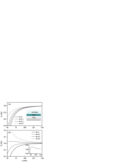
In many organic devices, the DNA film is generally deposited on a dielectric substrate. The Casimir pressure for a DNA thin film placed on a silica substrate is shown in Fig. 3(a), where the cladding background medium is the air. The result shows that the Casimir pressure is negative, and its magnitude increases by decreasing the DNA-film thickness. We note that the magnitude of the Casimir pressure is about 0.07 Pa for the dry DNA film at 100 nm, which declines considerably compared with the suspended configuration (about 0.6 Pa). In addition, the magnitude of the Casimir pressure for wet DNA film declines further with increasing volume fraction . The pressure is only about 0.04 Pa with volume fraction =0.4. Nonetheless, the Casimir pressure for a wet DNA deposited on a silica substrate is still much larger than the gravity of the DNA film. Overall, a thin DNA film and a low water volume fraction are preferred for stability of the DNA film.
As the DNA film is immersed in the water, some complicated or even reverse conclusions are obtained, in comparison with the air configurations. The Casimir pressure as a function of the thickness is shown in Fig. 3(b). The results show that the Casimir pressure is long-range negative at low volume fractions 0 and 0.1, and its magnitude decreases rapidly with increasing the DNA-film thickness. These properties suggest that a thin DNA film is favored for stability due to the attractive Casimir force, similar to the case of air in Fig. 3(a). However, the Casimir pressure for a large shows different features. For =0.4, the Casimir pressure is long-range positive, which means that a thin thickness is harmful to the stability of DNA films. For an intermediate value =0.2, the Casimir pressure turns from negative to positive with increasing the thickness , as shown in the inset of Fig. 3(b). Then, a maximum peak for the Casimir repulsion can be found near 60 nm, which contributes negatively to the stability. The Casimir pressure would decrease with increasing the thickness further.
The unusual behavior of Casimir pressure at the water background can be interpreted by the competition between the attractive and repulsive Casimir components. For a small , the permittivity of the wet DNA is larger than those of silica and water over a wide range of frequencies (). The dielectric permittivity of the DNA and silica are very close at zero frequency, resulting in a negligible contribution from the term . Therefore, the Casimir pressure is attractive according to the Eq. (7), and its magnitude increases rapidly with decreasing the DNA-film thickness, as demonstrated with =0 and 0.1 in Fig 3(b). For a large =0.4, the permittivity of the wet DNA is smaller(larger) than that of silica(water) for , resulting in repulsive Casimir force. At static frequency with =0, the permittivity of the wet DNA is larger (smaller) than that of silica (water), which also leads to repulsive Casimir force. Hence, the Casimir pressure would be long-range repulsive for a large . For an intermediate =0.2, the permittivity of the wet DNA is still larger than that of silica(water) for , resulting in attractive Casimir force at a small thickness . However, the contribution for =0 is still positive, resulting in repulsive Casimir force at a large value of . Due to the competition between the attractive and repulsive Casimir components, the peak for Casimir repulsion is expected at an intermediate thickness, as shown in the inset of Fig. 3(b).
III.3 The Casimir pressures for a metallic substrate
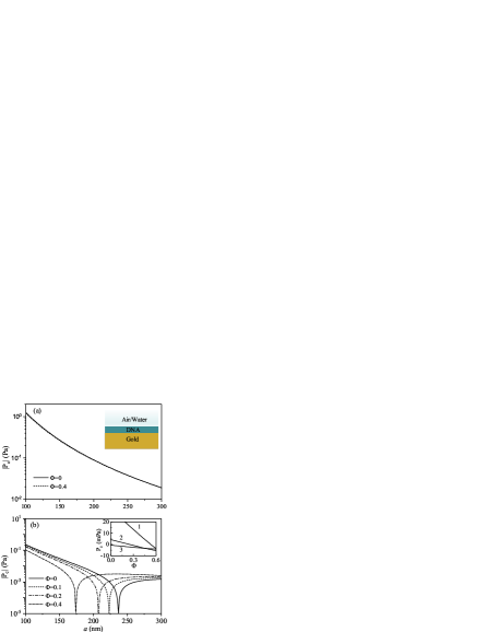
Now we consider the case of DNA films deposited on the metallic substrate. The magnitudes of the Casimir pressure as a function of thickness are shown in Fig. 4(a), where the cladding medium is the air. According to Eq.(7), we can predict that the Casimir pressure is long-range repulsive because is satisfied for (see, e.g., Ref.Munday et al. (2009)). The magnitude of Casimir pressure decreases monotonously with increasing the DNA-film thickness. As a result, the dispersion forces make the DNA film less stable for thin thickness. Note that the discrepancy of Casimir pressures acting on the DNA film is small between volume fractions =0 and 0.4.
The Casimir pressure acting on the DNA film immersed in the water exhibit different characteristic shown in Fig. 4(b). The Casimir pressure is repulsive at a thin thickness, while it becomes attractive for a large thickness. At a specific thickness, a stable Casimir equilibrium, i.e., the pressure equals to zero, is found. The critical thickness for the Casimir equilibrium can be modulated by the magnitude of . As the increases from 0 to 0.4, the critical thickness decreases correspondingly from about 237 to 174 nm. The interesting Casimir equilibrium at the water background can be understood by the contrast of permittivity at the Eq. (7). The repulsive relation is satisfied for , and the permittivity contrast between the water and wet DNA decreases with increasing the , resulting in a smaller Casimir repulsion. On the other side, the attractive Casimir interaction at a large thickness is attributed to the relation at the leading term .
The inset in Fig. 4(b) shows the Casimir pressure changed by the volume fraction of water with a fixed DNA-film thickness. We find that switching the sign of the Casimir pressure from positive to negative is achieved by increasing the volume fraction for thickness 150 nm and 200 nm. For thickness 250 nm, the Casimir pressure is negative and its magnitude increases by increasing the volume fraction . Hence, the DNA film deposited on the metallic substrate tends to be de-condensation in the water environment, according to the properties of its dispersion force.
IV Conclusions
In summary, the Casimir pressure of a DNA film is calculated in several configurations based on the Lifshitz theory. The Casimir pressure is attractive when a DNA film is suspended in the air or water, and its magnitude increases with decreasing the thickness of DNA film or/and the water volume fraction. Hence, the suspended DNA film trends to condensation due to the Casimir force. The Casimir pressure is hundreds of times larger than the gravity of the DNA film for a moderate thickness (e.g., 100 nm), manifesting the important role of the fluctuation-induced interactions. For DNA films deposited on the silica substrate, the Casimir pressure is attractive for the air background. Also, a thin DNA film and a low water fraction are favored for the stability. Instead, the Casimir pressure shows rich features in a water background. The Casimir pressure can be changed from attractive to repulsive by increasing the DNA-film thickness and the water fraction. At the end, the Casimir force of a DNA film deposited on a metallic substrate is explored. The Casimir pressure is dominant by the repulsive interactions at a small DNA-film thickness for both the air and water environment. For the setup immersed in a water environment, the Casimir pressure turns out to be attractive at a large DNA-film thickness, and a stable Casimir equilibrium can be found at a specific thickness. Our finding provides a theoretical guide for the adhesion stability, condensation, and de-condensation of DNA films, resulting from the fluctuation-induced dispersion forces.
Acknowledgements.
This work is supported by the National Natural Science Foundation of China (Grant No. 11804288 and No. 61974127), and Shanghai Pujiang Program (21PJ1411400). The research of L.X. Ge is further supported by Nanhu Scholars Program for Young Scholars of XYNU.References
- Feinberg et al. (1989) G. Feinberg, J. Sucher, and C.-K. Au, Phys. Rep. 180, 83 (1989).
- Rodriguez et al. (2011) A. W. Rodriguez, F. Capasso, and S. G. Johnson, Nature photonics 5, 211 (2011).
- Parsegian (2005) V. A. Parsegian, Van der Waals forces: a handbook for biologists, chemists, engineers, and physicists (Cambridge university press, 2005).
- Dagastine et al. (2002) R. R. Dagastine, D. C. Prieve, and L. R. White, J. Colloid Interface Sci. 249, 78 (2002).
- Bordag et al. (2009) M. Bordag, G. L. Klimchitskaya, U. Mohideen, and V. M. Mostepanenko, Advances in the Casimir effect, vol. 145 (OUP Oxford, 2009).
- Klimchitskaya et al. (2009) G. L. Klimchitskaya, U. Mohideen, and V. M. Mostepanenko, Rev. Mod. Phys. 81, 1827 (2009).
- Lee and Sigmund (2001) S.-w. Lee and W. M. Sigmund, J. Colloid Interface Sci. 243, 365 (2001).
- Gong et al. (2021) T. Gong, M. R. Corrado, A. R. Mahbub, C. Shelden, and J. N. Munday, Nanophotonics 10, 523 (2021).
- Somers et al. (2018) D. A. Somers, J. L. Garrett, K. J. Palm, and J. N. Munday, Nature 564, 386 (2018).
- Ge et al. (2020a) L. Ge, X. Shi, L. Liu, and K. Gong, Phys. Rev. B 102, 075428 (2020a).
- Munkhbat et al. (2021) B. Munkhbat, A. Canales, B. Küçüköz, D. G. Baranov, and T. O. Shegai, Nature 597, 214 (2021).
- Esteso et al. (2015) V. Esteso, S. Carretero-Palacios, and H. Míguez, J. Phys. Chem. C 119, 5663 (2015).
- Hough and White (1980) D. B. Hough and L. R. White, Adv. Colloid Interface Sci. 14, 3 (1980).
- Boinovich and Emelyanenko (2011) L. Boinovich and A. Emelyanenko, Adv. Colloid Interface Sci. 165, 60 (2011).
- Squarcini et al. (2022) A. Squarcini, J. M. Romero-Enrique, and A. O. Parry, Phys. Rev. Lett. 128, 195701 (2022).
- Li et al. (2022) Y. Li, K. A. Milton, I. Brevik, O. I. Malyi, P. Thiyam, C. Persson, D. F. Parsons, and M. Boström, Phys. Rev. B 105, 014203 (2022).
- Luengo-Márquez and MacDowell (2021) J. Luengo-Márquez and L. G. MacDowell, J. Colloid Interface Sci. 590, 527 (2021).
- Esteso et al. (2020) V. Esteso, S. Carretero-Palacios, L. G. MacDowell, J. Fiedler, D. F. Parsons, F. Spallek, H. Míguez, C. Persson, S. Y. Buhmann, I. Brevik, et al., Phys. Chem. Chem. Phys. 22, 11362 (2020).
- Fiedler et al. (2020) J. Fiedler, M. Bostro?m, C. Persson, I. Brevik, R. Corkery, S. Y. Buhmann, and D. F. Parsons, J. Phys. Chem. B 124, 3103 (2020).
- Lu and Podgornik (2015) B.-S. Lu and R. Podgornik, Phys. Rev. E 92, 022112 (2015).
- Blackwell et al. (2021) R. Blackwell, A. Hemmerle, A. Baer, M. Späth, W. Peukert, D. Parsons, K. Sengupta, and A.-S. Smith, J. Colloid Interface Sci. 598, 464 (2021).
- Baranov et al. (2019) M. A. Baranov, G. L. Klimchitskaya, V. M. Mostepanenko, and E. N. Velichko, Phys. Rev. E 99, 022410 (2019).
- Klimchitskaya et al. (2020) G. L. Klimchitskaya, V. M. Mostepanenko, and E. N. Velichko, Phys. Rev. B 102, 161405(R) (2020).
- Klimchitskaya et al. (2021) G. L. Klimchitskaya, V. M. Mostepanenko, and E. N. Velichko, Phys. Rev. B 103, 245421 (2021).
- Klimchitskaya et al. (2022) G. L. Klimchitskaya, V. M. Mostepanenko, and O. Y. Tsybin, Symmetry 14, 2196 (2022).
- Nykypanchuk et al. (2008) D. Nykypanchuk, M. M. Maye, D. Van Der Lelie, and O. Gang, Nature 451, 549 (2008).
- Chou et al. (2014) L. Y. Chou, K. Zagorovsky, and W. C. Chan, Nat. Nanotechnol. 9, 148 (2014).
- Rogers et al. (2016) W. B. Rogers, W. M. Shih, and V. N. Manoharan, Nat. Rev. Mater. 1, 1 (2016).
- Cui et al. (2022) F. Cui, S. Marbach, J. A. Zheng, M. Holmes-Cerfon, and D. J. Pine, Nat. Commun. 13, 2304 (2022).
- Campolongo et al. (2010) M. J. Campolongo, S. J. Tan, J. Xu, and D. Luo, Adv. Drug Deliv. Rev. 62, 606 (2010).
- Hu et al. (2018) Q. Hu, H. Li, L. Wang, H. Gu, and C. Fan, Che. Rev. 119, 6459 (2018).
- Weiden and Bastings (2021) J. Weiden and M. M. Bastings, Curr. Opin. Colloid Interface Sci. 52, 101411 (2021).
- Gu et al. (2021) W. Gu, F. Meng, R. Haag, and Z. Zhong, J. Control. Release 329, 676 (2021).
- Steckl (2007) A. J. Steckl, Nat. Photonics 1, 3 (2007).
- Bui et al. (2019) H. Bui, S. A. Díaz, J. Fontana, M. Chiriboga, R. Veneziano, and I. L. Medintz, Adv. Opt. Mater. 7, 1900562 (2019).
- Kawabe et al. (2000) Y. Kawabe, L. Wang, S. Horinouchi, and N. Ogata, Adv. Mater. 12, 1281 (2000).
- Zhang et al. (2012) Y. Zhang, P. Zalar, C. Kim, S. Collins, G. C. Bazan, and T.-Q. Nguyen, Adv. Mater. 24, 4255 (2012).
- Khazaeinezhad et al. (2017) R. Khazaeinezhad, S. Hosseinzadeh Kassani, B. Paulson, H. Jeong, J. Gwak, F. Rotermund, D.-I. Yeom, and K. Oh, Sci. Rep. 7, 1 (2017).
- Jung et al. (2017) W. Jung, H. Jun, S. Hong, B. Paulson, Y. S. Nam, and K. Oh, Opt. Mater. Express 7, 3796 (2017).
- Yakovchuk et al. (2006) P. Yakovchuk, E. Protozanova, and M. D. Frank-Kamenetskii, Nucleic Acids Res. 34, 564 (2006).
- Moazzami Gudarzi and Aboutalebi (2021) M. Moazzami Gudarzi and S. H. Aboutalebi, Sci. Adv. 7, eabg2272 (2021).
- Wittlin et al. (1986) A. Wittlin, L. Genzel, F. Kremer, S. Häseler, A. Poglitsch, and A. Rupprecht, Phys. Rev. A 34, 493 (1986).
- Inagaki et al. (1974) T. Inagaki, R. Hamm, E. Arakawa, and L. Painter, J. Chem. Phys. 61, 4246 (1974).
- Weidlich et al. (1987) T. Weidlich, S. Lindsay, and A. Rupprecht, Biopolymers: Original Research on Biomolecules 26, 439 (1987).
- Paulson et al. (2018) B. Paulson, I. Shin, H. Jeong, B. Kong, R. Khazaeinezhad, S. R. Dugasani, W. Jung, B. Joo, H.-Y. Lee, S. Park, et al., Sci. Rep. 8, 1 (2018).
- Ge et al. (2020b) L. Ge, X. Shi, Z. Xu, and K. Gong, Phys. Rev. B 101, 104107 (2020b).
- Esteso et al. (2016) V. Esteso, S. Carretero-Palacios, and H. Míguez, J. Appl. Phys. 119, 144301 (2016).
- Dzyaloshinskii et al. (1961) I. E. Dzyaloshinskii, E. M. Lifshitz, and L. P. Pitaevskii, Adv. Phys. 10, 165 (1961).
- Yang et al. (2010) Y. Yang, R. Zeng, H. Chen, S. Zhu, and M. S. Zubairy, Phys. Rev. A 81, 022114 (2010).
- Munday et al. (2009) J. N. Munday, F. Capasso, and V. A. Parsegian, Nature 457, 170 (2009).