Hybrid Swin Deformable Attention U-Net for Medical Image Segmentation
Abstract
Medical image segmentation is a crucial task in the field of medical image analysis. Harmonizing the convolution and multi-head self-attention mechanism is a recent research focus in this field, with various combination methods proposed. However, the lack of interpretability of these hybrid models remains a common pitfall, limiting their practical application in clinical scenarios. To address this issue, we propose to incorporate the Shifted Window (Swin) Deformable Attention into a hybrid architecture to improve segmentation performance while ensuring explainability. Our proposed Swin Deformable Attention Hybrid UNet (SDAH-UNet) demonstrates state-of-the-art performance on both anatomical and lesion segmentation tasks. Moreover, we provide a direct and visual explanation of the model focalization and how the model forms it, enabling clinicians to better understand and trust the decision of the model. Our approach could be a promising solution to the challenge of developing accurate and interpretable medical image segmentation models. (Our code can be made open source, once accepted.)
Index Terms:
Medical image segmentation, transformer, XAI, deformable attentionI Introduction
Accurate medical image segmentation is crucial in many clinical scenarios, and interpretability is essential for segmentation models as it enables clinicians to understand how the model arrives at its decision and provides insight into its working mechanism. However, the feature maps embedded in segmentation networks are often difficult to interpret due to their indirect correlation with the network optimization procedure. Therefore, segmentation methods that can simultaneously provide interpretation masks are required.
Gradient-assisted methods such as Gradient-weighted Class Activation Mapping (Grad-CAM) [1] and Layer-Wise Relevance Propagation [2] have been proposed as major solutions for interpretation visualization. However, these methods do not adequately capture fine-grained information regarding object boundaries, and as external extensions, they do not offer an intrinsic understanding of the decision-making process. The introduction of the multi-head self-attention (MSA) mechanism has partially mitigated this limitation [3, 4, 5, 6, 7, 8, 9]. These methods not only provide attention score heatmaps that enable internal interpretability of the model but also improve segmentation accuracy through the data-specific nature of the MSA [10].
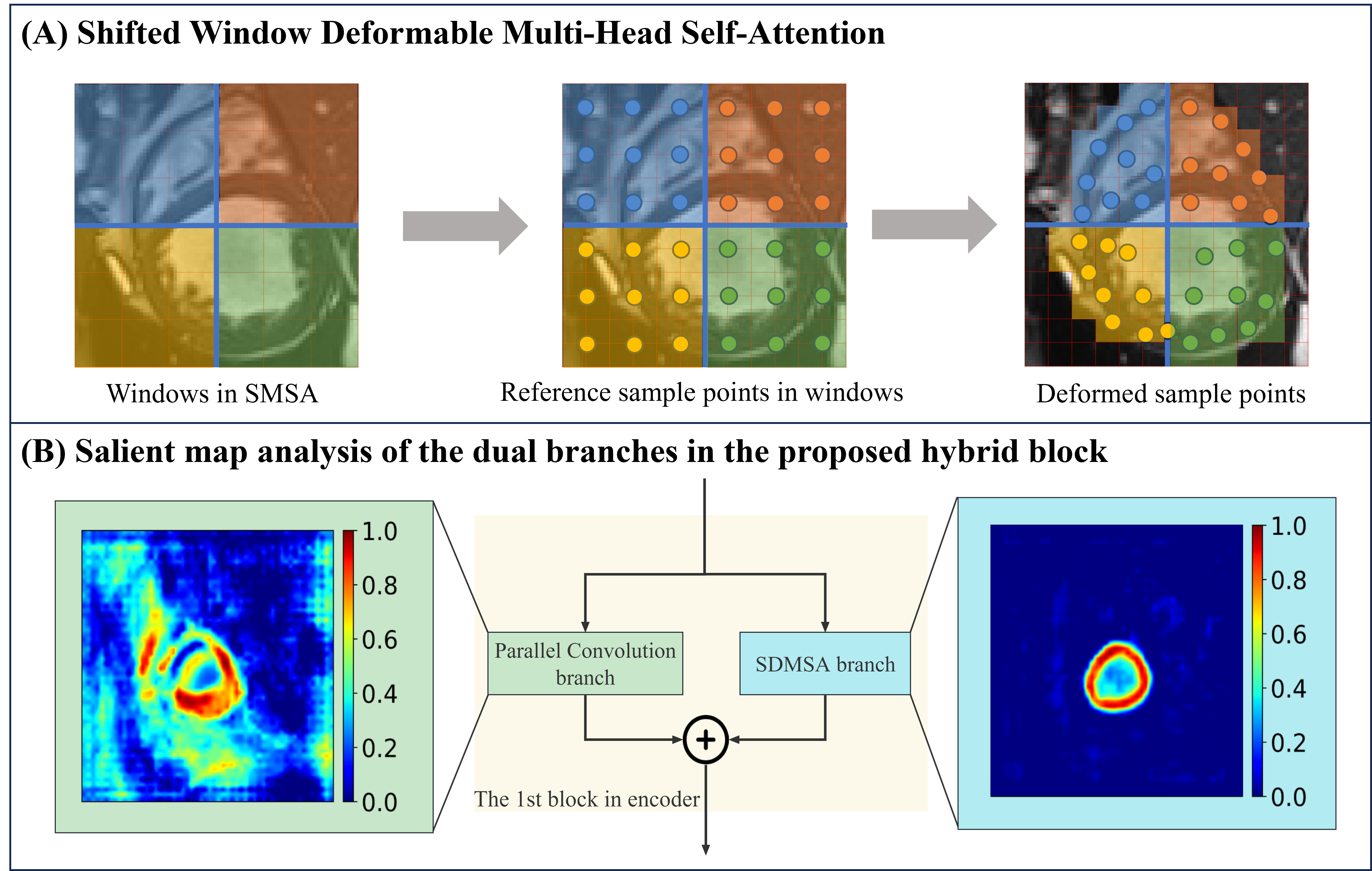
Despite the advantages of the attention mechanism, redundant attention sometimes occurs due to the lack of deformability of conventional attention modules [11]. For example, in a left ventricle (LV) walls segmentation task (Fig. 1 (A)), the Shifted Window (Swin) multi-head self-attention (SMSA) branch redundantly computes the MSA on all patches in each window due to the lack of ability to capture the complicated, irregularly distributed, and deformed LV wall area. This redundant attention raises questions regarding the interpretation and increases the computational cost.
To address this limitation, we develop a novel hybrid block containing a Swin Deformable MSA (SDMSA) module (Fig. 1 (A)) which enables the MSA mechanism to precisely focus on the segmentation target and remove redundancy. However, models with only the SDMSA in its basic block can result in the loss of detailed information, due to the patch-based mechanism and the lack of the local prior. Therefore, a computation-efficient convolution branch in parallel is designed to acquire detailed texture features [10]. As shown in Fig. 1 (B), the Parallel Convolution branch focuses more on the detailed features, while the SDMSA branch concentrates on the holistic shape structure.
We hypothesize that (1) the SDMSA module enables the model to focus more precisely on the segmentation targets and provides explainability; (2) our proposed model can achieve state-of-the-art performance on multiple medical image segmentation datasets, incorporating different clinical questions. The main contributions are listed as follows: (1) we propose a Swin Deformable Attention Hybrid UNet (SDAH-UNet) model for medical image segmentation; (2) our SDMSA with Parallel Convolution (SDAPC) block improves the segmentation performance based on the SDMSA module, which strengthens the focus of the model on the segmentation targets and can provide explanations; (3) in the experiment section, SDAH-UNet significantly outperforms state-of-the-art models, on automatic cardiac diagnosis challenge (ACDC) [13], and brain tumor segmentation 2020 (BraTS2020) [14].
II Methodology
II-A U-shape Architecture
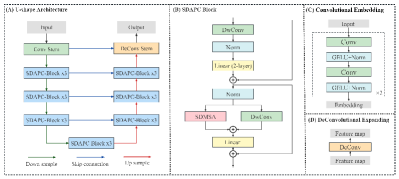
Our proposed model is an end-to-end model that takes an image slice as input and produces a corresponding segmentation mask as output. The proposed model follows a symmetrical design paradigm, with both the encoder and decoder consisting of three SDAPC blocks. A convolutional stem is added at the beginning of the encoder, and a deconvolutional stem is added at the end of the decoder. An additional SDAPC block is inserted in the bottleneck (Fig. 2 (A)).
II-B Convolutional Embedding Module and DeConvolutional Expanding Module
Drawing inspiration from recent studies [15, 8], we directly adopt successive convolutional layers with small kernels as the embedding module. This design aims to precisely encode pixel-level spatial information while minimizing computational complexity and maintaining a large receptive field. The embedding module comprises four convolutional layers, each followed by a Gaussian Error Linear Unit (GELU) [16] activation function and a layer normalization layer. The expanding module is comprised of a deconvolutional layer (Fig. 2 (C) and (D)). This simple yet effective architecture can be readily extended to support higher-resolution inputs.
II-C Swin Deformable Multi-Head Self-Attention with Parallel Convolution Block
Convolution and MSA have been shown to have distinct weighting mechanisms [8], filtering effects [10], and levels of focus on feature map details, which can be complementary to each other. To leverage the benefits of both mechanisms, our SDAPC block is designed in a parallel paradigm (Fig. 2 (B)). Partitioned by the two residual connections, the SDAPC block comprises two divisions. In the first division, the input first undergoes a Depthwise Convolution (DwConv) [17] layer, , followed by a standard 2-layer multi-layer perceptron (MLP), which can be described as
| (1) |
in which and are full connection layers. and denote the GELU and Layer Normalization layers.
In the second division, is passed to a paralleled DwConv layer, , and a SDMSA layer, after which their outputs are concatenated and passed through the final layer. Inspired by the design of ConvNeXt [18], the kernel size of DwConv is set as 7. The process can be described as
| (2) |
II-D Shifted Window Deformable Multi-Head Self-Attention
Followed by the Deformation Attention Transformer [11] and the Swin Deformable Attention U-Net Transformer [19], we introduce the deformation operation into the SMSA, resulting in the SDMSA. The input feature map is first split into non-overlapping windows and then divided into heads along the channels. After the partition, sub-feature maps are generated, where , stands for window size. For a sub-feature map , where and represent the window and the head respectively, we calculate the query , and set up a group of uniformly distributed reference sample points . The reference sample points are added with the offsets obtained from a two-layer convolution neural network , to generate the deformed sample points . A bilinear interpolation function is used to generate the sampled features. Then keys and values are calculated based on the sampled features. The SDMSA can be presented as follows
| (3) |
| (4) |
| (5) |
| (6) |
where , , and are the projection matrices for queries, keys, and values, respectively. represents the fixed positional bias [20]. The dimension of each head is represented as , where is the number of output channels and is the number of heads.
III Experiments
Experiments were conducted on two medical image segmentation tasks, i.e., ACDC [13] and BraTS2020 [14], which represent different clinical questions and MRI sequences to validate the efficacy of our proposed methods. The ACDC dataset is an anatomical segmentation task, with single MRI modality serving as the input. BraTS2020 is a lesion segmentation task, with multi-modal MRI data as the input. MRI volumetric data was split into slices for the model training.
III-A Implementation Details and Evaluation Methods
We conducted our experiments on an NVIDIA RTX3090 GPU with 24GB GPU RAM. Dice loss and cross-entropy loss were combined as the loss function.
During the training process, the batch size and input resolution were set to 24 and for both datasets. The initial learning rate was set to and decayed every 10,000 steps by 0.5 from the step. During the inference stage, our model makes predictions using a sliding window approach on both datasets[21]. We set 112, 0.5 times of the crop size (224), as the step size. For patch aggregation, the Gaussian importance weighting strategy was utilized, giving more weight to pixels in the center area.
The evaluation of performance in this study incorporates the utilization of two metrics: the Dice similarity coefficient (DSC) [22] and the Hausdorff95 metric (HD95) [23]. The statistical analysis conducted to ascertain the significance of the results involves the application of a paired sample t-test. For the purpose of evaluating the model size and time complexity, two quantitative measures were employed: the count of parameters (Params) and the rate of floating-point operations per second (FLOPs).
III-B Datasets
The ACDC data [13] is a collection of 100 cine MRI scans (1,902 slices). The data was randomly split into training, validation, and test sets. Within the ACDC dataset, short axis cine MRI scans are available for each patient, consisting of 12-35 frames, along with end-diastole (ED) and end-systole (ES) frames. The dataset provides segmentation targets for three foreground classes: right ventricle (RV), myocardium (MYO), and left ventricle (LV).
The BraTS2020 data [14] consists of 369 cases (57,195 slices) of brain MRI scans, each with four modalities: native T1-weighted, post-contrast T1-weighted, T2-weighted, and Fluid Attenuated Inversion Recovery scans. The data was randomly split into training, validation, and test sets. The segmentation accuracy was evaluated for the enhancing tumor region (ET, label 1), regions of the tumor core (TC, labels 1 and 4), and the whole tumor region (WT, labels 1, 2, and 4).
III-C The State-Of-The-Art Segmentation Performance
The quantitative results of the comparison on two general medical image segmentation datasets are reported in Table I and II. Results demonstrate that the proposed SDAH-UNet, with strong generalization ability, can be applied to multiple datasets including anatomical and lesion segmentation datasets. Table III demonstrates that SDAH-UNet exhibits a marginal increase in parameter count (attributed to CNNoffset and Parallel Convolution) compared to other SMSA-based models (SwinUNet [24], UNet-2022), while maintaining relatively lower FLOPs. This suggests that SDAH-UNet achieves state-of-the-art performance with a minimal increment of computational overhead.
| Methods | DSC | HD95 | DSC | ||
|---|---|---|---|---|---|
| AVG | RV | MYO | LV | ||
| UNet | 87.68 | 1.55 | 83.82 11.04† | 84.21 9.97† | 95.04 5.09† |
| nnUNet | 91.86 | 1.22 | 88.86 7.43 | 89.77 2.29 | 96.97 0.75 |
| SwinUNet | 88.08 | 1.61 | 85.24 8.48† | 84.45 7.76† | 94.56 4.64† |
| TransUNet | 89.48 | 1.41 | 87.21 9.23† | 85.73 6.01† | 95.52 3.39† |
| UNet-2022 | 90.42 | 1.41 | 87.36 9.09† | 87.78 5.09† | 96.13 3.28† |
| SDAH-UNet | 92.23 | 1.22 | 89.78 9.97 | 90.02 4.23 | 96.91 1.91 |
| Methods | DSC | HD95 | DSC | ||
|---|---|---|---|---|---|
| AVG | WT | TC | ET | ||
| UNet | 82.27 | 9.04 | 85.68 8.82† | 85.46 8.67† | 75.69 8.92† |
| nnUNet | 83.64 | 4.30 | 86.45 10.89† | 82.35 7.28† | 82.13 11.37† |
| SwinUNet | 84.56 | 9.25 | 87.47 7.10† | 85.50 9.08† | 80.71 8.21† |
| TransUNet | 84.64 | 6.02 | 87.87 9.03† | 85.88 9.73† | 80.19 8.88† |
| UNet-2022 | 84.76 | 5.52 | 87.95 9.03† | 85.97 9.61† | 80.35 8.68† |
| SDAH-UNet | 86.90 | 3.65 | 90.21 3.55 | 88.13 5.23 | 82.37 5.45 |
| Model | UNet | nnUNet | SwinUNet | TransUNet | UNet-2022 | SDAH-UNet |
| Params | 7.852M | 10.468M | 155.384M | 312.809M | 162.894M | 162.942M |
| FLOPs | 10.772G | 31.340G | 33.201G | 53.410G | 37.973G | 37.950G |
III-D The Accurate Interpretation
The visual explanations for decoder and encoder are shown in Fig. 3. For the ACDC dataset, all visual explanation methods were observed to effectively illustrate the focalization of the decoder. Although the deformation of sample points impaired the visualization of the feature map, the outline of the myocardium still remained discernible. In the case of the more challenging BraTS2020 dataset, the attention score heatmap only provided a general indication of the tumor location, and the gradient heatmap had excessive highlight areas.
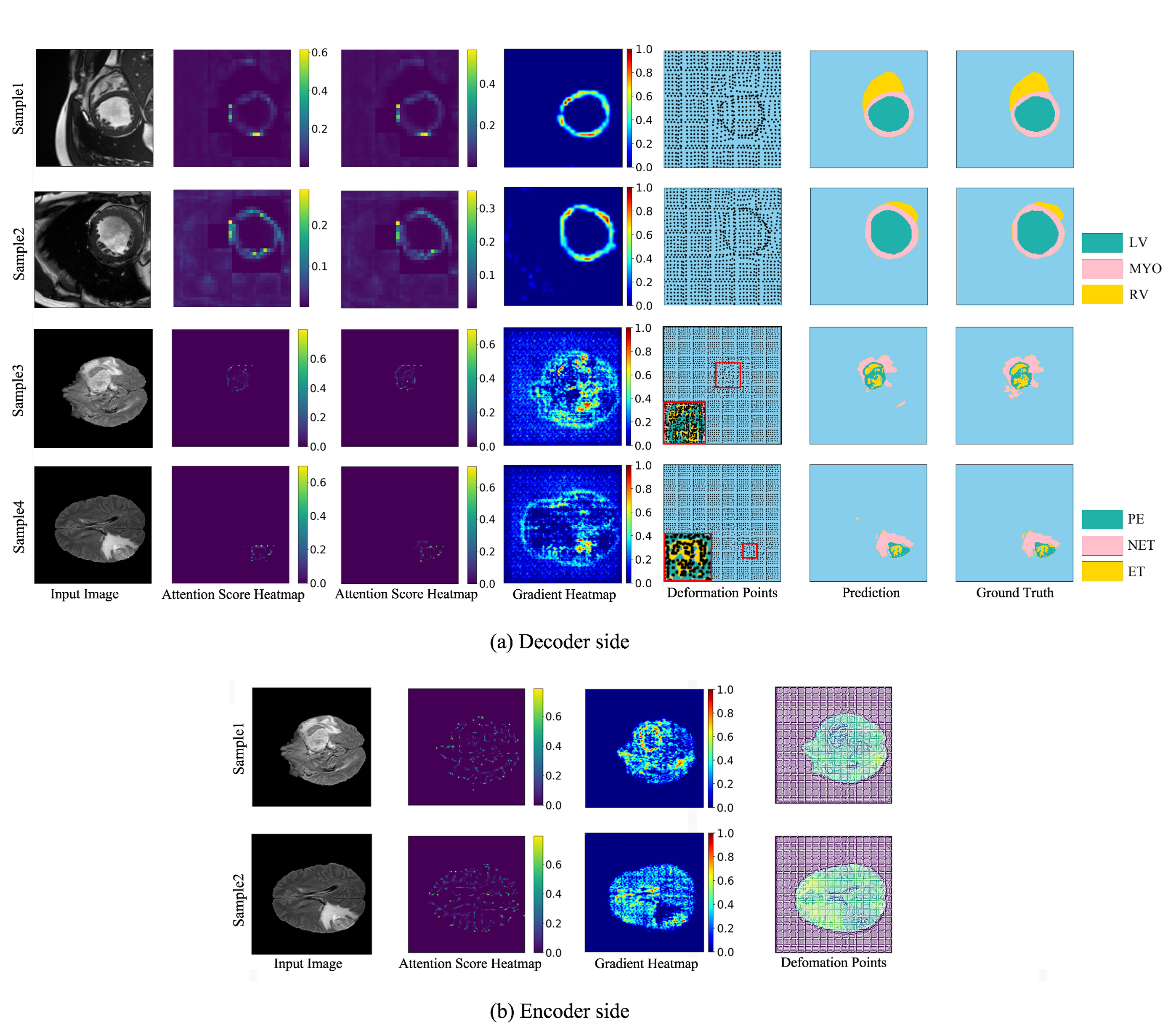
In contrast, the utilization of deformation points presented a clearer representation of the model focalization. Global area analysis supported this finding by revealing that only sample points sufficiently close to the tumor exhibited deformation and were clustered in the tumor area. Moreover, when examining the results in a zoomed-out window, it could be observed that sample points were concentrated in the enhanced tumor region, within the whole tumor area. In the encoder side, the attention score heatmap was found to only depict the approximate location of the tumor. The gradient heatmap has shown that, in sample 1, the model paid more attention to the highlighted area; however, for sample 2 the gradient heatmap has shown that the model ignores the highlighted tumor area. On the other hand, the deformation points illustrated that the model focused on both the highlighted areas and the boundaries.
This outcome supports our hypothesis that compared to other visual explanation methods, the deformation points can facilitate a better understanding of the focalization by offering a clearer and more robust indication of the model focalization. Moreover, it also indicates that, in the windows nearing the segmentation target, the MSA computation focuses more precisely on the information-dense area, which reduces the computation redundancy.
III-E Ablation Studies
We set two groups of ablation studies to investigate the influence of the number of the SDAPC blocks and the two branches of the SDAPC block.
Table. IV shows that the performance of the model improved when the N-block (block with SMSA module and Parallel Convolution) was gradually replaced by the SDAPC block. replacing the first N-block with the SDAPC block lead to nearly 1.2% performance enhancement for DSC. However, continuing to replace the N-blocks could not have a significant influence. Table. V illustrates that removing each branch from the SDAPC block leads to a decline in performance, nearly 2% DSC.
| Methods | DSC | HD95 | DSC | ||
|---|---|---|---|---|---|
| AVG | RV | MYO | LV | ||
| NNNN | 90.42 | 1.41 | 87.36 9.09 | 87.78 5.09 | 96.13 3.28 |
| DNNN | 91.61 | 1.22 | 89.30 5.80 | 88.79 2.82 | 96.74 0.88 |
| DDNN | 91.86 | 1.23 | 89.87 5.34 | 88.88 3.10 | 96.83 0.87 |
| DDDN | 91.99 | 1.22 | 90.02 6.21 | 89.35 3.01 | 96.61 1.83 |
| DDDD | 92.23 | 1.22 | 89.78 9.97 | 90.02 4.23 | 96.91 1.91 |
| Branch Type | DSC | HD95 | DSC | ||
|---|---|---|---|---|---|
| AVG | RV | MYO | LV | ||
| SDMSA-branch | 90.48 | 1.45 | 87.35 5.73 | 88.53 2.12 | 95.58 1.11 |
| Conv-branch | 90.26 | 1.98 | 87.06 6.41 | 88.35 3.25 | 95.38 1.32 |
| Dual-branch | 92.23 | 1.22 | 89.78 9.97 | 90.02 4.23 | 96.91 1.91 |
IV Conclusions
In this study, we have developed the Swin Deformable MSA with Parallel Convolution block, i.e., SDAPC block, and proposed the Swin Deformable Attention Hybrid UNet, i.e., SDAH-UNet, for explainable medical image segmentation. The experiments have proven that SDAH-UNet outperforms the state-of-the-art model on both anatomy and lesion segmentation . The ablation studies have illustrated that both the SDMSA module and the Parallel Convolution layer improved the segmentation performance of SDAH-UNet. Moreover, the deformation points can illustrate two-level of explanation: the model focalization and how the model forms the focalization. The explanations have also proven that the SDAPC block enables the model to focus more precisely on the segmentation targets, reduce MSA computation redundancy, and leads to performance enhancement.
References
- [1] R. R. Selvaraju, M. Cogswell, A. Das, R. Vedantam, D. Parikh, and D. Batra, “Grad-CAM: Visual Explanations from Deep Networks via Gradient-Based Localization,” in 2017 IEEE International Conference on Computer Vision (ICCV), 2017, pp. 618–626.
- [2] S. Bach, A. Binder, G. Montavon, F. Klauschen, K.-R. Müller, and W. Samek, “On Pixel-Wise Explanations for Non-Linear Classifier Decisions by Layer-Wise Relevance Propagation,” PLoS ONE, vol. 10, 2015.
- [3] J. Chen, Y. Lu, Q. Yu, X. Luo, E. Adeli, Y. Wang, L. Lu, A. L. Yuille, and Y. Zhou, “TransUNet: Transformers Make Strong Encoders for Medical Image Segmentation,” ArXiv, vol. abs/2102.04306, 2021.
- [4] G. Xu, X. Wu, X. Zhang, and X. He, “LeViT-UNet: Make Faster Encoders with Transformer for Medical Image Segmentation,” ArXiv, vol. abs/2107.08623, 2021.
- [5] A. Hatamizadeh, D. Yang, H. R. Roth, and D. Xu, “UNETR: Transformers for 3D Medical Image Segmentation,” 2022 IEEE/CVF Winter Conference on Applications of Computer Vision (WACV), pp. 1748–1758, 2021.
- [6] K. B. Park and J. Y. Lee, “SwinE-Net: hybrid deep learning approach to novel polyp segmentation using convolutional neural network and Swin Transformer,” Journal of Computational Design and Engineering, vol. 9, pp. 616–632, 4 2022.
- [7] Q. Sun, N. Fang, Z. Liu, L. Zhao, Y. Wen, and H. Lin, “HybridCTrm: Bridging CNN and Transformer for Multimodal Brain Image Segmentation,” Journal of Healthcare Engineering, vol. 2021, 2021.
- [8] J. Guo, H.-Y. Zhou, L. Wang, and Y. Yu, “UNet-2022: Exploring Dynamics in Non-isomorphic Architecture,” ArXiv, vol. abs/2210.15566, 2022.
- [9] Q. Liu, C. Kaul, J. Wang, C. Anagnostopoulos, R. Murray-Smith, and F. Deligianni, “Optimizing vision transformers for medical image segmentation,” in ICASSP 2023 - 2023 IEEE International Conference on Acoustics, Speech and Signal Processing (ICASSP), 2023, pp. 1–5.
- [10] N. Park and S. Kim, “How Do Vision Transformers Work?” in International Conference on Learning Representations, 2022.
- [11] Z. Xia, X. Pan, S. Song, L. E. Li, and G. Huang, “Vision Transformer with Deformable Attention,” in Proceedings of the IEEE/CVF conference on computer vision and pattern recognition, 2022, pp. 4794–4803.
- [12] K. Vinogradova, A. Dibrov, and E. W. Myers, “Towards Interpretable Semantic Segmentation via Gradient-weighted Class Activation Mapping,” in Proceedings of the AAAI Conference on Artificial Intelligence, vol. 34, 2020, pp. 13 943–13 944.
- [13] O. B. et al., “Deep Learning Techniques for Automatic MRI Cardiac Multi-Structures Segmentation and Diagnosis: Is the Problem Solved?” IEEE Transactions on Medical Imaging, vol. 37, pp. 2514–2525, 2018.
- [14] B. H. M. et al., “The Multimodal Brain Tumor Image Segmentation Benchmark (BRATS),” IEEE Transactions on Medical Imaging, vol. 34, pp. 1993–2024, 2015.
- [15] H.-Y. Zhou, J. Guo, Y. Zhang, L. Yu, L. Wang, and Y. Yu, “nnFormer: Interleaved Transformer for Volumetric Segmentation,” ArXiv, vol. abs/2109.03201, 2021.
- [16] D. Hendrycks and K. Gimpel, “Gaussian Error Linear Units (GELUs),” arXiv preprint arXiv:1606.08415, 2016.
- [17] F. Chollet, “Xception: Deep learning with depthwise separable convolutions,” in Proceedings of the IEEE conference on computer vision and pattern recognition, 2017, pp. 1251–1258.
- [18] Z. Liu, H. Mao, C. Wu, C. Feichtenhofer, T. Darrell, and S. Xie, “A ConvNet for the 2020s,” 2022 IEEE/CVF Conference on Computer Vision and Pattern Recognition (CVPR), pp. 11 966–11 976, 2022.
- [19] J. Huang, X. Xing, Z. Gao, and G. Yang, “Swin Deformable Attention U-Net Transformer (SDAUT) for Explainable Fast MRI,” in International Conference on Medical Image Computing and Computer-Assisted Intervention, 2022, pp. 538–548.
- [20] Z. Liu, Y. Lin, Y. Cao, H. Hu, Y. Wei, Z. Zhang, S. Lin, and B. Guo, “Swin Transformer: Hierarchical Vision Transformer using Shifted Windows,” 2021 IEEE/CVF International Conference on Computer Vision (ICCV), pp. 9992–10 002, 2021.
- [21] F. Isensee, J. Petersen, A. Klein, D. Zimmerer, P. F. Jaeger, S. Kohl, J. Wasserthal, G. Koehler, T. Norajitra, S. Wirkert, and K. H. Maier-Hein, “nnU-Net: Self-adapting Framework for U-Net-Based Medical Image Segmentation,” Nature methods, vol. 18(2), pp. 203–211, 9 2018.
- [22] L. R. Dice, “Measures of the Amount of Ecologic Association Between Species,” Ecology, vol. 26, no. 3, pp. 297–302, 1945.
- [23] T. Birsan and D. Tiba, “One Hundred Years Since the Introduction of the Set Distance by Dimitrie Pompeiu,” in System Modeling and Optimization: Proceedings of the 22nd IFIP TC7 Conference held from July 18–22, 2005, in Turin, Italy 22. Springer, 2006, pp. 35–39.
- [24] H. Cao, Y. Wang, J. Chen, D. Jiang, X. Zhang, Q. Tian, and M. Wang, “Swin-Unet: Unet-like Pure Transformer for Medical Image Segmentation,” in Proceedings of the European Conference on Computer Vision Workshops(ECCVW), 2022.