Self-organisation of spermatozoa in bulk suspensions
via unsteady elastohydrodynamic interactions
Abstract
We developed a mechanical model of spermatozoal swimming in bulk suspensions. We traced the spatiotemporal elastohydrodynamic interactions and found that spermatozoa engaged in self-organisation: flagellar undulatory motion generated a stable polar order. Conventional microswimmer theories use time-averaged stress fields to predict that the cellular structure is unstable. However, our present results reveal a novel self-organisation mechanism attributable to unsteady hydrodynamic interactions. We thus contribute to the theory of active soft matter dynamics.
Spermatozoal motility is essential for reproduction and is increasingly important given the rising human infertility rates in developed countries Fauci and Dillon (2006); Gaffney et al. (2011). Spermatozoal swimming is driven by the undulatory motions of flagella that generate a time-variant fluid flow around each cell Chakrabarti and Saintillan (2019); Fauci and McDonald (1995); Ishimoto et al. (2017); Smith et al. (2009). Spermatozoa have been conventionally categorised as pushers at low Reynolds numbers Ishimoto et al. (2017); Saintillan (2018); such theories generally employ time-averaged flows attributable to flagellar beating. The active stress created by pushers renders flow unstable Miles et al. (2019); Ishikawa et al. (2022), and often induces turbulence-like swarming Creppy et al. (2015); Ishikawa et al. (2011) during collective swimming of small groups Ishimoto and Gaffney (2018); Schoeller and Keaveny (2018); Tung et al. (2017). However, pushers form coherent structures in limited situations, such as within closed spaces E. et al. (2014). The importance of the spatiotemporal interactions between flow fields and cell motility has recently been emphasised Omori et al. (2022). For example, local interactions among swimmers that assume helical trajectories generate polar order Samatas and Lintuvuori (2023). Spermatozoal flagellar waveforms can vary by the physical environment Smith et al. (2009); Omori and Ishikawa (2019); Rikmenspoel (1984). Spatiotemporal variations in flagellar motion may result in hydrodynamic flagellar synchronisation Camalet and Julicher (2000); Chakrabarti and Sintillan (2019); Goldstein et al. (2016); Oriola et al. (2017); Riedel-Kruse et al. (2007); Sartori et al. (2016); Golestanian et al. (2011) or rheotaxis Miki and Clapham (2013); Kantsler et al. (2014); Ishimoto and Gaffney (2015); Omori and Ishikawa (2016).
Here, we show that spermatozoa form a polar order in a bulk suspension. The spatiotemporal elastohydrodynamic interactions in a suspension are analysed by developing a model that explicitly represents the elastic deformation of spermatozoal flagella, the driving force of dynein, and interactions with the intercellular fluid. Stable polar order is achieved irrespective of the number density, but the time required for establishment of the order decreases with density. Individual spermatozoa spontaneously assume a stable structure via repeated three-dimensional elastohydrodynamic interactions in a system with six degrees of freedom. To elucidate the mechanism by which the coherent structure forms, we compare our model to a coarse-grained model in which individual spermatozoa are assumed to be steady pushers, thus swimmers, the active stress generated by steady pusher does not vary over time. We show that polar order is not achieved by steady pushers but rather is formed via unsteady fluid-structure interactions. Our results suggest that the global structure formed via time-averaged interactions is quite different from that formed by ever-changing interactions, indicating the importance of unsteady interactions in collective active soft matter dynamics.
Modelling of spermatozoa and the problem setting– Consider the elastohydrodynamic interactions of spermatozoa swimming within their suspensions. The governing equation and the numerical methods used can be found in the supplemental material and our former studies Ito et al. (2019); Omori et al. (2020); Nanami et al. (2020), and are thus only briefly described here.
Assume that motile spermatozoa swim freely in a Newtonian liquid without walls. Given their small size, the effect of fluid inertia is negligible and fluid motion can be assumed to be governed by the Stokes equation. The flow field is then expressed by a boundary integral equation of slender body theory Tornberg and Shelley (2004) and is solved using a boundary element method Ito et al. (2019); Omori et al. (2020).
The flagellum of eukaryotic spermatozoa features a 9+2 doublet microtubule and is elastically deformable. The mean radius of a spermatozoal flagellum is very small compared to its length (e.g., for human spermatozoa Gaffney et al. (2011)) and the transverse shear stress of flagellar deformation is thus assumed to be negligible. The Euler-Bernoulli assumption is adopted when considering elastic flagellar deformation; the flagellum is modelled as a homogeneous, isotropic elastic body. The solid mechanics of flagellar deformation are solved using a finite element method and we simulate the fluid-structure interactions employing a boundary element-finite element coupling method Nanami et al. (2020).
Here, we model the driving forces of the molecular motor, dynein, that powers the flagellar beats. Assume that a balance is in play among the active force , the fluid viscous force , and the elastic force : . A reference waveformNanami et al. (2020) is input for all spermatozoa (but the phase varies individually) and is calculated from the flagellar shape using a finite element method. By determining by reference to the force balance, the flagellar velocity is given by the boundary integral equation. Although the reference is common to all spermatozoa, the flagellar waveform may vary via elastohydrodynamic interaction among individual flagella.
Spermatozoa are immersed in a cubic domain of length , where is the length of a flagellum. To apply triply periodic boundary conditions, we employ the Ewald summation of the Beenakker method Beenakker (1986). The number of spermatozoa in the domain varies from , to 144, to 216. A typical human spermatozoal flagellum is about 60 m Gaffney et al. (2011) in length, and the cell numbers thus translate into number densities of 5, 10 and 15 million cells/mL. For comparison, the spermatozoal density of normal human semen is over 15 million/mL.
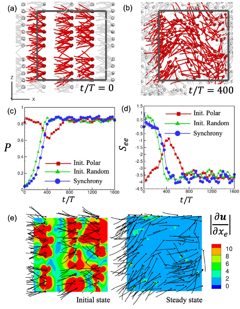
Self-organisation via elastohydrodynamic interactions– First, we investigate spermatozoal dynamics under high-density conditions (). To avoid among-spermatozoa contact, the spermatozoa are initially arranged in a grid with small fluctuations, but the swimming directions and flagellar beat phases are random (cf. Fig.1a, henceforth termed ‘of random order’). After the simulation commences, spermatozoa swim in the initially defined random directions, and coherent cell structures are therefore not observed (the average swimming direction is about 0.1). The bra-ket notation indicates the ensemble average of a physical quantity: . The spermatozoa repeatedly engage in mutual interactions, gradually change their swimming directions, and after a long period (400 cycles), a polar order is observed; all cells swim in almost the same direction (). This is maintained until the calculation ends at 800 cycles (Figs.1b, c and Movie S1).
Does such self-organisation develop only above a certain cell number density threshold, and does a phase change accompany a density variation? When the number density changes, self-organisation is observed even below the physiological conditions associated with male infertility (e.g., , equivalent to sperm/mL, in Fig.1c). However, the time required for re-orientation tends to decrease with increasing number density, and the relaxation time is density dependent. Define the relaxation time as the time required to form the polar order, thus the time to when attains 0.8, and, after normalising the time variation of by , the same exponential curve is obtained for all cases (see inset Fig.1c). Therefore, spermatozoal self-organisation does not evidence a phase change as the number density increases. The time scale over which the polar order emerges is next estimated (the details are in the supplement). The average is proportional to the number density (Fig.S5 in the supplement), suggesting that is basically proportional to . In addition, the expected value of given a random orientation of spermatozoa is Evans et al. (2011), is eventually scaled to , in good agreement with the simulation results (cf. Fig.1d).
Stability of the polar order structure– Although we found stable polar ordering, it has been suggested that coherent pusher structures are not maintained for long periods because of flow instability Alert et al. (2022); Ramaswamy (2019); Ishikawa (2009); Ishikawa et al. (2020). What factors stabilise the polar order? To answer this question, the initial configuration is varied to be near-polar in order. When spermatozoa are initially aligned in the same direction and the beating rhythm is almost the same (Fig.2a), waving instability Ishikawa et al. (2022) develops (cf. Fig.2b) and the mean swimming direction decreases from 0.8 to around 0.6 (at in Fig.2c). Although the coherent cell structure is temporarily disrupted, the swimming direction is again re-oriented to the polar state and is maintained for a very long time, as revealed by the random order results (cf. Fig.2c). Although these results indicate that spermatozoal suspensions are stable in a polar order, they also suggest that different energetic states may exist for the same -value.
Are these different swimming states attributable to changes in the stresslet? The ensemble average of the stresslet in the swimming direction is shown in Fig.2d. When the spermatozoa attain a stable state, the value of converges to about , and this is independent of the initial configuration. Notably, the convergence value is equivalent to the initial value of the polar order instability, indicating that cell structure stability cannot be based on the stresslet alone. We next investigated flow disturbance in the suspension, and the result is shown in Fig.2e. To assess the disturbance, we define the magnitude of the velocity gradient in the mean swimming direction as . Initially, spermatozoa are aligned in the same swimming direction, and the beating surfaces are near-identical. Thus, fluid motions around cells are greatly disturbed and a high region can be seen. Spermatozoa swim with six degrees of freedom in three-dimensional space; spermatozoa can spontaneously adopt an arrangement that is stable in terms of both the beating surface and the inter-spermatozoal distance, reducing flow disturbance in the steady state even at the same and .
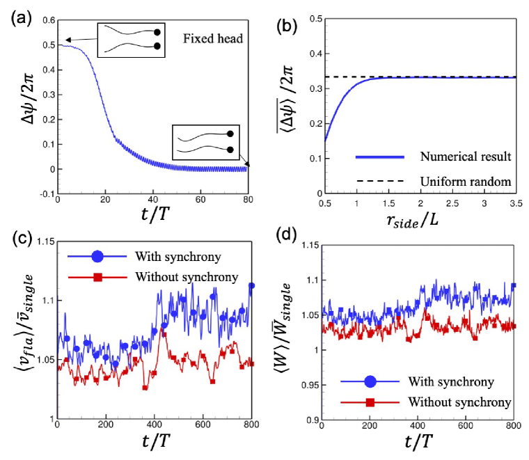
Effect of self-synchronisation– We next investigate the effect of flagellar synchronisation on collective swimming. We approach self-synchronisation via fluid motion. We thus add a curvature-dependent phase change model to the simulation, guided by Camalet and Julicher (2000); Chakrabarti and Saintillan (2019); Chakrabarti and Sintillan (2019); Goldstein et al. (2016); Oriola et al. (2017); Riedel-Kruse et al. (2007); Sartori et al. (2016) (see supplement and Nanami et al. (2020) for the details). The model is based on the geometric clutch hypothesis Lindemann (1994). The association-dissociation rate of the molecular motor dynein is regulated by the distance between the neighbouring microtubule, and the wave propagation of the active driving force is controlled by the waveform. Autonomous flagellar synchronisation is achieved by changes in both the flagellar waveform and frequency attributable to elastohydrodynamic interactions. For example, flagellar synchronisation of two fixed spermatozoa is shown in Fig. 3a.
To derive the phase difference in the suspension, the distance correlation difference is defined as , where is the distance between a spermatozoon perpendicular to the swimming direction of the th sperm and is the number of spermatozoa within from the th spermatozoon. Even in the free-swimming condition, the value is below the average that is expected (thus one third; see the supplement), suggesting that flagellar synchronisation is occurring where (cf. Fig.3b). The changes in the cell structure and the stresslet are small compared to the extent of flagellar synchronisation, which constitutes a stable polar order (see Figs.2c, d). The flagellar beating synchrony affects the flagellar dynamics more than does flagellar structure, increasing the beating speed and the work rate of spermatozoa when swimming (Figs.3c, d). These results are caused principally by frequency changes; there is little change in the mean beat amplitude on application of the synchronisation model (Fig.S12 in the supplement).
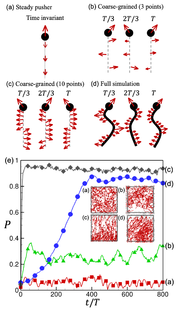
Mechanism of structure formation – Finally, we discuss how spermatozoa self-organise. We built coarse-grained models of spermatozoal swimming to investigate the effects of unsteady flagellar beating. The first model features a steady pusher (Fig.4a) for which the time-averaged driving force acts on the head and the counter-forces are distributed along the orientation vector axis to satisfy the force-free condition. Note that the stresslet magnitude of the coarse-grained pusher is the time-averaged value of the full model. The second model features a coarse-grained undulatory swimmer, similar to that of Ishimoto et al. (2017), for which time-varying driving forces are applied to representative points (Figs.4b, c). The time-varying driving force is calculated by reference to the full model. We then integrate the hydrodynamic forces acting on sections of the flagellum; we divide the flagellum into three or ten parts. Each spermatozoon is initially placed at random and the later swimming behaviour in suspension investigated (Fig. 4e). Self-organisation is not achieved by the pusher in the absence of temporal variation in flagellar beating. Undulatory swimmers with three-point forces evidence local collective swimming. As the spatial resolution of the driving force increases, thus for swimmers with ten-point forces, coherent structures are observed. The coarse-grained model neglects the fluid resistance of the flagellum, and the relaxation time is thus very short. These results indicate that unsteady fluid interactions are important when coherent structures of active swimmers are observed, and suggest that the superimposition of time-averaged stress fields does not effectively track the collective structure of unsteady self-propelled particles.
Conclusion– We developed a mechanical model of spermatozoal swimming; we explicitly expressed the elastohydrodynamic interactions among cells. We confirmed that spermatozoa enter a polar order in bulk suspension and that this structure is stable because each spermatozoon becomes autonomously stable in an individual configuration. Flagellar self-synchronisation via fluid motion affects the dynamics of flagellar beating, but the cell structure is less affected. The comparison with a coarse-grained steady pusher highlights the importance of unsteady hydrodynamic interactions in terms of polar order formation, revealing a novel self-organising mechanism of active swimmers. These results advance our knowledge on self-organisation of active matter and will contribute to analyses of biofluids and active fluids.
Acknowledgments– The authors acknowledge the support of JSPS KAKENHI (21H04999, 21H05308) and JST PRESTO (JPMJPR2142).
References
- Fauci and Dillon (2006) L. J. Fauci and R. Dillon, Annual Review of Fluid Mechanics 38, 371 (2006).
- Gaffney et al. (2011) E. Gaffney, H. Gadelha, D. Smith, J. Blake, and J. Kirkman-Brown, Annual Review of Fluid Mechanics 43, 501 (2011).
- Chakrabarti and Saintillan (2019) B. Chakrabarti and D. Saintillan, Physical Review Fluids 4, 043102 (2019).
- Fauci and McDonald (1995) L. J. Fauci and A. McDonald, Bulletin of Mathematical Biology 57, 679 (1995), ISSN 0092-8240.
- Ishimoto et al. (2017) K. Ishimoto, H. Gadêlha, E. A. Gaffney, D. J. Smith, and J. Kirkman-Brown, Physical Review Letters 118, 124501 (2017).
- Smith et al. (2009) D. J. Smith, E. A. Gaffney, H. Gadelha, N. Kapur, and J. C. Kirkman-Brown, Cell Motility and the Cytoskeleton 66, 220 (2009).
- Saintillan (2018) D. Saintillan, Annual Review of Fluid Mechanics 50, 563 (2018).
- Miles et al. (2019) C. J. Miles, A. A. Evans, M. J. Shelley, and S. E. Spagnolie, Physical Review Letters 122, 098002 (2019).
- Ishikawa et al. (2022) T. Ishikawa, T. Nghi Dang, and E. Lauga, Physical Review Fluids 7, 013104 (2022).
- Creppy et al. (2015) A. Creppy, O. Praud, X. Druart, P. L. Kohnke, and F. Plouraboue, Physical Review E 92, 032722 (2015).
- Ishikawa et al. (2011) T. Ishikawa, N. Yoshida, H. Ueno, M. Wideman, Y. Imai, and T. Yamaguchi, Physical Review Letters 107, 028102 (2011).
- Ishimoto and Gaffney (2018) K. Ishimoto and E. A. Gaffney, Scientific Reports 8, 15600 (2018).
- Schoeller and Keaveny (2018) S. F. Schoeller and E. E. Keaveny, Journal of Royal Society Interface. 15, 20170834 (2018).
- Tung et al. (2017) C. Tung, C. Lin, B. Harvey, A. G. Fiore, F. Ardon, M. Wu, and S. S. Suarez, Scientific Reports 7, 3152 (2017).
- E. et al. (2014) L. E., H. Wioland, and R. E. Goldstein, Proceedings of the National Academy of Sciences of the United States of America 111, 9733 (2014).
- Omori et al. (2022) T. Omori, K. Kikuchi, M. Schmitz, M. Pavlovic, C.-H. Chuang, and T. Ishikawa, Journal of Fluid Mechanics 930, A30 (2022).
- Samatas and Lintuvuori (2023) S. Samatas and J. Lintuvuori, Physical Review Letters 130, 024001 (2023).
- Omori and Ishikawa (2019) T. Omori and T. Ishikawa, Micromachine 10, 78 (2019).
- Rikmenspoel (1984) R. Rikmenspoel, Journal of Experimental Biology 108, 205 (1984).
- Camalet and Julicher (2000) S. Camalet and F. Julicher, New Journal of Physics 2, 24.1 (2000).
- Chakrabarti and Sintillan (2019) B. Chakrabarti and D. Sintillan, Physical Review Letters 123, 208101 (2019).
- Goldstein et al. (2016) R. E. Goldstein, E. Lauga, A. I. Pesci, and M. R. E. Proctor, Physical Review Fluids 1, 073201 (2016).
- Oriola et al. (2017) D. Oriola, H. Gadelha, and J. Casademunt, Royal Society Open Science 4, 160698 (2017).
- Riedel-Kruse et al. (2007) I. Riedel-Kruse, A. Hilfinger, J. Howard, and F. Julicher, HFSP Journal 1, 192 (2007).
- Sartori et al. (2016) P. Sartori, V. F. Geyer, A. Scholich, F. Julicher, and J. Howard, eLife. 5, e13258 (2016).
- Golestanian et al. (2011) R. Golestanian, J. M. Yeomans, and N. Uchida, Soft Matter 7, 3074 (2011).
- Miki and Clapham (2013) K. Miki and D. E. Clapham, Current Biology 23, 443 (2013).
- Kantsler et al. (2014) V. Kantsler, J. Dunkel, M. Blayney, and R. E. Goldstein, eLife 3, e02403 (2014).
- Ishimoto and Gaffney (2015) K. Ishimoto and E. A. Gaffney, Journal of The Royal Society Interface 12, 20150172 (2015).
- Omori and Ishikawa (2016) T. Omori and T. Ishikawa, Physical Review E 93, 032402 (2016).
- Ito et al. (2019) H. Ito, T. Omori, and T. Ishikawa, J. Fluid Mech. 874, 774 (2019).
- Omori et al. (2020) T. Omori, H. Ito, and T. Ishikawa, Proceedings of the National Academy of Sciences of the United States of America 117, 30201 (2020).
- Nanami et al. (2020) T. Nanami, T. Omori, and T. Ishikawa, Physics of Fluids 32, 101901 (2020).
- Tornberg and Shelley (2004) A.-K. Tornberg and M. J. Shelley, Journal of Computational Physics 196, 8 (2004).
- Beenakker (1986) C. W. J. Beenakker, Journal of Chemical Physics 85, 1581 (1986).
- Evans et al. (2011) A. A. Evans, T. Ishikawa, T. Yamaguchi, and E. Lauga, Physics of Fluids 23, 111702 (2011).
- Alert et al. (2022) R. Alert, J. Casademunt, and J.-F. Joanny, Annual Review of Condensed Matter Physics 13, 143 (2022).
- Ramaswamy (2019) S. Ramaswamy, Nature Reviews Physics 1, 640 (2019).
- Ishikawa (2009) T. Ishikawa, Journal of the Royal Society Interface 6, 815 (2009).
- Ishikawa et al. (2020) T. Ishikawa, T. Omori, and K. Kikuchi, APL Bioengineering 4, 041504 (2020).
- Lindemann (1994) C. B. Lindemann, Journal of Theoretical Biology 168, 175 (1994).