Probing the dynamics of solid density micro-wire targets after ultra-intense laser irradiation using a free-electron laser
Abstract
In this paper, we present an experiment that explores the plasma dynamics of a diameter carbon wire after being irradiated with a near-relativistic-intensity short pulse laser. Using an X-ray Free Electron Laser pulse to measure the small angle X-ray scattering signal, we observe that the scattering surface is bent and prone to instability over tens of picoseconds. The dynamics of this process are consistent with the presence of a sharp, propagating shock front inside the wire, moving at a speed close to the hole boring velocity.
Introdction
Modern laser technology allows the generation of ultra-intense optical light, compressed to a few femtoseconds and focused to a few micron spot sizes leading to intensities exceeding the threshold for relativistic electron motion of .
When a laser pulse is focused on a solid surface at these intensities, the atoms at the surface are ionized and a dense plasma is created.
The laser field interacts with electrons within a skin depth of a few tens of nanometers and can compress the surface and accelerate electron and ions to several .
The interaction of such high intensity lasers with solids has attracted considerable interest with the prospect of creating bright and compact particle accelerators Dalui et al. (2016); *Macchi2013; *Albert2021 and gamma-ray sources Stark et al. (2015).
Several physical processes occur before or after the laser pulse on a picosecond to nanosecond time scale leading for example to thermalization, diffusion, compression and ablationCraxton et al. (2015).
These processes generally take place in the regime of coupled plasmas, where atomic-scale collisions still play a role.
Processes on the sub-picosecond or even sub-femtosecond scale on the other hand usually are collective, giving rise to complex phenomena and waves.
One important aspect is the generation of a multitude of instabilities, influencing for example the coupling of the laser to the plasma, particle transport, and heating of the target.
So far, such surface plasma dynamics have been widely investigated using femtosecond optical laser pulses Sokolowski-Tinten et al. (1998), whereby various surface-sensitive methods based on optical shadowgraphymartin, interferometry Geindre et al. (1994); *Bocoum2015, and spectroscopy Mondal et al. (2010); Malvache et al. (2013); Hornung et al. (2021) have been applied. However, optical probes can only penetrate the plasma up to their critical density, i.e. a fraction of the solid density, and so cannot directly give access to the dynamics inside the solid density region.
X-ray diffraction can be employed to resolve physical processes above the critical density, such as for example non-thermal melting Siders et al. (1999), coherent lattice vibrations Sokolowski-Tinten et al. (2003), and ultrafast phase transitions McBride et al. (2019).
Recently, X-ray pulses from X-ray free electron lasers (XFEL) have been applied to investigate laser produced plasmas with femtosecond temporal resolution. Small-angle femtosecond X-ray Scattering (SAXS) has revealed density gradients of expanding solid-density plasmas with nanometer spatial and few femtosecond temporal resolution Kluge et al. (2014); Gorkhover et al. (2016); Kluge et al. (2018a), transient nano-jet emission from grating surfacesKluge et al. (2018a), as well as ultra-fast heating and ionization Kluge et al. (2016); *Kluge2017; *Gaus2021.
Here we use SAXS to study the dynamics inside solid density wires that turn into plasmas after high-intensity short-pulse laser irradiation. Our experimental data shows that the scattering surface is bent by a few microns and prone to instability development also on the scale of a few microns over the course of tens of picoseconds. The dynamics of this process are consistent with a sharp, propagating compression (shock) front inside the wire. With the novel SAXS method we measure the tilt angle of this front and observe that it is moving forward with a speed close to the hole boring (HB) velocity of up to .
Experimental setup
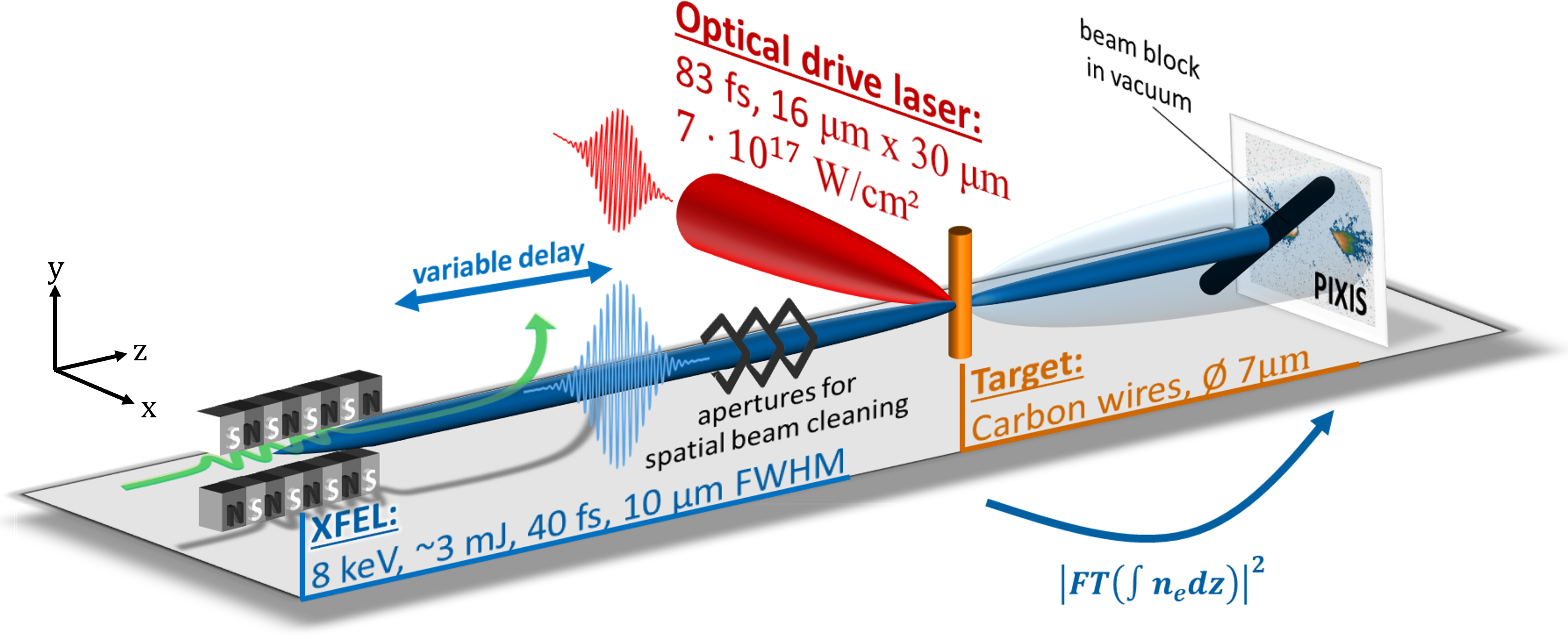
We report on an experiment probing the plasma dynamics of a diameter carbon wire after the irradiation with the near-relativistic high-intensity (HI) Titanium:Sapphire short-pulse near-infrared (wavelength ) laser at the Matter in Extreme Conditions (MEC) endstation at the Linear Coherent Light Source (LCLS) XFEL.
The HI laser pulse duration was and the spot size at FWHM was and in horizontal and vertical direction, respectively.
The laser pulse energy was measured to be up to before compression.
With transmission to the target and of the energy being in the FWHM area, the peak intensity on target can be calculated to approx. , equivalent to a normalized laser strength amplitude of .
The XFEL pulse had a diameter of and pulse duration of , both at FWHM.
The X-ray photon energy was , with approximately photons per pulse (the exact number on target varies from shot to shot due to different absorbers).
Temporal synchronization between the HI short-pulse laser and LCLS XFEL pulse was achieved with a precision of rms.
The carbon wire targets were irradiated under normal incidence by both the pump and the probe pulse, which were also perpendicular to each other, see Fig. 1.
For reasons detailed below, the XFEL axis was vertically offset by approximate along the wire towards the top.
In the following, we present a series of shots on these wires, varying the pump laser intensity between 50% and 100% (i.e. between and ), and the probe delay between and . We measure the small angle X-ray scattering (SAXS) signal with an PIXIS2048 X-ray detector with pixel size X-ray that was positioned behind the target. In this configuration, the signal is given in Born approximation (i.e. phase contrast only) by the Fourier transform of the electron density, , where is the scattering vector defined by the difference between the incoming XFEL wave vector and the scattered wave vector . Here, we only expect two streaks perpendicular to the wire surface, the interference pattern of scattered waves from the wire front and rear surface (i.e. ) cannot be resolved with the detector resolution (). The setup we employ here enables the study of a multitude of relevant physics following the relativistic laser irradiation Kluge et al. (2014, 2017), through the analysis of the change of the scattering streak’s signal intensity and slope, orientation, and structure, as discussed in the following.
SAXS Method
First, we would like to extract the temporal evolution of the plasma expansion, as we demonstrated before on grating targetsKluge et al. (2018a). Similar to the Debye-Waller analysis for thermal motion of particles, we can replace the displacement of scatterers due to thermal motion with the displacement of the plasma due to non-thermal melt/expansion into vacuum. The expansion scale can then simply be derived from the exponential roll-off of the scattering signal at large scattering angles Kluge et al. (2018a); Gorkhover et al. (2016)
| (1) |
where depends on the geometry of the sample. The exponent is expected for flat surfaces to be in the range of , e.g. for a cuboid, for a cylinder and for a sphereGuinier et al. (1955), or for a fractally rough surfaceBenedetti et al. (1993). The exponential term is the Debye-Waller factor.
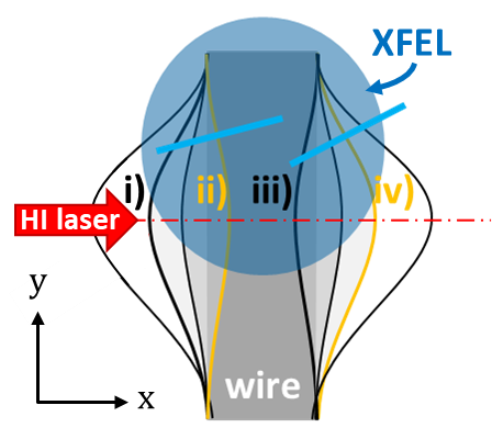
Secondly, we can expect the scattering streak of the wire to tilt if the wire is being deformed by the laser.
As depicted in Fig. 1(b), this can in principle happen due to (i) ablation into vacuum at the front following the thermal pressure set up by laser accelerated electrons or the bulk electrons heated by return current collisionsKluge et al. (2018b); Mora (2003); (ii) a traveling compression (hole boring or shock) front into the bulk launched by the laser pressureRobinson et al. (2009); Fiuza et al. (2012); (iii) a rarefaction (shock) wave into the wire at the rear surface Bulgakova (1999); (iv) ablation into vacuum at the rearMora (2003).
To determine the relevant mechanisms in the present setup, we employ simulations already published for similar conditions (cp. Fig. 3 of Gaus et al. (2021)).
The laser and XFEL parameters are the same while only the target material differs, being Si in the simulation instead of the carbon used here.
However, the effective scattering length densities are quite similar (for silicon it is approx. of that of carbon) and the simulations therefore describe also the present case qualitatively.
As can be seen in these simulations, over the course of only a few picoseconds the front surface is fully ablated (i.e. order of scalelength).
Such a diluted surface would not be possible to be detected due to the corresponding Debye-Waller factor that would be close to zero and would suppress the streak signal already at small , effectively making the streak vanish.
Simultaneously, a compression shock wave is launched by the laser pressure, which continues to travel through the target over 10s of picoseconds.
The shock front velocity is between (hydro simulation) and (simulation), i.e. around the hole boring velocity
| (2) |
where the piston parameter for a fully ionized plasma is , and are the ion and electron mass, respectively, and is the critical densityRobinson et al. (2009).
The HB velocity in carbon for the present case is computed to an only slightly larger value of , hence we can expect the compression wave to break out of the rear surface of the thick carbon wire after approx. .
Additionally, briefly after the laser irradiation some high energy electrons accelerated by the laser penetrate the full target ballistically, which excites a return current and consequently bulk target heating to some few .
Hence, the rear surface expands/ablates slightly due to the thermal pressure.
Both features, the shock front and the ablating rear surface, as well as the rarefaction wave countering the rear surface expansion, could in principle lead to a signal in form of a streak on our SAXS detector depending on their exact sharpness.
Finally, instabilities in the shock front or the rear surface, respectively, can lead to a break-up of the single wire scattering line into multiple lines, which in the case of a sinusoidal surface modulation would each reflect a certain orientation of the target normal at the point of inflection. Such instabilites can be, among others, Rayleigh-Taylor like instabilities leading to a surface rippling at relativistic intensitiesPalmer et al. (2012); Sgattoni et al. (2015); Kluge et al. (2015), filamentation of filamentation-two-stream instabilities in the target Bret et al. (2004); Metzkes et al. (2014) or at the target rear surfaceGöde et al. (2017), Weibel-like instabilities occurring during the plasma propagation in vacuum after laser accelerationQuinn et al. (2012), or the Z-pinch sausage (m=0) instability (especially at later times after few ps)Beg et al. (2004). The instabilities lead to a growth of initially small density fluctuations to form ripples even on an initially flat target surface.
In the regime relevant for this work, the dominant source of small angle X-ray scattering is coherent elastic Thomson scattering from electrons, but there is also a relevant diffractive contribution by bound-free transition absorption. The latter depends sensitively on the plasma ionization degree, i.e. the plasma temperature.
Results
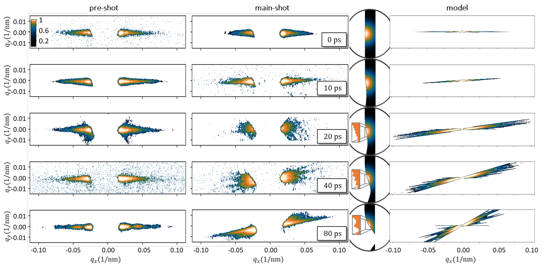
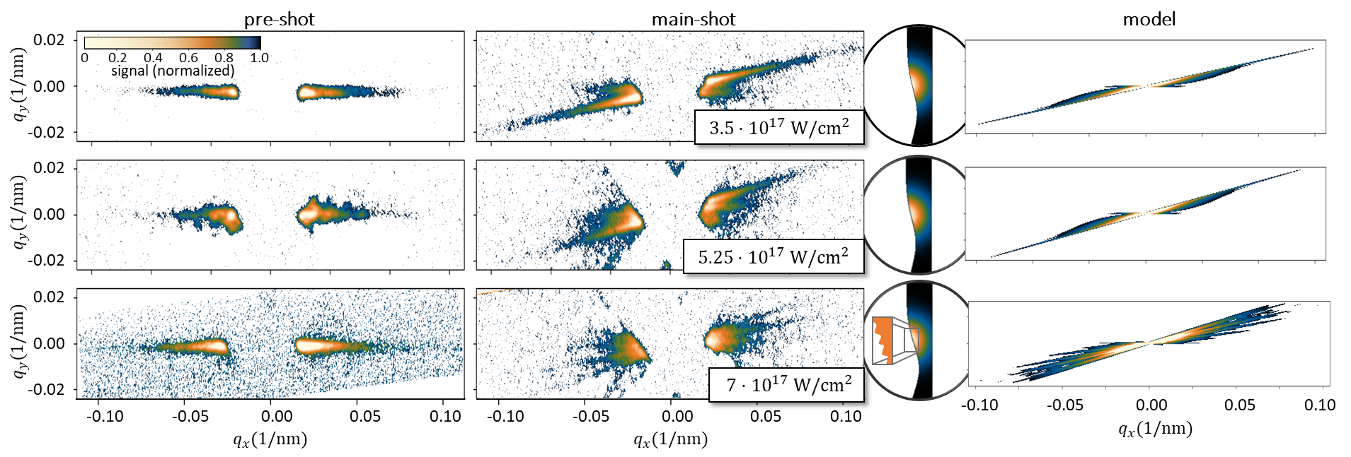
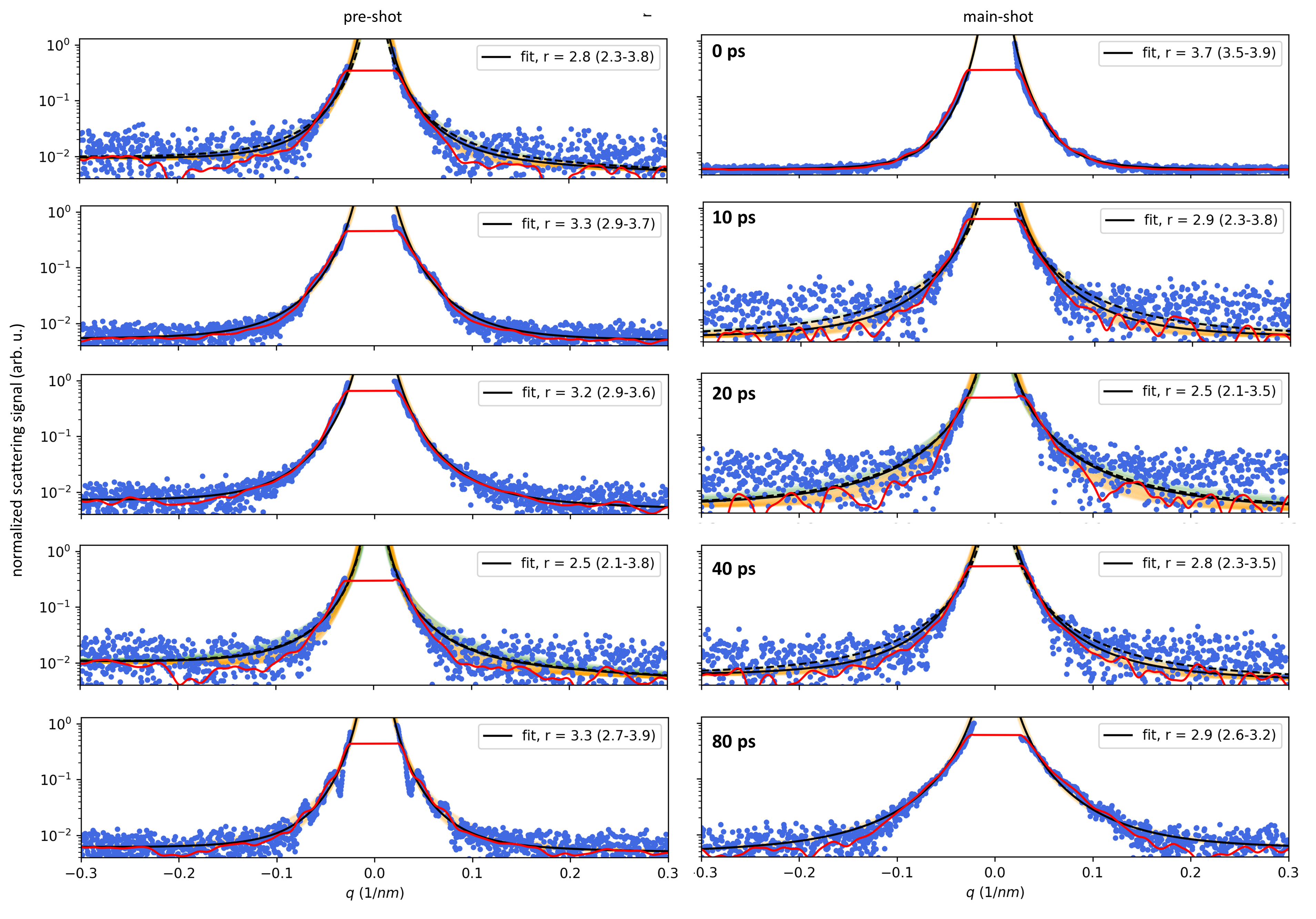
The scattering patterns recorded in this experiment are shown in Fig. 2 as a function of delay at full laser intensity, and in Fig. 3 as function of intensity at probe delay. For each shot we took two XFEL-only pre-shots as a reference, that we averaged for better statistics. We used this to determine the undriven target normal orientation and verify the surface quality and XFEL overlap. We also took an XFEL-only post-shot after the combined laser-XFEL main-shot, in order to be able to subtract in the pre- and main-shot the parasitic XFEL scattering on the beamstop and other optical components in the beamline. The figures show the background subtracted and normalized signal.
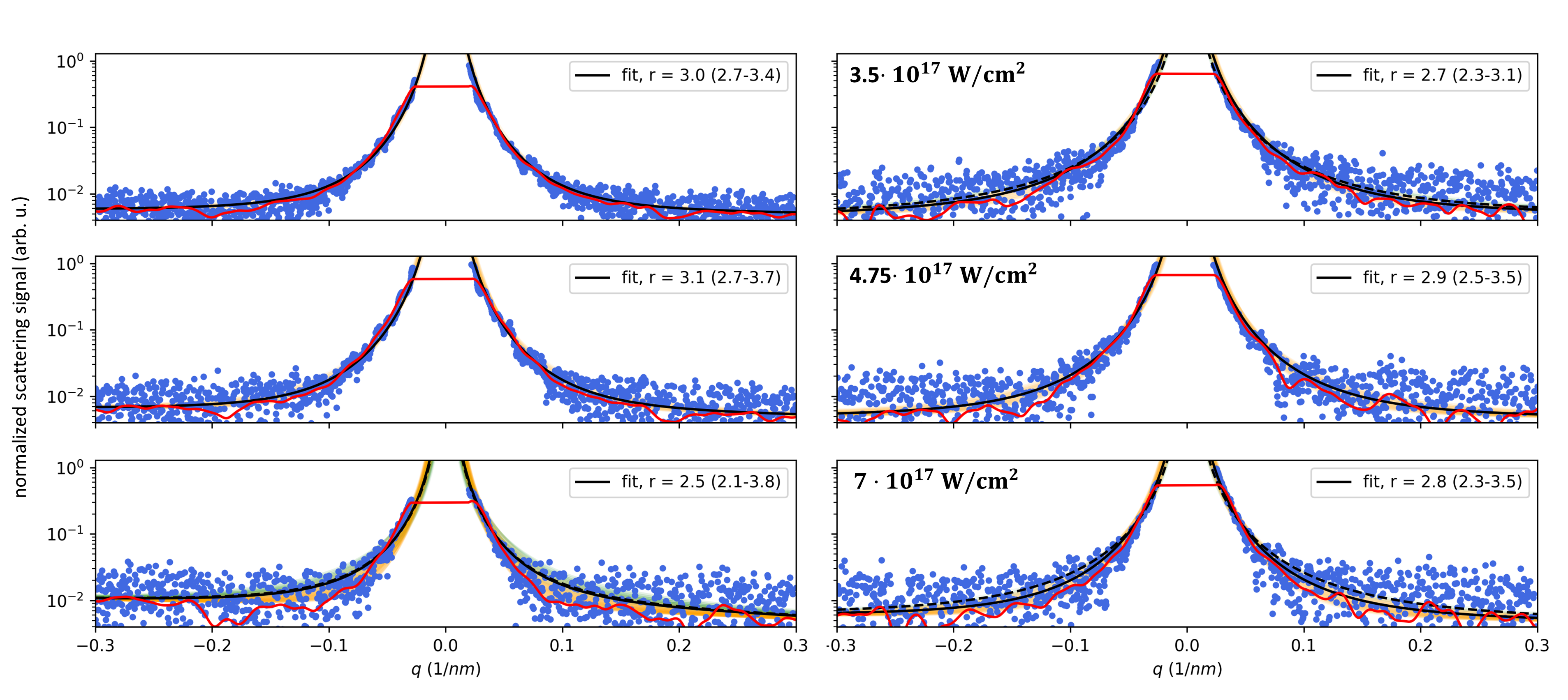
We start the analysis of the streaks by comparing their lengths, i.e the intensity fall-off with scattering angle (i.e. ), cf. Figs. 4 and 5. In none of the shots a Debye-Waller like exponential roll-off at large -values could be observed. Since the q-range with signal above the background is limited in most of the shots to , this means that the surface roughness would be less than
| (3) |
In reality this value is even much larger since photon number Poisson statistics, background uncertainty, and the uncertainty of and its fit correlation with add considerable fit uncertainty (the latter alone is approx. a factor of 2). This means that in our case due to the low dynamic range limiting us to very small q-value, the Debye-Waller factor cannot be discriminated from unity in all shots. Setting it to in Eqn. (1), the scattering intensity can thus simply be written as
| (4) |
where is the proportionality factor, comprises the radiation and detector background signal and should take on values between and .
We fitted all the streaks with with Eqn. (4) and find a value close to for all cases, see Fig. 4. This means that in all cases the shape of the scattering front remains that of a cylinder and is not significantly compressed by the laser towards flatter planar geometry.
Having found no evidence for expansion nor compression, we conclude that the visual impression of shorter or longer streaks is consistent with being only due to different signal levels instead of a change of or .
Secondly, we analyze the tilt angle of the streak in the main-shot relative to the pre-shot. Fig. 6 shows the tilt angles as a function of delay for full laser intensity (a), and as function of laser intensity at 40 ps delay (b). As described above, there are several mechanisms that in principle can cause a time-varying signal orientation. Some we can exclude based on our measurements by realizing that our XFEL spatial overlap was vertically shifted approx. above the laser interaction point (i.e. FWHM of the XFEL pulse spot size). This is why we only see one streak - the other streak in the opposite direction is simply not in the XFEL field of view. However, this allows us to infer the direction of the tilt of the scattering surface. In the present case, we conclude that the tilt must be in laser forward direction. This means that those processes that cause a tilted density contour tilt towards the laser can be excluded, i.e. front surface plasma expansion and rear surface rarefaction wave.
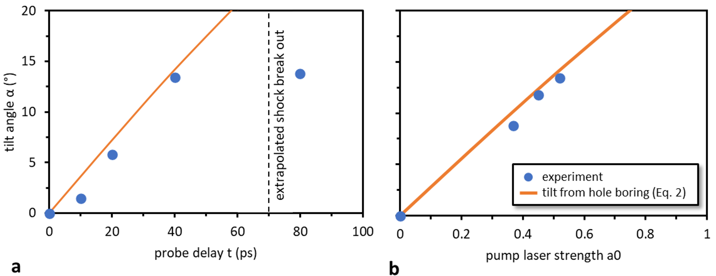
We are left with the possibilities of rear surface tilt of the target due to plasma expansion into vacuum, or a tilt of the front surface by hole boring or a compression (shock) front traveling forward, cf. Fig. 1(b) (orange lines). To connect the tilt angle of the streak with the plasma electron density contour more quantitative, we adopt a very simple model. We assume that the scattering surface is moving forward with a velocity that depends on the local laser intensity. In particular, the surface longitudinal position as a function of the transverse offset shall be Gaussian with the same FWHM as the laser amplitude focal spot size . Then it can easily be geometrically derived that the target position along the target normal is connected to the angle of the normal at the point of inflection (i.e. the tilt angle) via
| (5) |
From the delay scan of in Fig. 6a we can then directly obtain the peak forward velocity of until a probe delay of .
Obviously at delay the measured tilt does not increase anymore.
However, extrapolating the forward velocity, we would expect the scattering front to exit the rear of the wire at if launched from the front at (However, one needs to be careful with the interpretation, since we present only single shot results).
From the intensity scan in panel (b) we infer that the forward velocity is proportional to the laser strength parameter , i.e. the square root of the laser intensity, , with a proportionality constant of .
The forward velocity extracted from both the delay scan and the intensity scan is in remarkable agreement with the hole boring velocity from Eqn.(2), ,
and for the full intensity case agrees with the compression front velocity in the aforementioned simulation.
Finally, we point out that the tilted streak transitions into a set of multiple streaks at full intensity after . This is indicative for a modulation of the scattering surface. This filamentation starts around , is most visible at and is not visible at after the laser irradiation as strong anymore, though there is still a faint second streak visible. Potentially the instability is still present, but its spatial frequency decreased so that the field of view of the XFEL comprises less filaments. The growth of the instability apparently continues, as the angular spread between the streaks at larger delay values is larger than at earlier times, i.e. the modulation depth seems to be increasing with time. In most shots we additionally see remnants of the streak in horizontal direction, perpendicular to the undriven wire surface. We believe that this is primarily due to scattering of the outer wings of the XFEL spot at the rear surface or vertical distant, unperturbed regions.
Conclusions
In the experiment presented above we have employed SAXS to measure the target solid density response upon HI laser irradiation. We interpret our measurements with scattering at a compression front inside the target after it detached from the laser radiation pressure accelerated front surface. This follows from the following arguments: First, the simulation in Gaus et al. (2021) that predict a compression shock front persisting over 10s of ps. Secondly, the front and rear surface are expected to quickly smooth out due to plasma expansion, so that the streak cannot originate from the ablated wire surfaces. The tilt angle orientation of the measured SAXS streaks that indicates a forward deformation of the scattering surface, i.e. we can exclude the rear surface rarefaction wave. Rather, it is in agreement with a laser compression front following the lateral laser intensity shape. Moreover, the change of the tilt angle as a function of probe delay and pump intensity is in agreement with the hole boring velocity scaling. Surprisingly, apparently the scattering surface remains quite sharp over many picoseconds, since the observed streak lengths did not change.
At full laser intensity we observe the streak to filament at a probe time after HI irradiation. The number of visible streaks then decreases and the distance between streaks seems to increase between to . This indicates that if the signal was due to a sinusoidal-like modulation of the scattering surface, its period and amplitude would grow with time. With the limited data available from this small study, we cannot specify which instability type causes the splitting of the streak. However, apparently similar modulations on similar hydrodynamic time scales have been observed before by means of optical probes and hence at different spatial resolution and lower plasma densitymartin.
In this paper we reported on the plasma reaction after near-relativistic pump laser irradiation of a thin wire.
This study is limited to only a few shots, which means that we cannot give an estimate of the reproducibility.
Additionally, the fits of the streak to obtain are connected with large uncertainties and the fit of the surface expansion was not possible at all since the exponential roll-off due to the Debye-Waller factor was not visible (likely hidden in the background).
While absorption and phase contrast x-ray imaging Schropp et al. (2015) could also have been employed to visualize the dynamics of the bulk density on those scales, the spatial resolution of these methods is still limited to a few and is thus larger than the relevant scales during or shortly after the laser pump irradiation itself – which is our future aim in order to eventually probe directly the early dynamics.
For ultra-relativistic intensities, as they are relevant for many important applications such as ion acceleration, isochoric heating, or high harmonic generation, much faster and higher resolving probing is currently underdevelopment.
The next generation of high intensity XFEL experiments are currently starting at European XFEL, using shorter probe pulses, larger detector distance and dynamic range, and background radiation suppression with chromatic mirrors.
Then, the onset and early dynamics of the filamentation and plasma expansion could be studied and directly compared to existing models and particle-in-cell simulations.
Data availability
The data that support the findings of this study will be openly available once the peer-reviewed article is published.
Acknowledgements.
This work was partially supported by DOE Office of Science, Fusion Energy Science under FWP 100182. Use of the Linac Coherent Light Source (LCLS), SLAC National Accelerator Laboratory, is supported by the U.S. Department of Energy, Office of Science, Office of Basic Energy Sciences under Contract No. DE-AC02-76SF00515. The experiments were performed at the Matter at Extreme Conditions (MEC) instrument of LCLS, supported by the DOE Office of Science, Fusion Energy Science under contract No. SF00515. Christian Gutt acknoledges funding by DFG (GU 535/6-1). This work has also been supported by HIBEF (www.hibef.eu) and partially by H2020 Laserlab Europe V (PRISES) contract no. 871124, and by the German Federal Ministry of Education and Research (BMBF) under contract number 03Z1O511.References
- Dalui et al. (2016) Malay Dalui, M. Kundu, T. Madhu Trivikram, Krishanu Ray, and M. Krishnamurthy, “Manifestation of anharmonic resonance in the interaction of intense ultrashort laser pulses with microstructured targets,” Physics of Plasmas 23, 103101 (2016).
- Macchi et al. (2013) Andrea Macchi, Marco Borghesi, and Matteo Passoni, “Ion acceleration by superintense laser-plasma interaction,” Reviews of Modern Physics 85, 751–793 (2013).
- Albert et al. (2021) Félicie Albert, M E Couprie, Alexander Debus, Mike C Downer, Jérôme Faure, Alessandro Flacco, Leonida A Gizzi, Thomas Grismayer, Axel Huebl, Chan Joshi, M Labat, Wim P Leemans, Andreas R Maier, Stuart P D Mangles, Paul Mason, François Mathieu, Patric Muggli, Mamiko Nishiuchi, Jens Osterhoff, P P Rajeev, Ulrich Schramm, Jörg Schreiber, Alec G R Thomas, Jean-Luc Vay, Marija Vranic, and Karl Zeil, “2020 roadmap on plasma accelerators,” New Journal of Physics 23, 031101 (2021).
- Stark et al. (2015) David J. Stark, Chinmoy Bhattacharjee, Alexey V. Arefiev, Toma Toncian, R. D. Hazeltine, and S. M. Mahajan, “Relativistic Plasma Polarizer: Impact of Temperature Anisotropy on Relativistic Transparency,” Physical Review Letters 115, 025002 (2015).
- Craxton et al. (2015) R. S. Craxton, K. S. Anderson, T. R. Boehly, V. N. Goncharov, D. R. Harding, J. P. Knauer, R. L. McCrory, P. W. McKenty, D. D. Meyerhofer, J. F. Myatt, A. J. Schmitt, J. D. Sethian, R. W. Short, S. Skupsky, W. Theobald, W. L. Kruer, K. Tanaka, R. Betti, T. J. B. Collins, J. A. Delettrez, S. X. Hu, J. A. Marozas, A. V. Maximov, D. T. Michel, P. B. Radha, S. P. Regan, T. C. Sangster, W. Seka, A. A. Solodov, J. M. Soures, C. Stoeckl, and J. D. Zuegel, “Direct-drive inertial confinement fusion: A review,” Physics of Plasmas 22, 110501 (2015).
- Sokolowski-Tinten et al. (1998) K. Sokolowski-Tinten, J. Bialkowski, A. Cavalleri, D. Von der Linde, A. Oparin, J. Meyer-Ter-Vehn, and S. I. Anisimov, “Transient States of Matter during Short Pulse Laser Ablation,” Physical Review Letters 81, 224 (1998).
- Geindre et al. (1994) J. P. Geindre, A. Mysyrowicz, A. Dos Santos, P. Audebert, A. Rousse, G. Hamoniaux, A. Antonetti, F. Falliès, and J. C. Gauthier, “Frequency-domain interferometer for measuring the phase and amplitude of a femtosecond pulse probing a laser-produced plasma,” Optics Letters 19, 1997 (1994).
- Bocoum et al. (2015) Maïmouna Bocoum, Frederik Böhle, Aline Vernier, Aurélie Jullien, Jérôme Faure, and Rodrigo Lopez-Martens, “Spatial-domain interferometer for measuring plasma mirror expansion,” Optics Letters 40, 3009 (2015).
- Mondal et al. (2010) S. Mondal, Amit D. Lad, Saima Ahmed, V. Narayanan, J. Pasley, P. P. Rajeev, A. P.L. Robinson, and G. Ravindra Kumar, “Doppler spectrometry for ultrafast temporal mapping of density dynamics in laser-induced plasmas,” Physical Review Letters 105, 105002 (2010).
- Malvache et al. (2013) A. Malvache, A. Borot, F. Quéré, and R. Lopez-Martens, “Coherent wake emission spectroscopy as a probe of steep plasma density profiles,” Physical Review E - Statistical, Nonlinear, and Soft Matter Physics 87, 035101 (2013).
- Hornung et al. (2021) J. Hornung, Y. Zobus, S. Roeder, A. Kleinschmidt, D. Bertini, M. Zepf, and V. Bagnoud, “Time-resolved study of holeboring in realistic experimental conditions,” Nature Communications 2021 12:1 12, 1–7 (2021).
- Siders et al. (1999) C. W. Siders, A. Cavalleri, K. Sokolowski-Tinten, Cs. Toth, T. Guo, M. Kammler, M. Horn von Hoegen, K. R. Wilson, D. von der Linde, and C. P. J. Barty, “Detection of Nonthermal Melting by Ultrafast X-ray Diffraction,” Science 286, 1340 (1999).
- Sokolowski-Tinten et al. (2003) Klaus Sokolowski-Tinten, Christian Blome, Juris Blums, Andrea Cavalleri, Clemens Dietrich, Alexander Tarasevitch, Ingo Uschmann, Eckhard Förster, Martin Kammler, Michael Horn-von Hoegen, and Dietrich Von der Linde, “Femtosecond X-ray measurement of coherent lattice vibrations near the Lindemann stability limit,” Nature 2003 422:6929 422, 287–289 (2003).
- McBride et al. (2019) E. E. McBride, A. Krygier, A. Ehnes, E. Galtier, M. Harmand, Z. Konôpková, H. J. Lee, H. P. Liermann, B. Nagler, A. Pelka, M. Rödel, A. Schropp, R. F. Smith, C. Spindloe, D. Swift, F. Tavella, S. Toleikis, T. Tschentscher, J. S. Wark, and A. Higginbotham, “Phase transition lowering in dynamically compressed silicon,” Nature Physics 15, 89–94 (2019).
- Kluge et al. (2014) T. Kluge, C. Gutt, L. G. Huang, J. Metzkes, U. Schramm, M. Bussmann, and T. E. Cowan, “Using X-ray free-electron lasers for probing of complex interaction dynamics of ultra-intense lasers with solid matter,” Physics of Plasmas 21, 033110 (2014).
- Gorkhover et al. (2016) Tais Gorkhover, Sebastian Schorb, Ryan Coffee, Marcus Adolph, Lutz Foucar, Daniela Rupp, Andrew Aquila, John D. Bozek, Sascha W. Epp, Benjamin Erk, Lars Gumprecht, Lotte Holmegaard, Andreas Hartmann, Robert Hartmann, Günter Hauser, Peter Holl, Andre Hömke, Per Johnsson, Nils Kimmel, Kai Uwe Kühnel, Marc Messerschmidt, Christian Reich, Arnaud Rouzée, Benedikt Rudek, Carlo Schmidt, Joachim Schulz, Heike Soltau, Stephan Stern, Georg Weidenspointner, Bill White, Jochen Küpper, Lothar Strüder, Ilme Schlichting, Joachim Ullrich, Daniel Rolles, Artem Rudenko, Thomas Möller, and Christoph Bostedt, “Femtosecond and nanometre visualization of structural dynamics in superheated nanoparticles,” Nature Photonics 10, 93–97 (2016).
- Kluge et al. (2018a) Thomas Kluge, Melanie Rödel, Josefine Metzkes-Ng, Alexander Pelka, Alejandro Laso Garcia, Irene Prencipe, Martin Rehwald, Motoaki Nakatsutsumi, Emma E. McBride, Tommy Schönherr, Marco Garten, Nicholas J. Hartley, Malte Zacharias, Jörg Grenzer, Artur Erbe, Yordan M. Georgiev, Eric Galtier, Inhyuk Nam, Hae Ja Lee, Siegfried Glenzer, Michael Bussmann, Christian Gutt, Karl Zeil, Christian Rödel, Uwe Hübner, Ulrich Schramm, and Thomas E. Cowan, “Observation of Ultrafast Solid-Density Plasma Dynamics Using Femtosecond X-Ray Pulses from a Free-Electron Laser,” Physical Review X 8, 031068 (2018a).
- Kluge et al. (2016) T. Kluge, M. Bussmann, H.-K. Chung, C. Gutt, L. G. Huang, M. Zacharias, U. Schramm, and T. E. Cowan, “Nanoscale femtosecond imaging of transient hot solid density plasmas with elemental and charge state sensitivity using resonant coherent diffraction,” Physics of Plasmas 23, 033103 (2016).
- Kluge et al. (2017) T. Kluge, C. Rödel, M. Rödel, A. Pelka, E. E. McBride, L. B. Fletcher, M. Harmand, A. Krygier, A. Higginbotham, M. Bussmann, E. Galtier, E. Gamboa, A. L. Garcia, M. Garten, S. H. Glenzer, E. Granados, C. Gutt, H. J. Lee, B. Nagler, W. Schumaker, F. Tavella, M. Zacharias, U. Schramm, and T. E. Cowan, “Nanometer-scale characterization of laser-driven compression, shocks, and phase transitions, by x-ray scattering using free electron lasers,” Physics of Plasmas 24, 102709 (2017).
- Gaus et al. (2021) Lennart Gaus, Lothar Bischoff, Michael Bussmann, Eric Cunningham, Chandra B. Curry, Juncheng E, Eric Galtier, Maxence Gauthier, Alejandro Laso García, Marco Garten, Siegfried Glenzer, Jörg Grenzer, Christian Gutt, Nicholas J. Hartley, Lingen Huang, Uwe Hübner, Dominik Kraus, Hae Ja Lee, Emma E. McBride, Josefine Metzkes-Ng, Bob Nagler, Motoaki Nakatsutsumi, Jan Nikl, Masato Ota, Alexander Pelka, Irene Prencipe, Lisa Randolph, Melanie Rödel, Youichi Sakawa, Hans-Peter Peter Schlenvoigt, Michal Šmíd, Franziska Treffert, Katja Voigt, Karl Zeil, Thomas E. Cowan, Ulrich Schramm, Thomas Kluge, J. E. Juncheng, Eric Galtier, Maxence Gauthier, Alejandro Laso García, Marco Garten, Siegfried Glenzer, Jörg Grenzer, Christian Gutt, Nicholas J. Hartley, Lingen Huang, Uwe Hübner, Dominik Kraus, Hae Ja Lee, Emma E. McBride, Josefine Metzkes-Ng, Bob Nagler, Motoaki Nakatsutsumi, Jan Nikl, Masato Ota, Alexander Pelka, Irene Prencipe, Lisa Randolph, Melanie Rödel, Youichi Sakawa, Hans-Peter Peter Schlenvoigt, Michal Šmíd, Franziska Treffert, Katja Voigt, Karl Zeil, Thomas E. Cowan, Ulrich Schramm, and Thomas Kluge, “Probing ultrafast laser plasma processes inside solids with resonant small-angle x-ray scattering,” Physical Review Research 3, 043194 (2021).
- Guinier et al. (1955) André Guinier, Gérard Fournet, and Kenneth L Yudowitch, “Small-angle scattering of X-rays,” (1955).
- Benedetti et al. (1993) A. Benedetti, G. Fagherazzi, P. Riello, Y. W. Zeng, F. Pinna, and M. Signoretto, “Fractal properties of a partially crystalline zirconium oxide aerogel,” Journal of Applied Crystallography 26, 717–720 (1993).
- Kluge et al. (2018b) T. Kluge, M. Bussmann, U. Schramm, and T. E. Cowan, “Simple scaling equations for electron spectra, currents, and bulk heating in ultra-intense short-pulse laser-solid interaction,” Physics of Plasmas 25, 073106 (2018b).
- Mora (2003) P. Mora, “Plasma Expansion into a Vacuum,” Physical Review Letters 90, 185002 (2003).
- Robinson et al. (2009) A. P.L. Robinson, P. Gibbon, M. Zepf, S. Kar, R. G. Evans, and C. Bellei, “Relativistically correct hole-boring and ion acceleration by circularly polarized laser pulses,” Plasma Physics and Controlled Fusion 51, 024004 (2009).
- Fiuza et al. (2012) F. Fiuza, R. A. Fonseca, J. Tonge, W. B. Mori, and L. O. Silva, “Weibel-instability-mediated collisionless shocks in the laboratory with ultraintense lasers,” Physical Review Letters 108, 1–5 (2012).
- Bulgakova (1999) Nadezhda M Bulgakova, “Possibility of rarefaction shock wave under short pulse laser ablation of solids,” (1999).
- Palmer et al. (2012) C. A.J. Palmer, J. Schreiber, S. R. Nagel, N. P. Dover, C. Bellei, F. N. Beg, S. Bott, R. J. Clarke, A. E. Dangor, S. M. Hassan, P. Hilz, D. Jung, S. Kneip, S. P.D. Mangles, K. L. Lancaster, A. Rehman, A. P.L. Robinson, C. Spindloe, J. Szerypo, M. Tatarakis, M. Yeung, M. Zepf, and Z. Najmudin, “Rayleigh-Taylor instability of an ultrathin foil accelerated by the radiation pressure of an intense laser,” Physical Review Letters 108, 225002 (2012).
- Sgattoni et al. (2015) A. Sgattoni, S. Sinigardi, L. Fedeli, F. Pegoraro, and A. Macchi, “Laser-driven Rayleigh-Taylor instability: Plasmonic effects and three-dimensional structures,” Physical Review E - Statistical, Nonlinear, and Soft Matter Physics 91, 013106 (2015).
- Kluge et al. (2015) T. Kluge, J. Metzkes, K. Zeil, M. Bussmann, U. Schramm, and T. E. Cowan, “Two surface plasmon decay of plasma oscillations,” Physics of Plasmas 22, 64502 (2015).
- Bret et al. (2004) A. Bret, M. C. Firpo, and C. Deutsch, “Collective electromagnetic modes for beam-plasma interaction in the whole [Formula presented] space,” Physical Review E - Statistical Physics, Plasmas, Fluids, and Related Interdisciplinary Topics 70, 15 (2004).
- Metzkes et al. (2014) J. Metzkes, T. Kluge, K. Zeil, M. Bussmann, S. D. Kraft, T. E. Cowan, and U. Schramm, “Experimental observation of transverse modulations in laser-driven proton beams,” New Journal of Physics 16, 23008 (2014).
- Göde et al. (2017) S. Göde, C. Rödel, K. Zeil, R. Mishra, M. Gauthier, F.-E. Brack, T. Kluge, M.J. J. MacDonald, J. Metzkes, L. Obst, M. Rehwald, C. Ruyer, H.-P. P. Schlenvoigt, W. Schumaker, P. Sommer, T.E. E. Cowan, U. Schramm, S. Glenzer, and F. Fiuza, “Relativistic electron streaming instabilities modulate proton beams accelerated in laser-plasma interactions,” Physical Review Letters 118, 194801 (2017).
- Quinn et al. (2012) K. Quinn, L. Romagnani, B. Ramakrishna, G. Sarri, M. E. Dieckmann, P. A. Wilson, J. Fuchs, L. Lancia, A. Pipahl, T. Toncian, O. Willi, R. J. Clarke, M. Notley, A. MacChi, and M. Borghesi, “Weibel-induced filamentation during an ultrafast laser-driven plasma expansion,” Physical Review Letters 108, 135001 (2012).
- Beg et al. (2004) F. N. Beg, E. L. Clark, M. S. Wei, A. E. Dangor, R. G. Evans, A. Gopal, K. L. Lancaster, K. W. D. Ledingham, P. McKenna, P. A. Norreys, M. Tatarakis, M. Zepf, and K. Krushelnick, “High-Intensity-Laser-Driven ¡math display=”inline”¿ ¡mi¿Z¡/mi¿ ¡/math¿ Pinches,” Physical Review Letters 92, 095001 (2004).
- Schropp et al. (2015) Andreas Schropp, Robert Hoppe, Vivienne Meier, Jens Patommel, Frank Seiboth, Yuan Ping, Damien G. Hicks, Martha A. Beckwith, Gilbert W. Collins, Andrew Higginbotham, Justin S. Wark, Hae Ja Lee, Bob Nagler, Eric C. Galtier, Brice Arnold, Ulf Zastrau, Jerome B. Hastings, and Christian G. Schroer, “Imaging Shock Waves in Diamond with Both High Temporal and Spatial Resolution at an XFEL,” Scientific Reports 5, 11089 (2015).