MOC-AE: An Anatomically-Pathological-Based model for Clinical Decision Support System of tumoural brain images
Abstract
The present work proposes a Multi-Output Classification Autoencoder (MOC-AE) algorithm to extract features from brain tumour images. The proposed algorithm is able to focus on both the normal features of the patient and the pathological features present in the case, resulting in a compact and significant representation of each image. The architecture of MOC-AE combines anatomical information from the patient’s scan using an Autoencoder (AE) with information related to a specific pathology using a classification output with the same image descriptor. This combination of goals forces the network to maintain a balance between anatomical and pathological features of the case while maintaining the low cost of the labels being used.
The results obtained are compared with those of similar studies and the strengths and limitations of each approach are discussed. The results demonstrate that the proposed algorithm is capable of achieving state-of-the-art results in terms of both the anatomical and tumor characteristics of the recommended cases.
keywords:
MSC:
68T45, 68U10, 94A08, 65D17 \KWDcontent-based image retrieval, deep learning, autoencoder, feature extraction , clinical decision support system, comparative diagnostic, magnetic resonance imaging1 Introduction
The techniques used in medicine to treat pathologies are becoming more advanced each day and are capable of performing less invasive and more efficient treatments. However, before a patient can apply proper treatment, a correct diagnosis of the disease is necessary, which becomes an important task in this process.
Although medicine has radically evolved, diagnosis is still mostly a human process and the expert must be able to thoroughly evaluate the patient’s evidence and avoid making mistakes during the process. A late or incorrect diagnosis can lead to an increase in pathologies that, in cases such as cancer, can be fatal and irreversible [39].
Therefore, any improvement that a doctor can make during the diagnostic process may be vital and necessary to greatly improve treatment results, because early diagnosis improves treatment results, as it improves the result of the procedure [19, 38, 11]. At this point, computer algorithms are specially relevant, since they can be used as an additional tool for decision making, which is known as Clinical Decision Support System (CDSS). It is precisely in the current paradigm of health information science where these CDSSs can be used to process in real time large amounts of data.
In this sense, there are two approaches to process large amounts of medical images, e.g: X-rays, computed tomography scanners, magnetic resonance images, nuclear medicine images. The first solution are Content-Based Image Retrieval (CBIR) systems which retrieve the most similar images from a database using the information of the query image itself, extracting the most relevant features of the input and recommending the most similar images stored. While other approximation are the so-called Concept-Based Image Indexing systems, that use meta-information of the images to extract its content and be able of recommending similar samples from the database.
CBIR systems are focused on processing large amounts of data, Artificial Intelligence (AI) algorithms arise as the best solution to this problem. These frameworks are capable of obtaining the most similar images from a dataset given a certain query. Different solutions have been proposed during the last years combining CBIR with Deep Learning (DL) techniques [36, 21]. These systems make possible to use large amounts of information in an ordered manner, taking advantage of the information available without the necessity to manually use all information.
However, it is not widely considered that AI should replace health professionals in tasks they normally perform, but instead assist them in diagnosis and decision making. It should be taken into account that the human professional will always make the final decision, it should never be substituted by an AI. In the same way that surgical robots need an operator to perform their tasks, these tools based on AI should be considered diagnostic support systems, instead of decision makers. Aspects such as lack of explainability or precision [28], or inability to deal with outliers suggest that it is probably too early to completely delegate critical tasks to an AI, at least in the foreseeable future [23].
One of the techniques in which artificial intelligence has recently achieved better results is in Computer Vision (CV) [10]. Here, medical imaging data can be used by these algorithms to extract and process its information to help professionals perform certain tasks [7, 42]. Thus, this relationship between CV and medical imaging has led to the emergence of works that combine the latest advances of artificial intelligence in image feature extraction along with medical data [16, 18].
Autoencoders (AEs) [33] are machine learning models used in dimensionality reduction and feature extraction processes. In this work, it is proposed to use a novel AE variant, named as Multi-Output Classification Autoencoder (MOC-AE), that factorizes brain tumoural images, to then use the generated image descriptors to recommend cases with a similar pathology.
The architecture presented focuses on improving the retrieval accuracy of a standard AE without the need to use costly information, such as tumour area segmentation. MOC-AE is able to balance the normal anatomical features of each patient with the tumour features in a single descriptor. Thus, the model is able to recommend interesting cases taking into account relevant medical information, in this case, the tumour area.
The proposed MOC-AE architecture has two main advantages over previous similar CBIR models. First, the information required to develop the model training is much less costly compared to other approximations, due to the fact that the model only uses binary labels to learn the pathologies features of the images. This reduction in terms of cost does not imply worse results in image recommendation; as is studied in Section 5 the model results show improved performance in tumour similarity and a better balance between normal and abnormal features of each case.
The present work describes the process of training and evaluation of MOC-AE to extract features of the tumoural brain and its performance is demonstrated in the task of recommending cases with similar pathologies. The results obtained are compared with similar works and the strengths and drawbacks of each approximation are discussed.
The source code of the project, along with trained models and results are publicly available222https://purl.com/mocae_brats.
2 Related works
Different works published in the last decade use machine learning techniques to improve the diagnosis of doctors [8, 30, 11, 29]. Taking advantage of the latest AI research and applying it to the medical field, different works have obtained impressive results in tasks related to the medical field [9].
Regarding medical imaging algorithms, CDSSs is a prolific area where in recent years many articles have been published [27, 32, 31, 41]. These techniques combine the potential AI can achieve in extracting the most important features of medical images along with support systems that provide the doctor with the most relevant information in each case [17].
In this sense, CBIR arises as a solution where it is possible to make use of all data stored in large databases that, in another context, it would be impossible for a human to fully use. There are different works in this area, such as [25] where the categorisation of medical images from different sources is sought through CBIR using traditional algorithms to generate image descriptors. The recommendation of a similar descriptor is made using the nearest neighbour algorithm, as was done in other research based on descriptors CBIR, independently of medical imaging [36, 34, 25].
The work of [40] proposes a simple feature extraction method using Support Vector Machine (SVM) [4] and the Grey Level Co-occurrence Matrix as the main input of each image. A similar approximation was followed in [24] where SVM is used as the descriptor generator mechanism, in this case using different features of the liver images. Finally, using the weighted nearest neighbour [5] a classification of the query is produced.
The work presented in [21] proposes a CBIR scheme based on AEs to extract the most important features of brain tumour images. This work uses three different AE that generate three different image descriptors, one focused on the healthy features of the image, the other on the tumour area, and the last one uses the information from the entire image. Using these different outputs, researchers can disentangle the normal and abnormal characteristics of the query to provide a controlled recommendation. This work combines DL techniques along with traditional medical CBIR to recommend similar images of the database given a certain query. The authors then evaluated the accuracy of the recommendation using the Sorensen-Dice coefficient [6, 37], achieving a score of 0.695 in normal anatomy and 0.201 in abnormal.
Compared to previous research, the present work proposes a CBIR system for tumour segmentation using DL algorithms. The proposed MOC-AE improves the results of previous work while using less costly information to train the model.
3 Multimodal Brain Tumor Segmentation Challenge dataset
Multimodal Brain Tumor Segmentation Challenge (BraTS) 2020 dataset [26, 1, 2] was used to perform CBIR, which contains two different divisions. MICCAI_BraTS_Training contains information on 369 different cases; this subset also contains manually segmented regions of the tumoural areas of each case. Additionally, MICCAI_BraTS_Validation contains 125 non-labelled scanners.
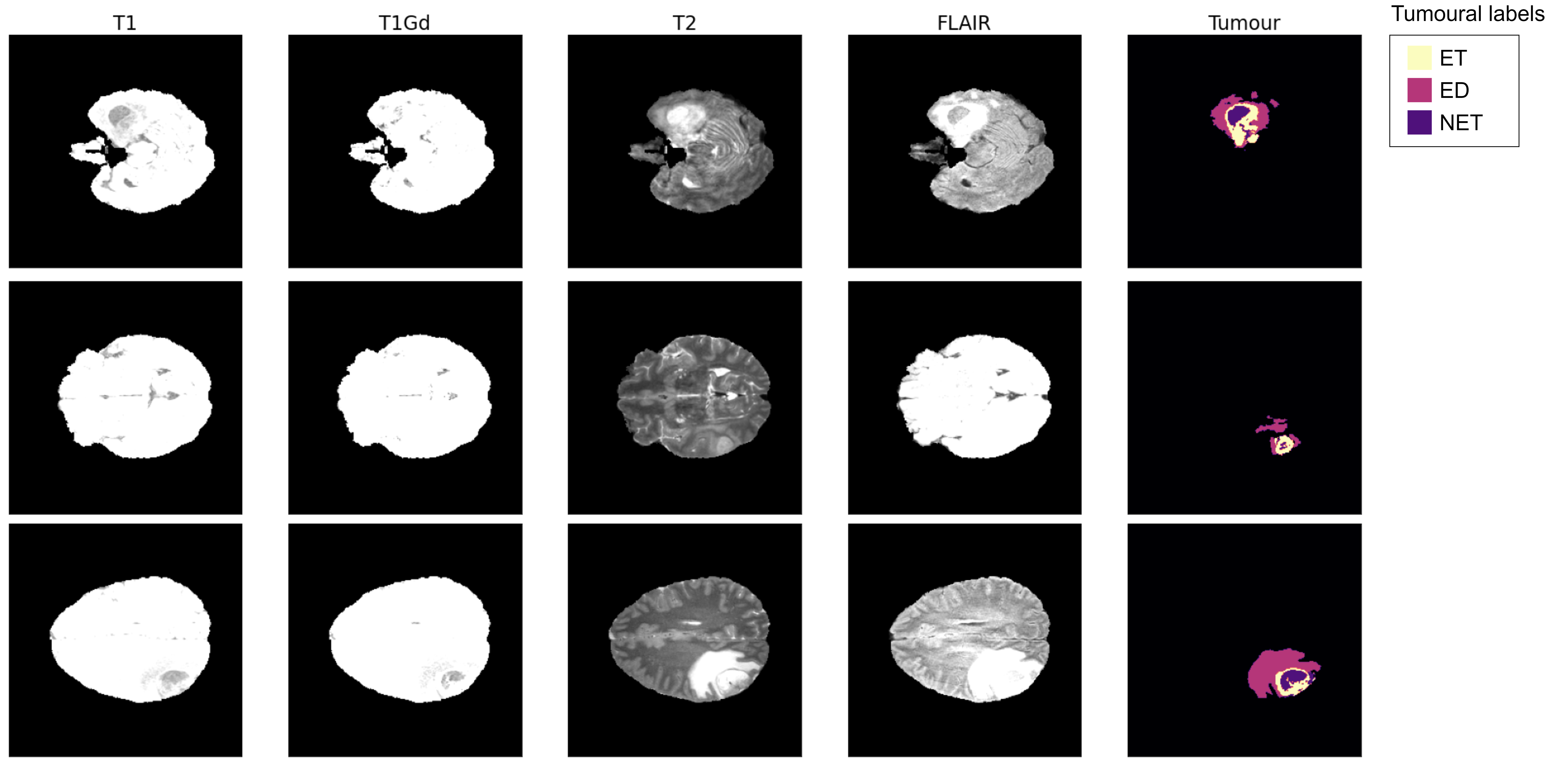
Each scanner is available as four different Neuroimaging Informatics Technology Initiative (NIfTI) files: Native scanner (T1), Post-contrast T1 weighted (T1Gd), T2 weighted (T2) and T2 Fluid Attenuated Inversion Recovery (T2-Flair). Segmentation of each scan is divided into three labels, Gd-enhancing tumour (ET), Pleritumoural edema (ED) and Non-enhancing tumour core (NET), manually segmented and approved by neuroradiologists.
Due to the fact that for the training and evaluation of the proposed algorithm, the labelled information for each scanner is necessary, it will only be used in the MICCAI_BraTS_Training partition. This dataset will be divided into a training and evaluation set.
Figure 1 contains samples of the information present in the dataset. It must be noted that each image represents a full 3 dimensional scan of a patient.
To test the cases, we study the similarity of segmentation between the query and the retrieved images will be studied. In the case of tumoural characteristics of each patient, similarity measurement is performed using the tumour segmentation information present in the original dataset. However, to compare the similarity of the healthy areas of each patient, we have the same approximation of [21] where the brain of each patient is divided anatomically. This process must be performed to compare the results of the proposed model with those of the work of [21] and, to obtain the same information they used, each scanner must be preprocessed.
3.1 Dataset preprocessing
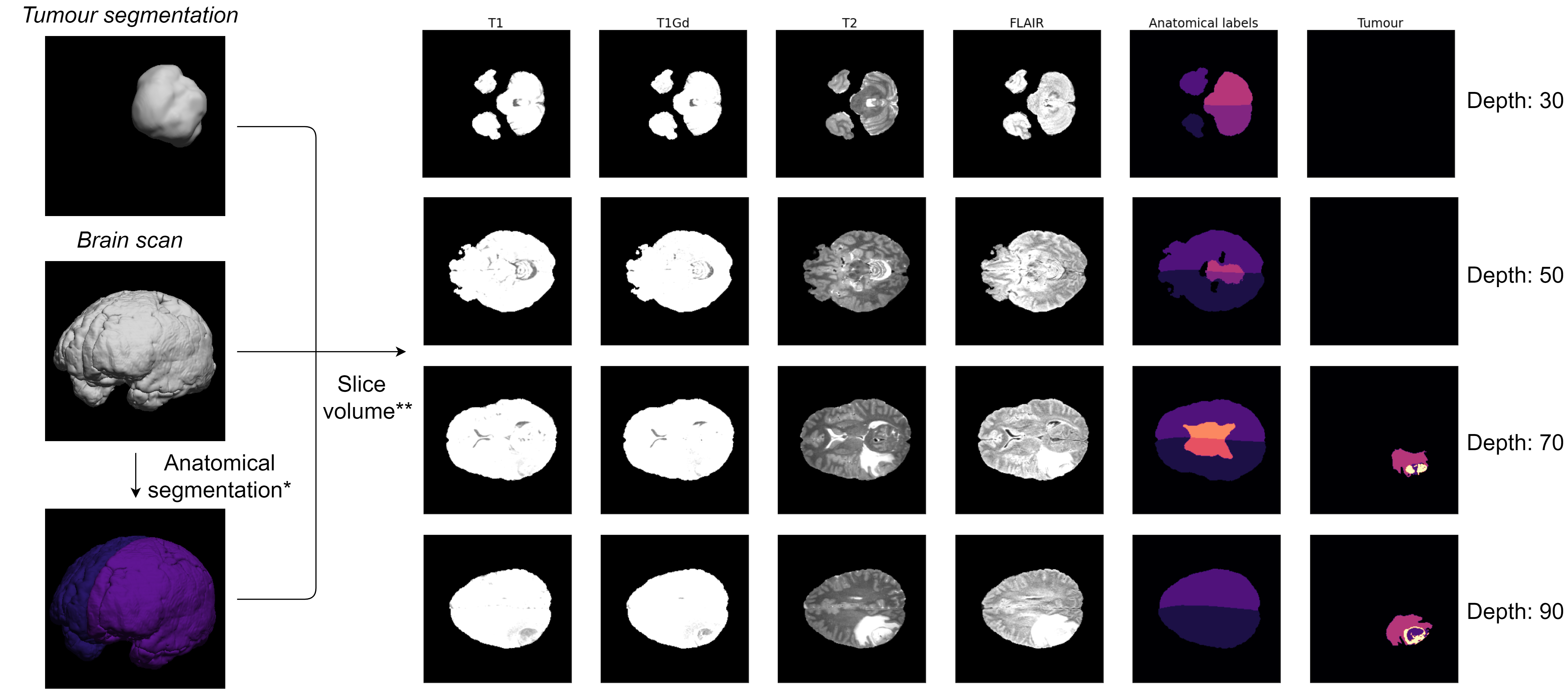
Each 3 dimensional scanner consists on a scanner of 240x240x155 pixels of information, but the input of the proposed method is a two dimensional image. In order to obtain the images from the 3 dimensional data that are stored in the BraTS 2020 dataset the scanners must be sliced in layers. Originally, the data dimension was 240x240x155, which is sliced on the third axis to generate 240x240 images by taking the information about each layer separately. Furthermore, each image is normalized between the range [-1, 1] to be treated with Artificial Neural Networks (ANNs).
In addition, each healthy image is labelled with six normal anatomical labels: left and right cerebrum, left and right cerebellum and left and right ventricle. This division is achieved using BrainSuite 19a software [35]. This program is able to obtain a voxel segmentation of the cerebrum, cerebellum and ventricle of each case, making it possible to use this information to evaluate the similarity between the query and the retrieved cases.
Figure 2 has a sample of the new labels generated for each image. As can be seen after segmentation of the anatomical labels, each brain is divided into six different areas, as was done in [21].
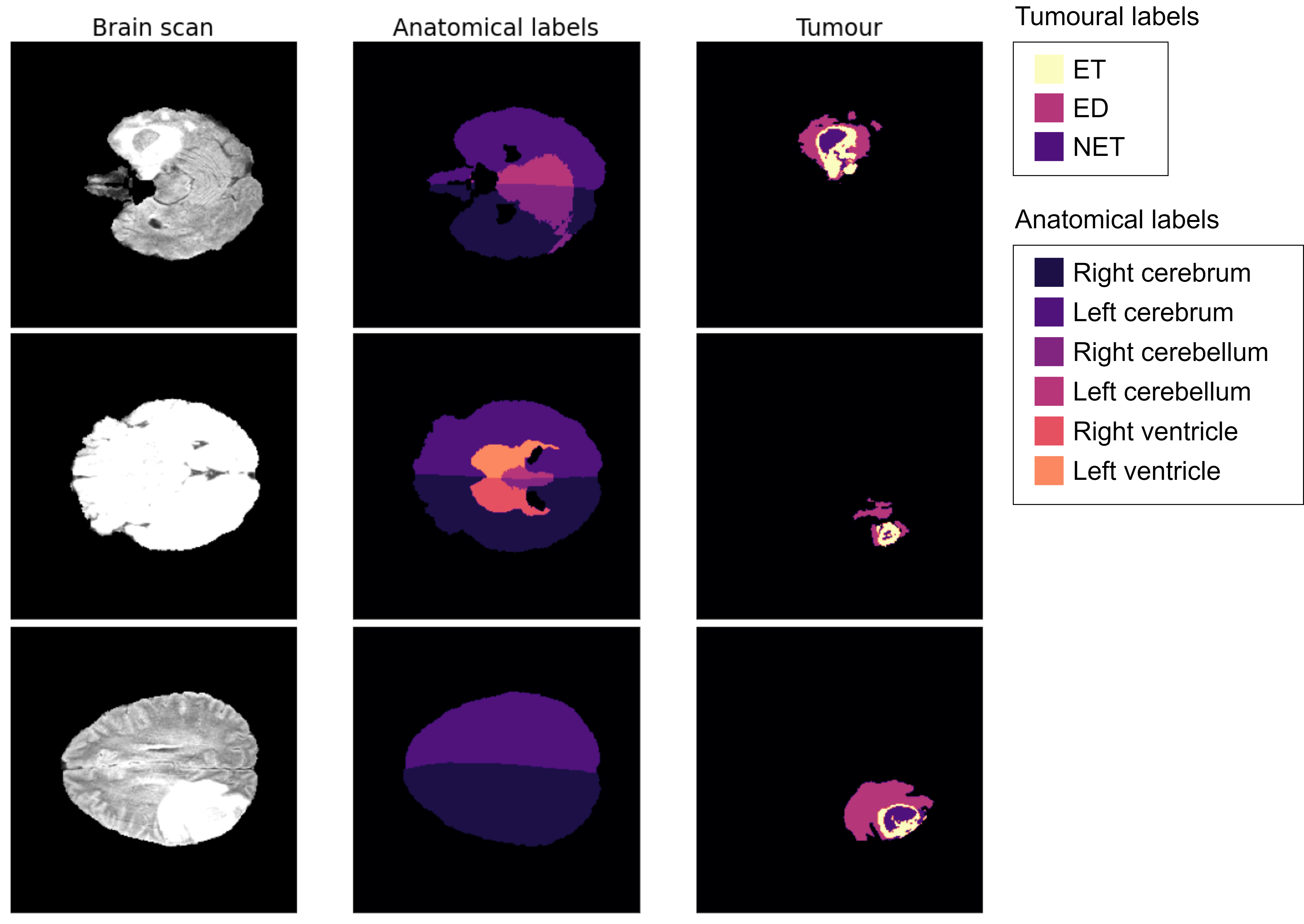
Figure 3 shows a brief scheme of the preprocessing process, from the NIfTI files to a 2 dimensional images of each case, obtaining in addition a segmentation of the anatomical labels of each patient.
4 Multi-Output Classification Autoencoder
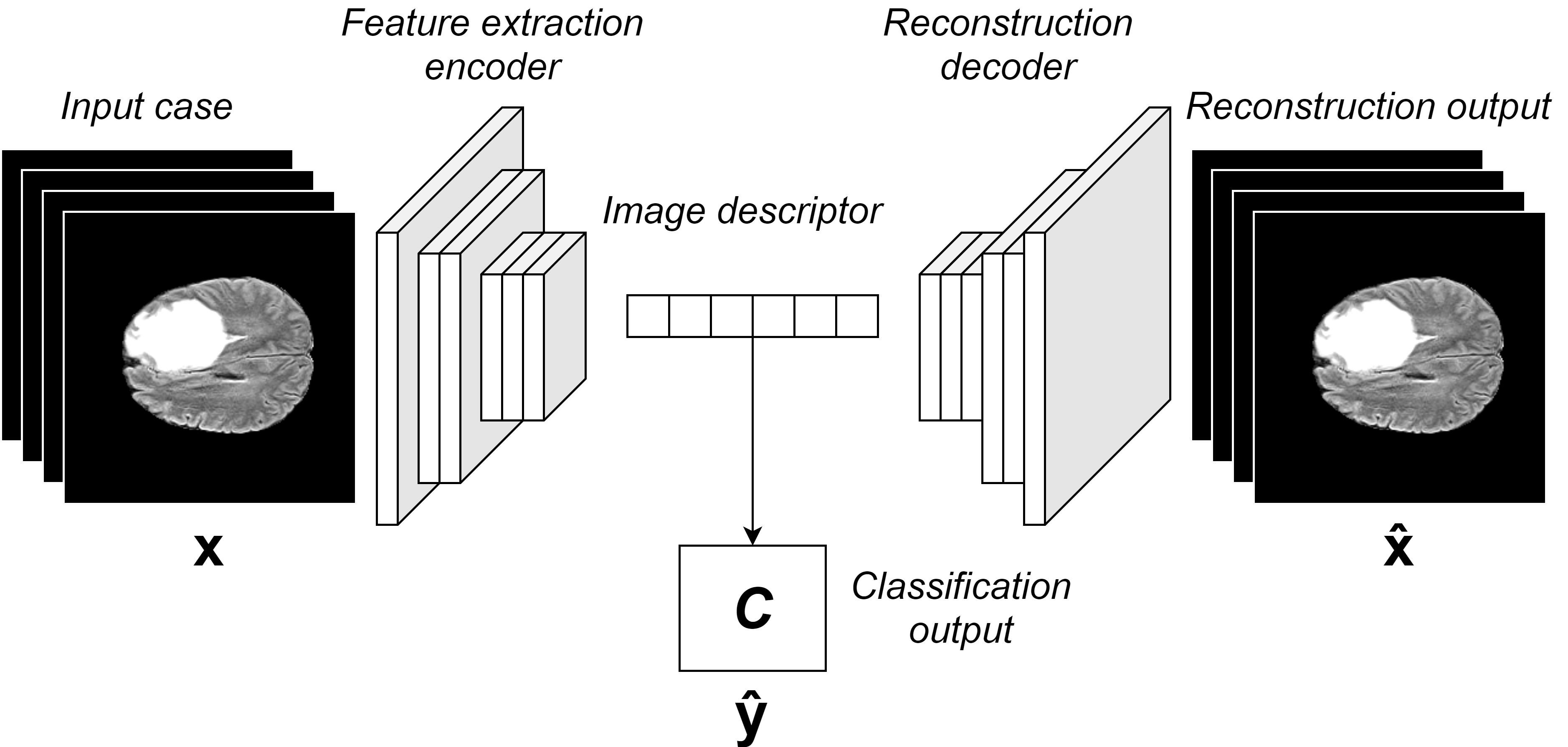
In Figure 4 the schematic of the proposed method can be seen. According to the figure, the architecture presented combines two different approaches: an AE network that extracts the structural information of each image and a binary classifier that is responsible for extracting the tumour information from each case. This dual-objective architecture enhances the features represented in the descriptor, which is obtained from the latent vector.
On the one hand, in MOC-AE an AE is used following the same approximation as [36] where a CBIR system is designed using the latent space of an AE as image descriptors. This simple scheme makes it possible to extract the composition features of an image by forcing the network to reduce the dimensionality of the input images. One of the main advantages of this method is that it does not require any label to work, as a result of the self-supervised learning scheme of AEs [22]. Using an AE as the main base for image descriptor generation, the network can learn latent representations of the input image.
The main drawback of using the latent space of an AE as a descriptor is that it considers with the same importance every portion of the input image. Furthermore, there is no control over the information represented in the latent space.
To solve this problem, it is proposed to add an auxiliary classifier that shares the descriptor with AE. This addition to the network will attempt to maintain the information from the patient’s tumour. Here, it is important to note that this learning scheme is focused on trying to keep certain features of the input information, in order to disentangle the healthy and tumoural information of the patient in the image descriptor. This solution is based on the work of [21] where three different AEs were used to disentangle tumour and normal information from each case.
Using the classifier, the network is forced to learn the information of the tumour to be able to classify the cases where a tumour is present. At the same time, AE forces the descriptor to maintain the structural characteristics of each patient. This dual-objective forces the network to focus on some features that are present in this case.
This architecture is focused on medical CBIR due to the fact that it makes possible to make the network focus on the possible pathologies of each image. With regard to the work of [21], this scheme does not require a segmented dataset. In the proposed method, the binary classifier only needs information about the presence of a tumour in the image, but segmentation of the tumoural region of the image is not necessary.
As stated in [22], in the field of medicine, the process of annotating the data is particularly costly because manual annotation must be performed by specialists in the field. Therefore, the possibility of developing a CBIR system capable of focusing in the most relevant areas of the image while maintaining the relatively low cost of the labels used is a characteristic that is particularly desired.
4.1 Model training scheme
The proposed architecture must combine two different learning processes using the same parameters. As stated above, this shared information forces the network to combine the normal and pathological features of each image.
Regarding the training of the model, two different outputs will use two different losses, which must be combined to train the model. The AE output is responsible for reconstructing the information in the input image, while the classifier must differentiate between healthy and regions with the presence of a tumour in it.
First, the input image must be reconstructed by the AE generating , both the input and the reconstructed image will be compared using the L2 norm.
| (1) |
This reconstruction loss function will force the latent space to learn the spatial features of the input image.
At the same time, the classification head of the proposed model will be trained using the binary cross-entropy loss function, formulated as follows:
| (2) |
The head loss classification function focuses on differentiating healthy and tumoural images, focusing on detecting the presence of tumours in the input image. When seeking this objective, the model will be forced to maintain tumoural features in the latent space.
Both losses are combined to train the model using the following equation:
| (3) |
where both terms are normalization coefficients to balance both losses.
The loss function is minimised using the Adam [20] optimizer. The best values of the parameters were found using the grid search in the training phase. The range of values tested varied between and , requiring that the sum of both must be of . The best performance was found to be achieved with and .
4.2 Network architecture definition
The proposed network composition is Convolutional Neural Network (CNN) using the Residual blocks presented in the ResNet architecture [12]. The same principles as those used in ResNet were used to design the Encoder and Decoder networks of the model. But in particular, it was decided to use complete pre-activation blocks as was proposed in [13] that generally produce the best results. The AE generates the latent vector by reducing the information to a space of 500 dimensions that corresponds to the image descriptor.
Regarding the classifier, it uses the latent vector information of 500 positions and generates a binary classification, using an intermediate dense layer of 64 neurons.
Figure 5 shows details of the model architecture. The input data that the Encoder receives are 4 slices of each case, corresponding to the different scanners (T1, T1Gd, T2, T2-Flair). Each residual block contains two separable convolutions [3] along with Batch Normalization [15] and Dropout [14] layers. The Rectified Linear Unit (ReLU) activation function was used in the hidden layers, while the hyperbolic tangent and the sigmoid activation were used in the reconstruction and classification outputs, respectively.
4.3 CBIR algorithm
CBIR system aims to obtain, from the database of documented clinical cases, the patient most similar to the query. In other words, each time a scan is received, the algorithm CBIR finds in the database the most similar cases in the database by comparing the healthy and tumoural structures of the query.
To compare each query with the rest of the documented cases, the image descriptor is generated. This descriptor corresponds to the latent space of MOC-AE generated using the trained Encoder network. To compare the query image with the rest of the database, we use the Euclidean distance, following the same approximation as [21, 40, 34, 36].
The recommendation system will also use the classification learning of the network in order to retrieve similar cases from the database. Because of the inherent ability of the network of classifying a certain query as tumoural or not this information will be used to enhance the recommendation. If the query is classified as tumoural by the MOC-AE with certain reliability it will only search among other tumoural images. This way, when the network is sure that a case contains a tumour it will only search on other tumoural cases. In particular, it is decided to put a threshold of 90% of confidence from where search on tumoural cases.
5 Results
The experimental results of the proposed system CBIR are shown in this section. The proposed algorithm performance will be analysed by its training results and by comparing it with similar works, this way evaluating its individual performance and measuring how it performs with respect state-of-the-art similar systems.
As mentioned in Section 3 the dataset from the input of the experiment consists of 369 cases divided into 155 slices each. Thus, a total of 57.195 images were used. However, 10% of the whole dataset (5.720 images) is reserved for testing purposes.
5.1 Empirical evaluation of the CBIR system
It is decided to empirically evaluate the results of the CBIR recommendation. To compare the results of the model, Figure 6 shows the results of different queries used as input to the system. As can be seen, the model is capable of retrieving similar images in both anatomical and tumoural aspects.
5.2 Comparison results
Table 1 shows the results of the proposed algorithm compared with the results obtained in [21] in terms of the Sorensen-Dice coefficient [6, 37]. In particular, the table measures the consistency between the anatomy of the healthy and tumoural features of the image. This coefficient varies between 1 and 0, where the higher the value, the more similar are the query and the retrieved images. To calculate the Sorensen-Dice coefficient of the proposed method, the same procedure will be followed as [21], where the queries used are the slices of each case with the largest tumoural area. Three different coefficients are analysed, first, the normal Dice measures the similarity of the healthy features of each patient, using the six anatomical labels obtained with the procedure explained in Section 3.1 and calculating the multi-label Sorensen-Dice coefficient, the tumoural Dice uses the segmentation information of each image, measuring the similarity of the tumoural sections of each case and finally the entire Dice combines the normal and tumoural Dice to balance the result and provide a more complete measurement.
| Model | Normal Dice | Tumoural Dice | Entire Dice |
|---|---|---|---|
| MOC-AE | 0.6340.220 | 0.3100.276 | 0.4720.175 |
| Kobayashi et al. Normal latent space | 0.7300.196 | 0.0720.098 | 0.4010.108 |
| Kobayashi et al. Abnormal latent space | 0.5050.235 | 0.2890.120 | 0.3970.137 |
| Kobayashi et al. Entire latent space | 0.6950.208 | 0.2010.158 | 0.4480.123 |
The presented results outperform the results of [21], especially in terms of tumour detection. Thus, it is shown that MOC-AE can achieve state-of-the-art results in tumour recommendation. In addition, it should be remembered that the main strength of MOC-AE is that it does not require segmentation information of the cases to train the network. With respect to the compared work, MOC-AE shows a global improvement in terms of patient tumoural features and a performance comparable to that of Kobayashi et al. in anatomical features. The best tumoural performance is achieved with the proposed method, while the best anatomical similarity is obtained in [21]. Finally, the best balance between normal and abnormal features of each patient is achieved with MOC-AE. Therefore, the proposed MOC-AE is able to balance the anatomical and tumour areas of each patient, to retrieve more similar images from the database.
6 Discussion
CDSSs represent critical algorithms that could significantly improve the diagnostic tasks of doctors. The proposed MOC-AE achieves state-of-the-art results in both the anatomical and tumoural features of recommended cases. When factorizing medical images using MOC-AE, the model can focus on both the normal features of the patient and the pathologies present in the case, generating a compact and significant representation of each image. These image descriptors can be used to recommend similar cases from a database, helping the doctor diagnose the task.
The architecture presented in the current work is able to combine features present in the image with labels annotated by professionals. One of the main strengths that differentiates MOC-AE from similar CBIR models is that it does not require costly information on the label such as tumour segmentation. The MOC-AE learn characteristics of each patient combining recommendation and classification outputs, thus generating an enriched image descriptor using only binary label information. In an area where the cost of producing high-quality labels is especially costly, it is considered crucial to develop a model that is able to produce state-of-the-art results with a low-cost associated.
The results of the model improve the previous work by obtaining better results in both retrieving cases with similar pathologies and balancing both anatomical and abnormal features of each case. In view of the results, MOC-AE is considered the best alternative to develop a CDSS specialized in medical imaging recommendation.
7 Conclusions
In the present paper, a novel CBIR neural network called MOC-AE is presented to recommend the most similar cases given a query, taking particular attention to the anatomical and pathological features of the patient at the same time. The proposed model combines in an unique descriptor the most relevant characteristics of the case, being able to extract from a database the most similar cases.
This solution is capable of improving the results of state-of-the-art similar approaches, improving the results regarding the similarity of the tumoural area and balancing better healthy and abnormal features of the patient. Thus, the proposed system arises as the best solution to create a CDSS in tumoural brain recommendation.
At the same time, with respect to other works, the presented MOC-AE reduces the cost of the training process, only being necessary labels for the presence of the tumour. This cost reduction is particularly relevant when treating with medical images, where the cost of manually annotating costly information requires the time and effort of specialised professionals in the area. With MOC-AE this information is drastically reduced, while the results are improved.
In conclusion, the use of MOC-AE in tumoural CBIR is a promising option to improve efficiency and accuracy in comparative diagnostic and tumoural pathologies treatment.
References
- Bakas et al. [2017] Bakas, S., Akbari, H., Sotiras, A., Bilello, M., Rozycki, M., Kirby, J.S., Freymann, J.B., Farahani, K., Davatzikos, C., 2017. Advancing the cancer genome atlas glioma mri collections with expert segmentation labels and radiomic features. Scientific data 4, 1–13.
- Bakas et al. [2018] Bakas, S., Reyes, M., Jakab, A., Bauer, S., Rempfler, M., Crimi, A., Shinohara, R.T., Berger, C., Ha, S.M., Rozycki, M., et al., 2018. Identifying the best machine learning algorithms for brain tumor segmentation, progression assessment, and overall survival prediction in the brats challenge. arXiv preprint arXiv:1811.02629 .
- Chollet [2017] Chollet, F., 2017. Xception: Deep learning with depthwise separable convolutions, in: Proceedings of the IEEE conference on computer vision and pattern recognition, pp. 1251–1258.
- Cortes and Vapnik [1995] Cortes, C., Vapnik, V., 1995. Support-vector networks. Machine learning 20, 273–297.
- Cover and Hart [1967] Cover, T., Hart, P., 1967. Nearest neighbor pattern classification. IEEE Transactions on Information Theory 13, 21–27. doi:10.1109/TIT.1967.1053964.
- Dice [1945] Dice, L.R., 1945. Measures of the amount of ecologic association between species. Ecology 26, 297–302.
- Esteva et al. [2021] Esteva, A., Chou, K., Yeung, S., Naik, N., Madani, A., Mottaghi, A., Liu, Y., Topol, E., Dean, J., Socher, R., 2021. Deep learning-enabled medical computer vision. NPJ digital medicine 4, 1–9.
- Fatima et al. [2017] Fatima, M., Pasha, M., et al., 2017. Survey of machine learning algorithms for disease diagnostic. Journal of Intelligent Learning Systems and Applications 9, 1.
- Gardezi et al. [2019] Gardezi, S.J.S., Elazab, A., Lei, B., Wang, T., 2019. Breast cancer detection and diagnosis using mammographic data: Systematic review. Journal of medical Internet research 21, e14464.
- Guo et al. [2022] Guo, M.H., Xu, T.X., Liu, J.J., Liu, Z.N., Jiang, P.T., Mu, T.J., Zhang, S.H., Martin, R.R., Cheng, M.M., Hu, S.M., 2022. Attention mechanisms in computer vision: A survey. Computational Visual Media , 1–38.
- Haq et al. [2021] Haq, N.F., Moradi, M., Wang, Z.J., 2021. A deep community based approach for large scale content based x-ray image retrieval. Medical Image Analysis 68, 101847.
- He et al. [2016a] He, K., Zhang, X., Ren, S., Sun, J., 2016a. Deep residual learning for image recognition, in: Proceedings of the IEEE conference on computer vision and pattern recognition, pp. 770–778.
- He et al. [2016b] He, K., Zhang, X., Ren, S., Sun, J., 2016b. Identity mappings in deep residual networks, in: European conference on computer vision, Springer. pp. 630–645.
- Hinton et al. [2012] Hinton, G.E., Srivastava, N., Krizhevsky, A., Sutskever, I., Salakhutdinov, R.R., 2012. Improving neural networks by preventing co-adaptation of feature detectors. arXiv preprint arXiv:1207.0580 .
- Ioffe and Szegedy [2015] Ioffe, S., Szegedy, C., 2015. Batch normalization: Accelerating deep network training by reducing internal covariate shift, in: International conference on machine learning, PMLR. pp. 448–456.
- Ji et al. [2021] Ji, W., Yu, S., Wu, J., Ma, K., Bian, C., Bi, Q., Li, J., Liu, H., Cheng, L., Zheng, Y., 2021. Learning calibrated medical image segmentation via multi-rater agreement modeling, in: Proceedings of the IEEE/CVF Conference on Computer Vision and Pattern Recognition, pp. 12341–12351.
- Jiang et al. [2016] Jiang, M., Zhang, S., Huang, J., Yang, L., Metaxas, D.N., 2016. Scalable histopathological image analysis via supervised hashing with multiple features. Medical image analysis 34, 3–12.
- Karimi et al. [2021] Karimi, D., Vasylechko, S.D., Gholipour, A., 2021. Convolution-free medical image segmentation using transformers, in: International Conference on Medical Image Computing and Computer-Assisted Intervention, Springer. pp. 78–88.
- Kimber-Trojnar et al. [2021] Kimber-Trojnar, Ż., Pilszyk, A., Niebrzydowska, M., Pilszyk, Z., Ruszała, M., Leszczyńska-Gorzelak, B., 2021. The potential of non-invasive biomarkers for early diagnosis of asymptomatic patients with endometriosis. Journal of Clinical Medicine 10, 2762.
- Kingma and Ba [2014] Kingma, D.P., Ba, J., 2014. Adam: A method for stochastic optimization. arXiv preprint arXiv:1412.6980 .
- Kobayashi et al. [2021] Kobayashi, K., Hataya, R., Kurose, Y., Miyake, M., Takahashi, M., Nakagawa, A., Harada, T., Hamamoto, R., 2021. Decomposing normal and abnormal features of medical images for content-based image retrieval of glioma imaging. Medical image analysis 74, 102227.
- Krishnan et al. [2022] Krishnan, R., Rajpurkar, P., Topol, E.J., 2022. Self-supervised learning in medicine and healthcare. Nature Biomedical Engineering , 1–7.
- Krittanawong [2018] Krittanawong, C., 2018. The rise of artificial intelligence and the uncertain future for physicians. European journal of internal medicine 48, e13–e14.
- Kumar et al. [2016] Kumar, A., Dyer, S., Kim, J., Li, C., Leong, P.H., Fulham, M., Feng, D., 2016. Adapting content-based image retrieval techniques for the semantic annotation of medical images. Computerized Medical Imaging and Graphics 49, 37–45.
- Lehmann et al. [2005] Lehmann, T.M., Güld, M.O., Deselaers, T., Keysers, D., Schubert, H., Spitzer, K., Ney, H., Wein, B.B., 2005. Automatic categorization of medical images for content-based retrieval and data mining. Computerized Medical Imaging and Graphics 29, 143–155.
- Menze et al. [2014] Menze, B.H., Jakab, A., Bauer, S., Kalpathy-Cramer, J., Farahani, K., Kirby, J., Burren, Y., Porz, N., Slotboom, J., Wiest, R., et al., 2014. The multimodal brain tumor image segmentation benchmark (brats). IEEE transactions on medical imaging 34, 1993–2024.
- Musen et al. [2021] Musen, M.A., Middleton, B., Greenes, R.A., 2021. Clinical decision-support systems, in: Biomedical informatics. Springer, pp. 795–840.
- Ossa et al. [2022] Ossa, L.A., Starke, G., Lorenzini, G., Vogt, J.E., Shaw, D.M., Elger, B.S., 2022. Re-focusing explainability in medicine. Digital health 8.
- Quellec et al. [2010] Quellec, G., Lamard, M., Cazuguel, G., Cochener, B., Roux, C., 2010. Wavelet optimization for content-based image retrieval in medical databases. Medical image analysis 14, 227–241.
- Ralbovsky and Lednev [2020] Ralbovsky, N.M., Lednev, I.K., 2020. Towards development of a novel universal medical diagnostic method: Raman spectroscopy and machine learning. Chemical Society Reviews 49, 7428–7453.
- Rama Krishna and Sirajuddin [2022] Rama Krishna, S., Sirajuddin, M., 2022. A role of emerging technologies in the design of novel framework for covid-19 data analysis and decision support system. Understanding COVID-19: The role of computational intelligence , 313–337.
- Rani et al. [2021] Rani, P., Kumar, R., Ahmed, N.M., Jain, A., 2021. A decision support system for heart disease prediction based upon machine learning. Journal of Reliable Intelligent Environments 7, 263–275.
- Rumelhart et al. [1985] Rumelhart, D.E., Hinton, G.E., Williams, R.J., 1985. Learning internal representations by error propagation. Technical Report. California Univ San Diego La Jolla Inst for Cognitive Science.
- Shakarami and Tarrah [2020] Shakarami, A., Tarrah, H., 2020. An efficient image descriptor for image classification and cbir. Optik 214, 164833.
- Shattuck and Leahy [2002] Shattuck, D.W., Leahy, R.M., 2002. Brainsuite: an automated cortical surface identification tool. Medical image analysis 6, 129–142.
- Siradjuddin et al. [2019] Siradjuddin, I.A., Wardana, W.A., Sophan, M.K., 2019. Feature extraction using self-supervised convolutional autoencoder for content based image retrieval, in: 2019 3rd International Conference on Informatics and Computational Sciences (ICICoS), IEEE. pp. 1–5.
- Sorensen [1948] Sorensen, T.A., 1948. A method of establishing groups of equal amplitude in plant sociology based on similarity of species content and its application to analyses of the vegetation on danish commons. Biol. Skar. 5, 1–34.
- Sullivan et al. [2021] Sullivan, F.M., Mair, F.S., Anderson, W., Armory, P., Briggs, A., Chew, C., Dorward, A., Haughney, J., Hogarth, F., Kendrick, D., et al., 2021. Earlier diagnosis of lung cancer in a randomised trial of an autoantibody blood test followed by imaging. European Respiratory Journal 57.
- Tan and Fielding [2006] Tan, Y.K., Fielding, J.W., 2006. Early diagnosis of early gastric cancer. European journal of gastroenterology & hepatology 18, 821–829.
- Tarjoman et al. [2013] Tarjoman, M., Fatemizadeh, E., Badie, K., 2013. An implementation of a cbir system based on svm learning scheme. Journal of Medical Engineering & Technology 37, 43–47.
- Tuppad and Patil [2022] Tuppad, A., Patil, S.D., 2022. Machine learning for diabetes clinical decision support: a review. Advances in Computational Intelligence 2, 1–24.
- Ward et al. [2021] Ward, T.M., Mascagni, P., Ban, Y., Rosman, G., Padoy, N., Meireles, O., Hashimoto, D.A., 2021. Computer vision in surgery. Surgery 169, 1253–1256.
Declaration of interest
The authors report no conflict of interest. The authors alone are responsible for the content and writing of this paper
Acknowledgements
The authors thank the research group KNOwledge Discovery and Information Systems (KNODIS) for the compute infrastructure.
Supplementary Material
The source code of the project, along with trained models and results are publicly available and can be consulted in https://purl.com/mocae_brats.