Structure-based drug discovery with deep learning
Abstract
Artificial intelligence (AI) in the form of deep learning bears promise for drug discovery and chemical biology, e.g., to predict protein structure and molecular bioactivity, plan organic synthesis, and design molecules de novo. While most of the deep learning efforts in drug discovery have focused on ligand-based approaches, structure-based drug discovery has the potential to tackle unsolved challenges, such as affinity prediction for unexplored protein targets, binding-mechanism elucidation, and the rationalization of related chemical kinetic properties. Advances in deep learning methodologies and the availability of accurate predictions for protein tertiary structure advocate for a renaissance in structure-based approaches for drug discovery guided by AI. This review summarizes the most prominent algorithmic concepts in structure-based deep learning for drug discovery, and forecasts opportunities, applications, and challenges ahead.
Keywords
Artificial intelligence De novo design Machine learning Medicinal chemistry Structural biology
Introduction
Deep learning – a subfield of artificial intelligence (AI) based on multi-layer neural networks1 – has gained remarkable traction in science and technology, e.g., to advance mathematics 2, 3, investigate galaxies 4, and generate realistic images5. Chemistry and biology have witnessed several AI breakthroughs, for instance, in protein structure prediction6, 7, chemical synthesis planning8, 9, and atomistic simulations10, 11. Drug discovery has particularly benefited from the advent of deep learning12, 13, with success in molecule prioritization and automated de novo design14, 15, 16, 17. Here, deep learning can accelerate the navigation of the extremely vast chemical space of drug-like molecules18 in search for potential therapeutics, and complement resource- and time-intensive high-throughput screening campaigns 19. Most deep learning studies have focused on ligand-based approaches 12, which leverage solely the structural information of small molecule ligands to provide predictions. For these applications, numerous systematic studies 20, 21 and experimental proofs-of-concept16, 17, 22 have been published. On the other hand, structure-based deep learning approaches – which leverage information on the target protein – have not found parallel interest yet.
Structure-based drug discovery (SBDD) methods augmented with AI are arguably a more complex and a higher-potential endeavor compared to their ligand-based counterparts. Numerous marketed drugs have been identified by ‘traditional’ SBDD (e.g., HIV-1 protease inhibitors23, the thymidylate synthase inhibitor raltitrexed 24, and the antibiotic norfloxacin 25). Accelerating SBDD with deep learning can help address existing drug discovery challenges, such as polypharmacology by design 26, selectivity optimization 27, activity cliff prediction 28, and target deorphanization 29. Deep learning does not require explicit feature engineering and can thus be applied to learn directly from molecular representations of both ligands and proteins. This is particularly relevant for SBDD, where engineering numerical features for complex molecular entities like proteins 30 is inevitably more laborious than for small molecules31. Therefore, deep learning for SBDD bears an untapped potential to capture highly non-linear structure-activity relationships and has recently started to show its promise. Accurate protein structure prediction efforts like AlphaFold6, 7 are expected to further accelerate computer-assisted SBDD. Deep learning for SBDD is still in its infancy but is moving forward at a fast pace, and its relevance in the years to come is expected to increase.
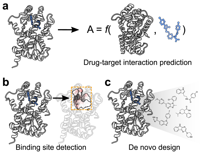
This review provides a comprehensive overview of how deep learning can be leveraged for SBDD, to incorporate protein information at different levels of complexity (e.g., aminoacid sequence, and/or tertiary structure). After addressing how proteins can be represented for deep learning, we address current state-of-the-art methods for structure-based drug discovery, with a particular focus on drug-target interaction prediction, binding site detection, and de novo design (Fig. 1). Finally, we discuss current limitations and research gaps, along with foreseen future directions and opportunities. A glossary of selected terms can be found in Box 1.
[t]
Representing proteins for deep learning
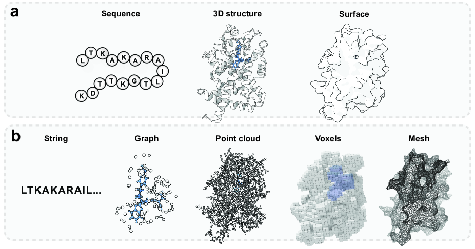
The design of deep learning approaches for SBDD is inherently more intricate than those that are ligand-based, due to the need to represent protein information at different levels of complexity. Proteins are large polypeptide chains, organized in a ‘structural hierarchy’36: (a) primary structure, referring to the sequential arrangement of amino acids along the polypeptide chain, (b) secondary structure, capturing the occurrence of alpha-helices and beta-pleated sheets along the protein sequence, and (c) tertiary structure, capturing how proteins fold in the three-dimensional space. Such complexity is reflected in the various protein representations used for deep learning (Fig. 2):
-
•
Primary (amino acid) sequence. The amino acid sequence is specified starting from the amino-terminal end (N-terminus) and ending at the carboxyl-terminal (C-terminus) end. For deep learning purposes, the primary sequence is often represented as a character string, where each letter represents one of the twenty naturally-occurring amino acids (e.g., ‘AIR’ corresponds to alanine, isoleucine, and arginine). These representations are at the core of established protein ‘featurization’ techniques, such as ProtVec 37, SeqVec 38, and ProtTrans 39. Although less frequently encountered 40, 41, the primary sequence can also be represented as a graph, whose nodes are amino acids (featurized by type or corresponding physicochemical features) and whose edges capture their adjacency in the chain.
-
•
Tertiary (three-dimensional) structure. The three-dimensional shape of a protein (tertiary structure) is determined by the interactions among its side chains, and features a certain degree of conformational plasticity 42. The protein structure contains key information for SBDD, since it relates to protein function 43, 44, and it determines ligand binding 45. Moreover, inducing conformational changes is often the goal of drug discovery42. Several ways exist to learn from tertiary structures with deep learning. Early approaches 46, 47 have used grid-based voxel representations (see Box 1) to capture the spatial distribution of the protein’s physicochemical or pharmacophore properties. While these representations are suited to well-established deep learning architectures (e.g., convolutional neural networks, CNNs), many voxels representing empty space do not carry relevant information, and they involve increased computational costs at higher spatial grid resolutions. Other approaches48 represent proteins as molecular graphs, where each atom is a node and each bond is an edge. Depending on the chosen level of coarse-graining, often only backbone atoms are chosen to correspond to nodes, while edges often represent geometrical proximity in the coordinate space rather than direct chemical bonds 49. Edges and bonds can be characterized by additional geometrical and/or physicochemical properties.
-
•
Protein surface. The protein surface is usually defined as the separation between solvent-accessible and inaccessible regions 50, 51 (Fig. 2a) and it plays a key role in the protein interactions with (macro)molecular entities. Protein surfaces are usually represented as either meshes (i.e., a set of polygons capturing the location of the surface, whose vertices can be described by a 2D grid or a 3D graph structure) or point clouds (i.e., graphs whose nodes describe the location of the surface at a certain resolution). Although often computed from the 3D structure, the surface representation might better reflect the physicochemical features responsible for the interaction with other (macro)molecules, as well as aspects of protein function that go beyond sequence similarity.
The chosen protein representation for deep learning affects the type, quality, and quantity of chemical information captured. It will also affect the type of suitable deep learning strategies (Box 2) and the corresponding advantages and drawbacks. Additionally, the number of data available for machine learning depends on the chosen protein representation (Table 1). Primary sequences are easy to obtain but lack information about the spatial configuration of atoms, particularly relevant to determine the binding pose of ligands. On the other hand, 3D protein structures contain potentially richer information for drug discovery, but they are more difficult to obtain experimentally and thus relatively scarce (although the cost has been steadily decreasing with the advent of experimental techniques like cryogenic electron microscopy 52). In this context, AI breakthroughs in protein structure prediction like AlphaFold6 bear promise to bridge the gap in data availability, by making thousands of predicted protein structures available in an unprecedented effort53, 54.
Small molecule ligands can be represented in analogous ways to protein structures. The most commonly used representations are: (a) molecular strings (e.g., SMILES strings35), which capture 2D information (atom occurrence and connectivity), (b) 2D and 3D molecular graphs (based on the availability of experimentally determined or computed conformational information), and (c) molecular surfaces. An in-depth description of small molecule representations and corresponding deep learning approaches can be found in a recent work 55.
| Dataset | Description | No. entries | Link (if available) |
| Protein Data Bank (PDB)56 | Structural data of biological macromolecules. | 189,735 structures | rcsb.org |
| scPDB57 | Druggable binding sites and ligands extracted from the PDB. | 4782 protein structures and 6326 ligands. | bioinfo-pharma.u-strasbg.fr/scPDB |
| BioLip58 | Semi-manually curated ligand–protein interactions. | 573,225 entries. | zhanggroup.org/BioLiP |
| PDBbind59 | Protein-ligand co-complexes and associated affinities extracted from the PDB. | 23,496 complexes. | pdbbind.org.cn |
| UniProt59 | Protein sequence and functional information. | > 60 million sequences. | uniprot.org |
| AlphaFold Protein Structure Database53 | Predictions of protein structures by AlphaFold v 2.0. 6 | 992,316 predicted structures. | alphafold.ebi.ac.uk |
| AlphaFill 54 | Common ligands and cofactor transplants for AlphaFold models. | 12,029,789 transplants. | alphafill.eu |
| Binding MOAD60, 61 | X-ray crystal structures with bound ligands and experimental binding affinities. | 41,409 protein-ligand complexes, and 15,223 binding measurements. | bindingmoad.org |
| Directory of Useful Decoys (DUD-E)62 | Directory of decoys designed to benchmark molecular docking programs. | 22,886 active molecules and affinities on 102 targets; 59 decoys per compound. | dude.docking.org |
| BindingDB 63 validation sets | Binding affinities of protein-ligand pairs curated from the literature. | 1200 series with at least 1 cocrystal available in each. | bindingdb.org |
| BigBind64 | Associated protein structures to ChEMBL 65 assay data via Pocketome. 66 | 818,995 activities with associated protein structures. | Brocidiacono et al., 2022 |
| KIBA67 | Bioactivity measurements of compounds against kinases. | 246,088 measurements. | Tang et al., 2014 |
| Davis68 | Binding affinities ( values) of inhibitors against kinases. | 30,056 measurements. | Davis et al., 2011 |
Deep learning for structure-based drug discovery
This section aims to provide a concise overview of SBDD approaches fueled by deep learning. SBDD will be considered in its broader sense, i.e., not only limited to 3D protein structure, but also including sequence and surface representations. We focus on three key tasks (Fig. 1), namely binding site detection, drug-target interaction prediction, and structure-based de novo design. For each task, selected deep learning approaches are described through the lenses of the protein representation they rely on (sequence, structure, or surface). A summary of selected deep learning studies is reported in Table 2.
| Task | Description | Protein representation |
| Drug-target interaction prediction | Predict the interaction between one or more proteins and one or more ligands. | Amino-acid sequence 69, 70, 71, 72, 73, 74 3D structure 75, 46, 76, 77, 78 |
| Docking | Determination of a ligand pose within a target binding site. | 3D structure 79, 80, 81 |
| Binding site detection | Identification and/or localization of functional protein binding sites. | Amino-acid sequence 82, 70, 83, 84 3D structure 47, 85, 86, 87, 88 Surface 89, 90, 91 |
| De novo design | Generation of ligands with desired properties conditioned on a protein. | Amino-acid sequence 92, 93 3D structure 94, 95, 96, 97, 98, 99, 100, 101, 102, 103, 104 |
Drug-target interaction prediction
The identification of interactions between molecules and macromolecular targets is a key step in drug discovery, drug repurposing, and off-target activity prediction. Drug-target interaction (DTI) prediction aims to predict the bioactivity (e.g., binding affinity) of a given set of molecules on one or more macromolecular targets, by leveraging both protein and ligand information. Given the complexity of ligand-protein interactions and of engineering suitable molecular features for DTI, it is no surprise that this topic has found a widespread application of deep learning techniques 105. In what follows, deep learning models developed for DTI prediction are categorized on the basis of the protein representation they rely on.
-
•
Sequence-based approaches. Sequence-based DTI prediction models use amino acid sequences in combination with ligand representations to perform a prediction. One of the earliest approaches, DeepDTA69, applied 1-dimensional CNNs to simultaneously learn from string representations of both ligands (in the form of SMILES) and protein sequence, by first creating separated embeddings and then concatenating them to perform a prediction. Later works have replaced CNNs with other methods, such as recurrent neural networks (Box 2),70, 106 and attention-based70, 106, 107 or transformer architectures71, 108, 72. Several works have addressed how to improve the representation of protein sequences, e.g., by incorporating evolutionary information,109, 110 or protein sequence composition descriptors 73, 111. Ligands are usually represented as strings (e.g., SMILES 69, 110 or DeepSMILES 112, 109), binary fingerprints,73, 113 frequently occurring substructures109, or molecular graphs.111, 114, 115
-
•
3D structure-based approaches. These models leverage atom coordinates, usually of co-crystallized protein-ligand complexes, for training. Early approaches projected 3D protein-ligand complexes into grids featurized with physio-chemical properties, and subsequently applied CNNs for binding affinity prediction75, 46, 116. Later works extended this idea by including more sophisticated features, e.g., inter-molecular interaction fingerprints76 and computed molecular energies117. 3D grid-based approaches have also been used for lead optimization by predicting relative binding free energies linked to small modifications of ligand structures118, 119. More recent approaches have replaced grid-based representations with graphs 48, 120, 77, 78, allowing to explicitly represent atom neighborhoods and connectivity, and apply roto-translational invariant graph neural networks for binding affinity prediction.
-
•
Surface-based approaches. Surface-based approaches have found limited application for DTI prediction. OctSurf121 represents both binding pockets and ligands as surfaces, by partitioning the 3D space recursively into octants and considering only portions containing van der Waals surface points. Non-empty octants, along with their physicochemical and geometric features, are then used as the input to a CNN. Other approaches, such as HoloProt122, merge 3D structure (graph) and surface (point cloud) information for task-specific training, e.g., enzyme-catalyzed reaction classification and binding affinity prediction.
Another topic of recent interest by the deep learning community is protein-ligand docking, which aims to predict the putative binding pose of a ligand upon binding to a macromolecular target (Box 1). Although these methods do not aim to predict the affinity between a ligand-protein pair directly, they can be used as a proxy to elucidate potential mechanisms of interaction. Deep learning has been mostly applied to ligand pose optimization while considering a rigid target structure, although recent approaches have started taking side-chain flexibility into account 123. Early approaches used protein-ligand interaction fingerprints124, 125, while successive approaches have leveraged either a voxelized version of the protein structure combined with CNNs 126, 127 or graph-based representations with message-passing neural networks128, 129, 130, 131 in lieu of classical scoring functions. Finally, several approaches have attempted to directly predict the ligand binding pose in an end-to-end fashion 132, 79, 80, without the need for a classical search algorithm by exploiting advances in equivariant deep learning.
Deep learning has undoubtedly accelerated DTI prediction, thanks to the possibility to represent and learn from protein-ligand complexes more efficiently. However, simpler models based on well-established descriptors might reach comparable performance 133, due to undesired memorization and hidden bias in ligand-protein interaction data134, 135, 136, 137. Moreover, no relationship has been observed between the complexity of protein and ligand representations and the accuracy of the resulting deep learning models 133. Thus, more attention should be put on strategies for model evaluation and data selection/splitting procedures to ensure a reliable prediction of DTIs with deep learning 133, 138, 139.
Binding site detection
The identification of ‘druggable’ binding sites in proteins plays a pivotal role in SBDD, from hit identification and molecule screening to mechanism formulation140. Over the years, a plethora of methods have been developed for binding site detection140, 141, 142, 143, 144, e.g., via interatomic gap volumes142 or regions of buried pocket surfaces145. Recently, deep learning methods learning directly from ‘raw’ representations of proteins have gained increasing traction to detect binding sites. These approaches can be grouped by the molecular representations they rely on, i.e., protein sequence, 3D structure, and surface, as described below:
-
•
Sequence-based models. Binding site detection can be performed by predicting which residues of the amino-acid sequence are involved in ligand binding, although sequence-based approaches have found limited application. Early methods approached binding site detection as a ‘side-product’ of binding affinity prediction, by using explainable AI techniques to highlight relevant residues for a model’s prediction 82, 70, 83. Few works have addressed binding site detection only 146, 147. Recently, sequence-based binding site detection has been jointly modeled with drug-target affinity prediction, leading to improved performance on both tasks 84.
-
•
3D structure-based models, which use the spatial information of proteins to detect likely binding sites. Early approaches represented the protein structure with voxels featurized with pharmacophore-like properties, along with convolutional neural networks47, 85. Subsequent works have refined structure-based binding site detection with additional techniques from the computer vision domain, e.g., image segmentation.86, 87. BiteNet148 extended ‘static’ CNN-based approaches by incorporating conformational ensembles of proteins. The approach was later adapted to predict protein-peptide binding sites 149. A recent approach combined spatial properties with amino acid sequence information to predict protein-protein interaction sites and is applicable to ligand binding site prediction88.
-
•
Surface-based models. Voxelized representations of protein coordinates have several drawbacks,91, 150 (e.g., carrying non-informative voxels that are either deeply buried and not accessible by a ligand or represent empty space, and information coarse-graining due to discretization of the input protein space) and might lead to worse results than working with surfaces alone150. For this reason, several methods based on protein surfaces have been developed over the years. These approaches rely on the representation of protein structures as continuous shapes characterized by geometric and physicochemical features to perform a prediction. Geodesic CNNs have been used to determine interaction fingerprints of molecular surfaces, and to predict protein and ligand binding sites 89. The approach was later expanded to obtain fully-learnable protein representations.90 DeepSurf91 discretizes the solvent accessible surface using a combination of K-means clustering and and density reduction91.
A recent analysis of computational approaches for protein-ligand binding site recognition 151 has shown DeepSurf91 to perform remarkably well. Moreover, non-machine-learning algorithms resulted competitive alternatives to deep learning 151. All analyzed methods struggled on shallow binding sites, due to the higher frequency of deep grooves used for model training 151. Despite the recent progress of binding pocket detection, room for improvement remains, e.g., to increase pocket coverage and detect sub-pockets151, and to predict allosteric binding sites 152.
Protein-based de novo design
De novo design refers to the generation of novel chemical entities possessing desired properties from scratch 153 and is among the most challenging tasks in computer-assisted drug discovery. Computational algorithms are faced with an incredibly vast ‘chemical universe’, whose cardinality has been estimated between 1024 and 10100 molecules 154, 18, 155. In this context, ‘brute-force’ molecule assembly or enumeration approaches are computationally unfeasible. In recent years, generative deep learning has shown great promise for de novo drug design 15, 17, 156 and to complement traditional approaches based on human-engineered rules 157, 158, 24, 159.
Generative deep learning approaches for de novo design are usually applied to produce molecules in the form of molecular graphs160, 161, 162 or strings15, 16, 17 (e.g., SMILES). While most de novo design approaches are ligand-based 156, 163, 164, 15, 165, 166, structure-based approaches have recently emerged as a promising research direction, due to their potential to design molecules interacting with pharmacologically-relevant targets on demand.
-
•
Sequence-based approaches. Sequence-based de novo design approaches usually cast the problem into a machine translation task, where high-affinity protein-ligand pairs are considered as sentences in different languages to be matched. To this end, sequence and SMILES strings for proteins and ligands are used, respectively. The first-in-kind approach 92 trained a transformer architecture to ‘translate’ amino-acid sequences into the SMILES strings of the corresponding ligand. This approach can be used for sequence-conditioned de novo design. A recent work used a transformer-based pipeline 93 that combined language models that were pre-trained on large corpora of proteins 167 and small molecules 168.
-
•
3D structure-based approaches. De novo design conditioned on the tertiary structure information can usually generate molecules in the form of 3D ligands (molecular graphs) or strings (e.g., SMILES). In the former case, 3D representations of protein-ligand complexes are used as the input to generate novel 3D molecular graphs. As one of the earliest approaches, LigVoxel94 relied on 3D grids to generate spatial ‘blobs’ of ligand properties such as occupancy, aromaticity, and hydrogen-bond donor/acceptors that match the protein pocket. Later works used equivariant diffusion networks103, 104, variational autoencoders95, and reinforcement learning97, 96 to directly generate ligand conformations for the binding pocket. Recently, equivariant neural networks coupled with point-cloud representations have been used for molecule optimization, via pocket-based fragment expansion 169. Compared to 3D graphs, molecular strings are usually easier to generate and might match or outperform graph-based models 170. A pioneering work of this category leverages generative adversarial networks171 to produce SMILES strings for protein pockets, where a pocket is featurized and fed into a generator network with a conditioning ligand99. A subsequent model adopts graph neural networks to represent active sites and generates targeted SMILES strings101, whereas another model uses a pharmacophore model to represent ligands and condition the generation for the targeted pocket100. Recently 102, a recurrent neural network model has been coupled with ligand–protein interaction fingerprints (determined on ligand docking poses) for conditioned ligand generation in the form of SMILES strings.
While ligand-based de novo design pipelines using deep learning have been experimentally validated in multiple instances 17, 172, 173, structure-based de novo design has to date not been applied prospectively. This highlights an important gap in the potential for acceptance of structure-based de novo design algorithms.
Gaps, opportunities, and outlook
Deep learning for structure-based drug discovery is gaining increasing traction, as evidenced by the exponential increase in the number of published approaches over the last few years. Breakthroughs in protein structure prediction53, 7 not only exemplify the potential of deep learning in the molecular sciences but are expected to further propel structure-based drug discovery with AI. SBDD is arguably more challenging than its ligand-based counterpart, due to the number and structural complexity of chemical entities involved, and aspects like target conformational flexibility 175. Most of the current SBDD algorithms are agnostic to dynamical information and, in certain instances, might not outperform simpler methods 151, 133. However, SBDD has an untapped potential to tackle drug discovery for new, uncharted protein targets. Such a ‘zero-’ and ‘few-shot’ learning potential makes AI-driven SBDD a high-risk/high-gain endeavor, expected to advance future drug discovery.
An area of continuous development that bears promise for SBDD is geometric deep learning176, 34. Geometric deep learning attempts to unify neural networks from the perspective of symmetry and topology. Roto-translational invariance/equivariance is particularly relevant for the three-dimensional representations of molecular systems, which is beneficial to limit function search space during training 55. Geometric deep learning is expected to boost AI’s capability to model molecular complexes and their interactions89, 177, as well as molecular design104, 178. Diffusion models 179 – a family of generative models inspired by non-equilibrium thermodynamics – are also gaining increasing popularity in deep learning thanks to their generative capabilities, and have found pioneering applications in the molecular sciences, too 104, 180, 81. These approaches have reached state-of-the-art in several deep learning applications and are expected to propel SBDD in the future.
A current bottleneck of AI-driven SBDD is constituted by the available training data. While protein sequence information and experimental assay data are largely available, high-quality 3D data of co-crystallized proteins and ligands with accompanying properties is largely missing. Furthermore, obtaining high-quality protein structure information is resource-intensive and challenging for several drug targets181, e.g., disordered and membrane proteins. Furthermore, non- and poorly binding molecules are often strongly under-reported in medicinal chemistry datasets. As a result, available three-dimensional datasets are often highly biased in their content 134, 135, 136, 137, 182, which has historically led to poor generalizability 183, 184, 185, 186. Several studies have attempted to alleviate this issue, via data curation185, 187, bias-controlled training98, and debiasing138, 188. Bridging the gap between the different types of available information will be an active task in upcoming years, with some recent work pointing in this direction already 64.
Finally, the application of deep learning in well-established fields like chemical biology and medicinal chemistry might at times be met with skepticism by experimentalists. These well-grounded concerns commonly originate from the black-box nature of deep learning. Additionally, robust performance benchmarks and evaluation datasets are currently missing, especially for de novo design studies. Moreover, to the best of our knowledge, no deep learning approaches for SBDD have been validated in a prospective setting yet. To foster broader acceptance of structure-based deep learning, we need to ‘open the box’ and validate methods experimentally. We envision that more sophisticated applications of explainable AI189, 190 will aid in identifying underlying structure-activity relationships and binding modes, and bridging the gap between theory and real-world applications.
Conclusions
In recent years, deep learning has taken drug discovery by storm, offering new opportunities for more efficient exploration of chemical space. Ligand binding site detection, drug-target interaction prediction, and structure-based de novo design can be valuable tools in early drug discovery, especially for unexplored macromolecular targets. As a whole, these approaches bear great promise to extend upon the successes of ligand-based methods. However, structure-based methods have not yet proven their applicability in prospective scenarios. Overcoming such barriers will mostly depend on additional efforts in data collection and curation, as well as on methodologies that efficiently exploit relationships between assay and structural data.
Acknowledgements
F.G. acknowledges the support from the Institute for Complex Molecular Systems (ICMS, TU/e) and the Centre for Living Technologies (Alliance TU/e, WUR, UU, UMC Utrecht).
Conflict of interest
None to declare.
Author contribution
Conceptualization: F.G., with contributions from all authors; Investigation: all authors; Visualization: D.v.T., R.O., F.G.; Writing – original draft: F.G., R.O., D.v.T.; Writing – review & editing: all authors.
List of abbreviations
AI: Artificial intelligence.
CNN: Convolutional neural network.
DTI: Drug-target interaction prediction.
GNN: Graph Neural Network.
SBDD: Structure-based drug discovery.
SMILES: Simplified molecular input line entry system.
RNN: Recurrent Neural Network.
2D (3D): Two- (three-)dimensional.
References
- [1] LeCun, Y., Bengio, Y. & Hinton, G. Deep learning. \JournalTitleNature 521, 436–444 (2015).
- [2] Davies, A. et al. Advancing mathematics by guiding human intuition with ai. \JournalTitleNature 600, 70–74 (2021).
- [3] Fawzi, A. et al. Discovering faster matrix multiplication algorithms with reinforcement learning. \JournalTitleNature 610, 47–53 (2022).
- [4] Ho, M. et al. The dynamical mass of the coma cluster from deep learning. \JournalTitleNature Astronomy 6, 936–941 (2022).
- [5] Gregor, K., Danihelka, I., Graves, A., Rezende, D. & Wierstra, D. Draw: A recurrent neural network for image generation. In International conference on machine learning, 1462–1471 (PMLR, 2015).
- [6] Jumper, J. et al. Highly accurate protein structure prediction with alphafold. \JournalTitleNature 596, 583–589 (2021).
- [7] Baek, M. et al. Accurate prediction of protein structures and interactions using a three-track neural network. \JournalTitleScience 373, 871–876 (2021).
- [8] Segler, M. H., Preuss, M. & Waller, M. P. Planning chemical syntheses with deep neural networks and symbolic AI. \JournalTitleNature 555, 604–610 (2018).
- [9] Coley, C. W., Barzilay, R., Jaakkola, T. S., Green, W. H. & Jensen, K. F. Prediction of organic reaction outcomes using machine learning. \JournalTitleACS central science 3, 434–443 (2017).
- [10] Batatia, I., Kovács, D. P., Simm, G. N., Ortner, C. & Csányi, G. Mace: Higher order equivariant message passing neural networks for fast and accurate force fields. \JournalTitlearXiv preprint arXiv:2206.07697 (2022).
- [11] Batzner, S. et al. E (3)-equivariant graph neural networks for data-efficient and accurate interatomic potentials. \JournalTitleNature communications 13, 1–11 (2022).
- [12] Jiménez-Luna, J., Grisoni, F., Weskamp, N. & Schneider, G. Artificial intelligence in drug discovery: recent advances and future perspectives. \JournalTitleExpert Opinion on Drug Discovery 16, 949–959 (2021).
- [13] Chen, H., Engkvist, O., Wang, Y., Olivecrona, M. & Blaschke, T. The rise of deep learning in drug discovery. \JournalTitleDrug Discovery Today 23, 1241–1250 (2018).
- [14] Stokes, J. M. et al. A deep learning approach to antibiotic discovery. \JournalTitleCell 180, 688–702.e13 (2020).
- [15] Segler, M. H. S., Kogej, T., Tyrchan, C. & Waller, M. P. Generating focused molecule libraries for drug discovery with recurrent neural networks. \JournalTitleACS Central Science 4, 120–131 (2018).
- [16] Yuan, W. et al. Chemical space mimicry for drug discovery. \JournalTitleJournal of Chemical Information and Modeling 57, 875–882 (2017).
- [17] Merk, D., Friedrich, L., Grisoni, F. & Schneider, G. De novo design of bioactive small molecules by artificial intelligence. \JournalTitleMolecular Informatics 37, 1700153 (2018).
- [18] Dobson, C. M. et al. Chemical space and biology. \JournalTitleNature 432, 824–828 (2004).
- [19] Schneider, G. Automating drug discovery. \JournalTitleNature reviews drug discovery 17, 97–113 (2018).
- [20] Wu, Z. et al. Moleculenet: a benchmark for molecular machine learning. \JournalTitleChemical science 9, 513–530 (2018).
- [21] Brown, N., Fiscato, M., Segler, M. H. & Vaucher, A. C. Guacamol: benchmarking models for de novo molecular design. \JournalTitleJournal of chemical information and modeling 59, 1096–1108 (2019).
- [22] Zhavoronkov, A. et al. Deep learning enables rapid identification of potent ddr1 kinase inhibitors. \JournalTitleNature biotechnology 37, 1038–1040 (2019).
- [23] Wlodawer, A. & Vondrasek, J. Inhibitors of hiv-1 protease: a major success of structure-assisted drug design. \JournalTitleAnnual review of biophysics and biomolecular structure 27, 249–284 (1998).
- [24] Anderson, A. C. The process of structure-based drug design. \JournalTitleChemistry & biology 10, 787–797 (2003).
- [25] Rutenber, E. E. & Stroud, R. M. Binding of the anticancer drug zd1694 to e. coli thymidylate synthase: assessing specificity and affinity. \JournalTitleStructure 4, 1317–1324 (1996).
- [26] Reddy, A. S. & Zhang, S. Polypharmacology: drug discovery for the future. \JournalTitleExpert review of clinical pharmacology 6, 41–47 (2013).
- [27] Kawasaki, Y. & Freire, E. Finding a better path to drug selectivity. \JournalTitleDrug Discovery Today 16, 985–990 (2011).
- [28] van Tilborg, D., Alenicheva, A. & Grisoni, F. Exposing the limitations of molecular machine learning with activity cliffs. \JournalTitleJournal of Chemical Information and Modeling 62, 5938–5951 (2022).
- [29] Civelli, O. et al. G protein–coupled receptor deorphanizations. \JournalTitleAnnual review of pharmacology and toxicology 53, 127 (2013).
- [30] Kurgan, L. & Miri Disfani, F. Structural protein descriptors in 1-dimension and their sequence-based predictions. \JournalTitleCurrent Protein and Peptide Science 12, 470–489 (2011).
- [31] Todeschini, R. & Consonni, V. Molecular Descriptors for Chemoinformaticss (Wiley-VCH, 2009).
- [32] Morris, G. M. & Lim-Wilby, M. Molecular docking. In Molecular modeling of proteins, 365–382 (Springer, 2008).
- [33] Foster, D. Generative deep learning: teaching machines to paint, write, compose, and play (O’Reilly Media, 2019).
- [34] Bronstein, M. M., Bruna, J., Cohen, T. & Veličković, P. Geometric deep learning: Grids, groups, graphs, geodesics, and gauges. \JournalTitlearXiv preprint arXiv:2104.13478 (2021).
- [35] Weininger, D. Smiles, a chemical language and information system. 1. introduction to methodology and encoding rules. \JournalTitleJournal of chemical information and computer sciences 28, 31–36 (1988).
- [36] Branden, C. I. & Tooze, J. Introduction to protein structure (Garland Science, 2012).
- [37] Asgari, E. & Mofrad, M. R. Continuous distributed representation of biological sequences for deep proteomics and genomics. \JournalTitlePloS one 10, e0141287 (2015).
- [38] Heinzinger, M. et al. Modeling aspects of the language of life through transfer-learning protein sequences. \JournalTitleBMC bioinformatics 20, 1–17 (2019).
- [39] Elnaggar, A. et al. Prottrans: towards cracking the language of life’s code through self-supervised deep learning and high performance computing. \JournalTitlearXiv preprint arXiv:2007.06225 (2020).
- [40] Borgwardt, K. M. et al. Protein function prediction via graph kernels. \JournalTitleBioinformatics 21, i47–i56 (2005).
- [41] Ingraham, J., Garg, V., Barzilay, R. & Jaakkola, T. Generative models for graph-based protein design. \JournalTitleAdvances in neural information processing systems 32 (2019).
- [42] Teague, S. J. Implications of protein flexibility for drug discovery. \JournalTitleNature reviews Drug discovery 2, 527–541 (2003).
- [43] Orengo, C. A. et al. The CATH Database provides insights into protein structure/function relationships. \JournalTitleNucleic Acids Research 27, 275–279, DOI: 10.1093/nar/27.1.275 (1999). https://academic.oup.com/nar/article-pdf/27/1/275/4065073/27-1-275.pdf.
- [44] Orellana, L. Large-scale conformational changes and protein function: breaking the in silico barrier. \JournalTitleFrontiers in molecular biosciences 6, 117 (2019).
- [45] Di Cera, E. Mechanisms of ligand binding. \JournalTitleBiophysics Reviews 1, 011303 (2020).
- [46] Ragoza, M., Hochuli, J., Idrobo, E., Sunseri, J. & Koes, D. R. Protein–ligand scoring with convolutional neural networks. \JournalTitleJournal of chemical information and modeling 57, 942–957 (2017).
- [47] Jiménez, J., Doerr, S., Martínez-Rosell, G., Rose, A. S. & De Fabritiis, G. DeepSite: protein-binding site predictor using 3D-convolutional neural networks. \JournalTitleBioinformatics 33, 3036–3042 (2017).
- [48] Feinberg, E. N. et al. Potentialnet for molecular property prediction. \JournalTitleACS central science 4, 1520–1530 (2018).
- [49] Kong, X., Huang, W. & Liu, Y. Conditional antibody design as 3d equivariant graph translation. \JournalTitlearXiv preprint arXiv:2208.06073 (2022).
- [50] Lee, B. & Richards, F. M. The interpretation of protein structures: estimation of static accessibility. \JournalTitleJournal of molecular biology 55, 379–IN4 (1971).
- [51] Connolly, M. L. Analytical molecular surface calculation. \JournalTitleJournal of applied crystallography 16, 548–558 (1983).
- [52] Renaud, J.-P. et al. Cryo-em in drug discovery: achievements, limitations and prospects. \JournalTitleNature reviews Drug discovery 17, 471–492 (2018).
- [53] Varadi, M. et al. Alphafold protein structure database: Massively expanding the structural coverage of protein-sequence space with high-accuracy models. \JournalTitleNucleic acids research 50, D439–D444 (2022).
- [54] Hekkelman, M. L., de Vries, I., Joosten, R. P. & Perrakis, A. Alphafill: enriching the alphafold models with ligands and co-factors. \JournalTitlebioRxiv (2021).
- [55] Atz, K., Grisoni, F. & Schneider, G. Geometric deep learning on molecular representations. \JournalTitleNature Machine Intelligence 3, 1023–1032 (2021).
- [56] Berman, H., Henrick, K. & Nakamura, H. Announcing the worldwide protein data bank. \JournalTitleNature Structural & Molecular Biology 10, 980–980 (2003).
- [57] Desaphy, J., Bret, G., Rognan, D. & Kellenberger, E. sc-PDB: a 3D-database of ligandable binding sites—10 years on. \JournalTitleNucleic Acids Research 43, D399–D404 (2014).
- [58] Yang, J., Roy, A. & Zhang, Y. BioLiP: a semi-manually curated database for biologically relevant ligand–protein interactions. \JournalTitleNucleic acids research 41, D1096–D1103 (2012).
- [59] Wang, R., Fang, X., Lu, Y. & Wang, S. The pdbbind database: Collection of binding affinities for protein- ligand complexes with known three-dimensional structures. \JournalTitleJournal of medicinal chemistry 47, 2977–2980 (2004).
- [60] Benson, M. L. et al. Binding MOAD, a high-quality protein–ligand database. \JournalTitleNucleic Acids Research 36, D674–D678 (2007).
- [61] Smith, R. D. et al. Updates to binding moad (mother of all databases): Polypharmacology tools and their utility in drug repurposing. \JournalTitleJournal of Molecular Biology 431, 2423–2433 (2019). Computation Resources for Molecular Biology.
- [62] Mysinger, M. M., Carchia, M., Irwin, J. J. & Shoichet, B. K. Directory of useful decoys, enhanced (dud-e): better ligands and decoys for better benchmarking. \JournalTitleJournal of medicinal chemistry 55, 6582–6594 (2012).
- [63] Liu, T., Lin, Y., Wen, X., Jorissen, R. N. & Gilson, M. K. Bindingdb: A web-accessible database of experimentally determined protein–ligand binding affinities. \JournalTitleNucleic Acids Research 35, D198–D201 (2007).
- [64] Brocidiacono, M. et al. Bigbind: Learning from nonstructural data for structure-based virtual screening. \JournalTitleChemRxiV (2022).
- [65] Gaulton, A. et al. Chembl: a large-scale bioactivity database for drug discovery. \JournalTitleNucleic acids research 40, D1100–D1107 (2012).
- [66] Kufareva, I., Ilatovskiy, A. V. & Abagyan, R. Pocketome: an encyclopedia of small-molecule binding sites in 4d. \JournalTitleNucleic acids research 40, D535–D540 (2012).
- [67] Tang, J. et al. Making sense of large-scale kinase inhibitor bioactivity data sets: a comparative and integrative analysis. \JournalTitleJournal of Chemical Information and Modeling 54, 735–743 (2014).
- [68] Davis, M. I. et al. Comprehensive analysis of kinase inhibitor selectivity. \JournalTitleNature biotechnology 29, 1046–1051 (2011).
- [69] Öztürk, H., Özgür, A. & Ozkirimli, E. Deepdta: deep drug–target binding affinity prediction. \JournalTitleBioinformatics 34, i821–i829 (2018).
- [70] Karimi, M., Wu, D., Wang, Z. & Shen, Y. Deepaffinity: interpretable deep learning of compound–protein affinity through unified recurrent and convolutional neural networks. \JournalTitleBioinformatics 35, 3329–3338 (2019).
- [71] Shin, B., Park, S., Kang, K. & Ho, J. C. Self-attention based molecule representation for predicting drug-target interaction. In Machine Learning for Healthcare Conference, 230–248 (PMLR, 2019).
- [72] Gaspar, H. A. et al. Proteochemometric models using multiple sequence alignments and a subword segmented masked language model. \JournalTitleChemRxiv preprint: 14604720.v1 (2021).
- [73] Wen, M. et al. Deep-learning-based drug–target interaction prediction. \JournalTitleJournal of proteome research 16, 1401–1409 (2017).
- [74] Cheng, Z., Zhao, Q., Li, Y. & Wang, J. Iifdti: predicting drug–target interactions through interactive and independent features based on attention mechanism. \JournalTitleBioinformatics (2022).
- [75] Wallach, I., Dzamba, M. & Heifets, A. Atomnet: a deep convolutional neural network for bioactivity prediction in structure-based drug discovery. \JournalTitlearXiv preprint arXiv:1510.02855 (2015).
- [76] Zheng, L., Fan, J. & Mu, Y. Onionnet: a multiple-layer intermolecular-contact-based convolutional neural network for protein–ligand binding affinity prediction. \JournalTitleACS omega 4, 15956–15965 (2019).
- [77] Lim, J. et al. Predicting drug–target interaction using a novel graph neural network with 3d structure-embedded graph representation. \JournalTitleJournal of chemical information and modeling 59, 3981–3988 (2019).
- [78] Cho, H., Lee, E. K. & Choi, I. S. Layer-wise relevance propagation of interactionnet explains protein–ligand interactions at the atom level. \JournalTitleScientific reports 10, 1–11 (2020).
- [79] Stärk, H., Ganea, O., Pattanaik, L., Barzilay, R. & Jaakkola, T. Equibind: Geometric deep learning for drug binding structure prediction. In International Conference on Machine Learning, 20503–20521 (PMLR, 2022).
- [80] Zhang, Y., Cai, H., Shi, C., Zhong, B. & Tang, J. E3bind: An end-to-end equivariant network for protein-ligand docking. \JournalTitlearXiv preprint arXiv:2210.06069 (2022).
- [81] Corso, G., Stärk, H., Jing, B., Barzilay, R. & Jaakkola, T. Diffdock: Diffusion steps, twists, and turns for molecular docking. \JournalTitlearXiv preprint arXiv:2210.01776 (2022).
- [82] Tsubaki, M., Tomii, K. & Sese, J. Compound–protein interaction prediction with end-to-end learning of neural networks for graphs and sequences. \JournalTitleBioinformatics 35, 309–318 (2019).
- [83] Gligorijević, V. et al. Structure-based protein function prediction using graph convolutional networks. \JournalTitleNature communications 12, 1–14 (2021).
- [84] Lee, I. & Nam, H. Sequence-based prediction of protein binding regions and drug–target interactions. \JournalTitleJournal of cheminformatics 14, 1–15 (2022).
- [85] Jiang, M., Li, Z., Bian, Y. & Wei, Z. A novel protein descriptor for the prediction of drug binding sites. \JournalTitleBMC bioinformatics 20, 1–13 (2019).
- [86] Stepniewska-Dziubinska, M. M., Zielenkiewicz, P. & Siedlecki, P. Improving detection of protein-ligand binding sites with 3d segmentation. \JournalTitleScientific reports 10, 1–9 (2020).
- [87] Kandel, J., Tayara, H. & Chong, K. T. PUResNet: prediction of protein-ligand binding sites using deep residual neural network. \JournalTitleJournal of cheminformatics 13, 1–14 (2021).
- [88] Tubiana, J., Schneidman-Duhovny, D. & Wolfson, H. J. Scannet: An interpretable geometric deep learning model for structure-based protein binding site prediction. \JournalTitleNature Methods 1–10 (2022).
- [89] Gainza, P. et al. Deciphering interaction fingerprints from protein molecular surfaces using geometric deep learning. \JournalTitleNature Methods 17, 184–192 (2020).
- [90] Sverrisson, F., Feydy, J., Correia, B. E. & Bronstein, M. M. Fast end-to-end learning on protein surfaces. In Proceedings of the IEEE/CVF Conference on Computer Vision and Pattern Recognition, 15272–15281 (2021).
- [91] Mylonas, S. K., Axenopoulos, A. & Daras, P. Deepsurf: a surface-based deep learning approach for the prediction of ligand binding sites on proteins. \JournalTitleBioinformatics 37, 1681–1690 (2021).
- [92] Grechishnikova, D. Transformer neural network for protein-specific de novo drug generation as a machine translation problem. \JournalTitleScientific reports 11, 1–13 (2021).
- [93] Uludoğan, G., Ozkirimli, E., Ulgen, K. O., Karalı, N. & Özgür, A. Exploiting pretrained biochemical language models for targeted drug design. \JournalTitleBioinformatics 38, ii155–ii161 (2022).
- [94] Skalic, M., Varela-Rial, A., Jiménez, J., Martínez-Rosell, G. & De Fabritiis, G. Ligvoxel: inpainting binding pockets using 3d-convolutional neural networks. \JournalTitleBioinformatics 35, 243–250 (2019).
- [95] Ragoza, M., Masuda, T. & Koes, D. R. Generating 3d molecules conditional on receptor binding sites with deep generative models. \JournalTitleChemical science 13, 2701–2713 (2022).
- [96] Li, Y., Pei, J. & Lai, L. Structure-based de novo drug design using 3d deep generative models. \JournalTitleChemical science 12, 13664–13675 (2021).
- [97] McNaughton, A. D., Bontha, M. S., Knutson, C. R., Pope, J. A. & Kumar, N. De novo design of protein target specific scaffold-based inhibitors via reinforcement learning. \JournalTitlearXiv preprint arXiv:2205.10473 (2022).
- [98] Chan, L., Kumar, R., Verdonk, M. & Poelking, C. 3d pride without 2d prejudice: Bias-controlled multi-level generative models for structure-based ligand design. \JournalTitlearXiv preprint arXiv:2204.10663 (2022).
- [99] Skalic, M., Sabbadin, D., Sattarov, B., Sciabola, S. & De Fabritiis, G. From target to drug: generative modeling for the multimodal structure-based ligand design. \JournalTitleMolecular pharmaceutics 16, 4282–4291 (2019).
- [100] Zhu, H., Zhou, R., Tang, J. & Li, M. Pgmg: A pharmacophore-guided deep learning approach for bioactive molecular generation. \JournalTitlearXiv preprint arXiv:2207.00821 (2022).
- [101] Krishnan, S. R. et al. De novo structure-based drug design using deep learning. \JournalTitleJournal of Chemical Information and Modeling 62, 5100–5109 (2021).
- [102] Zhang, J. & Chen, H. De novo molecule design using molecular generative models constrained by ligand–protein interactions. \JournalTitleJournal of Chemical Information and Modeling (2022).
- [103] Schneuing, A. et al. Structure-based drug design with equivariant diffusion models. \JournalTitlearXiv preprint arXiv:2210.13695 (2022).
- [104] Igashov, I. et al. Equivariant 3d-conditional diffusion models for molecular linker design. \JournalTitlearXiv preprint arXiv:2210.05274 (2022).
- [105] Bagherian, M. et al. Machine learning approaches and databases for prediction of drug–target interaction: a survey paper. \JournalTitleBriefings in Bioinformatics 22, 247–269, DOI: 10.1093/bib/bbz157 (2020). https://academic.oup.com/bib/article-pdf/22/1/247/35935006/bbz157.pdf.
- [106] Abbasi, K. et al. Deepcda: deep cross-domain compound–protein affinity prediction through lstm and convolutional neural networks. \JournalTitleBioinformatics 36, 4633–4642 (2020).
- [107] Zhao, Q. et al. Attentiondta: drug-target binding affinity prediction by sequence-based deep learning with attention mechanism. \JournalTitleIEEE/ACM Transactions on Computational Biology and Bioinformatics (2022).
- [108] Monteiro, N. R., Oliveira, J. L. & Arrais, J. P. Dtitr: End-to-end drug–target binding affinity prediction with transformers. \JournalTitleComputers in Biology and Medicine 147, 105772 (2022).
- [109] Öztürk, H., Ozkirimli, E. & Özgür, A. Widedta: prediction of drug-target binding affinity. \JournalTitlearXiv preprint arXiv:1902.04166 (2019).
- [110] Özçelik, R., Öztürk, H., Özgür, A. & Ozkirimli, E. Chemboost: A chemical language based approach for protein–ligand binding affinity prediction. \JournalTitleMolecular Informatics 40, 2000212 (2021).
- [111] Feng, Q., Dueva, E., Cherkasov, A. & Ester, M. Padme: A deep learning-based framework for drug-target interaction prediction. \JournalTitlearXiv preprint arXiv:1807.09741 (2018).
- [112] O’Boyle, N. & Dalke, A. Deepsmiles: an adaptation of smiles for use in machine-learning of chemical structures. \JournalTitleChemRxiv preprint: 7097960.v1 (2018).
- [113] Lee, I., Keum, J. & Nam, H. Deepconv-dti: Prediction of drug-target interactions via deep learning with convolution on protein sequences. \JournalTitlePLoS computational biology 15, e1007129 (2019).
- [114] Nguyen, T. et al. Graphdta: Predicting drug–target binding affinity with graph neural networks. \JournalTitleBioinformatics 37, 1140–1147 (2021).
- [115] Wang, J. & Dokholyan, N. V. Yuel: Improving the generalizability of structure-free compound–protein interaction prediction. \JournalTitleJournal of Chemical Information and Modeling 62, 463–471 (2022).
- [116] Jiménez, J., Skalic, M., Martinez-Rosell, G. & De Fabritiis, G. K deep: protein–ligand absolute binding affinity prediction via 3d-convolutional neural networks. \JournalTitleJournal of chemical information and modeling 58, 287–296 (2018).
- [117] Hassan-Harrirou, H., Zhang, C. & Lemmin, T. Rosenet: improving binding affinity prediction by leveraging molecular mechanics energies with an ensemble of 3d convolutional neural networks. \JournalTitleJournal of chemical information and modeling 60, 2791–2802 (2020).
- [118] Jiménez-Luna, J. et al. Deltadelta neural networks for lead optimization of small molecule potency. \JournalTitleChemical science 10, 10911–10918 (2019).
- [119] McNutt, A. T. & Koes, D. R. Improving g predictions with a multitask convolutional siamese network. \JournalTitleJournal of Chemical Information and Modeling 62, 1819–1829 (2022).
- [120] Torng, W. & Altman, R. B. Graph convolutional neural networks for predicting drug-target interactions. \JournalTitleJournal of chemical information and modeling 59, 4131–4149 (2019).
- [121] Liu, Q. et al. Octsurf: Efficient hierarchical voxel-based molecular surface representation for protein-ligand affinity prediction. \JournalTitleJournal of Molecular Graphics and Modelling 105, 107865 (2021).
- [122] Somnath, V. R., Bunne, C. & Krause, A. Multi-scale representation learning on proteins. \JournalTitleAdvances in Neural Information Processing Systems 34, 25244–25255 (2021).
- [123] Qiao, Z., Nie, W., Vahdat, A., Miller III, T. F. & Anandkumar, A. Dynamic-backbone protein-ligand structure prediction with multiscale generative diffusion models. \JournalTitlearXiv preprint arXiv:2209.15171 (2022).
- [124] Pereira, J. C., Caffarena, E. R. & Dos Santos, C. N. Boosting docking-based virtual screening with deep learning. \JournalTitleJournal of chemical information and modeling 56, 2495–2506 (2016).
- [125] Gentile, F. et al. Deep docking: a deep learning platform for augmentation of structure based drug discovery. \JournalTitleACS central science 6, 939–949 (2020).
- [126] Wang, X., Terashi, G., Christoffer, C. W., Zhu, M. & Kihara, D. Protein docking model evaluation by 3d deep convolutional neural networks. \JournalTitleBioinformatics 36, 2113–2118 (2020).
- [127] McNutt, A. T. et al. Gnina 1.0: molecular docking with deep learning. \JournalTitleJournal of cheminformatics 13, 1–20 (2021).
- [128] Zhang, H., Liao, L., Saravanan, K. M., Yin, P. & Wei, Y. Deepbindrg: a deep learning based method for estimating effective protein–ligand affinity. \JournalTitlePeerJ 7, e7362 (2019).
- [129] Morrone, J. A., Weber, J. K., Huynh, T., Luo, H. & Cornell, W. D. Combining docking pose rank and structure with deep learning improves protein–ligand binding mode prediction over a baseline docking approach. \JournalTitleJournal of chemical information and modeling 60, 4170–4179 (2020).
- [130] Méndez-Lucio, O., Ahmad, M., del Rio-Chanona, E. A. & Wegner, J. K. A geometric deep learning approach to predict binding conformations of bioactive molecules. \JournalTitleNature Machine Intelligence 3, 1033–1039 (2021).
- [131] Stafford, K. A., Anderson, B. M., Sorenson, J. & van den Bedem, H. Atomnet poseranker: Enriching ligand pose quality for dynamic proteins in virtual high-throughput screens. \JournalTitleJournal of chemical information and modeling 62, 1178–1189 (2022).
- [132] Masters, M., Mahmoud, A. H., Wei, Y. & Lill, M. A. Deep learning model for flexible and efficient protein-ligand docking. In ICLR2022 Machine Learning for Drug Discovery (2022).
- [133] Volkov, M. et al. On the frustration to predict binding affinities from protein–ligand structures with deep neural networks. \JournalTitleJournal of Medicinal Chemistry (2022).
- [134] Peng, K., Obradovic, Z. & Vucetic, S. Exploring bias in the protein data bank using contrast classifiers. In Biocomputing 2004, 435–446 (World Scientific, 2003).
- [135] Chaput, L., Martinez-Sanz, J., Saettel, N. & Mouawad, L. Benchmark of four popular virtual screening programs: construction of the active/decoy dataset remains a major determinant of measured performance. \JournalTitleJournal of cheminformatics 8, 1–17 (2016).
- [136] Chen, L. et al. Hidden bias in the dud-e dataset leads to misleading performance of deep learning in structure-based virtual screening. \JournalTitlePloS one 14, e0220113 (2019).
- [137] Tran-Nguyen, V.-K., Jacquemard, C. & Rognan, D. Lit-pcba: An unbiased data set for machine learning and virtual screening. \JournalTitleJournal of chemical information and modeling 60, 4263–4273 (2020).
- [138] Özçelik, R. et al. Debiaseddta: Improving the generalizability of drug-target affinity prediction models. \JournalTitlearXiv preprint arXiv:2107.05556 (2022).
- [139] Zhu, H., Yang, J. & Huang, N. Assessment of the generalization abilities of machine-learning scoring functions for structure-based virtual screening. \JournalTitleJournal of Chemical Information and Modeling DOI: 10.1021/acs.jcim.2c01149 (2022). PMID: 36268980, %****␣main.bbl␣Line␣1150␣****https://doi.org/10.1021/acs.jcim.2c01149.
- [140] Pérot, S., Sperandio, O., Miteva, M. A., Camproux, A.-C. & Villoutreix, B. O. Druggable pockets and binding site centric chemical space: a paradigm shift in drug discovery. \JournalTitleDrug discovery today 15, 656–667 (2010).
- [141] Zhao, J., Cao, Y. & Zhang, L. Exploring the computational methods for protein-ligand binding site prediction. \JournalTitleComputational and Structural Biotechnology Journal 18, 417–426 (2020).
- [142] Laskowski, R. A. Surfnet: a program for visualizing molecular surfaces, cavities, and intermolecular interactions. \JournalTitleJournal of molecular graphics 13, 323–330 (1995).
- [143] Brady, G. P. & Stouten, P. F. Fast prediction and visualization of protein binding pockets with pass. \JournalTitleJournal of computer-aided molecular design 14, 383–401 (2000).
- [144] Capra, J. A., Laskowski, R. A., Thornton, J. M., Singh, M. & Funkhouser, T. A. Predicting protein ligand binding sites by combining evolutionary sequence conservation and 3d structure. \JournalTitlePLoS computational biology 5, e1000585 (2009).
- [145] Weisel, M., Proschak, E. & Schneider, G. Pocketpicker: analysis of ligand binding-sites with shape descriptors. \JournalTitleChemistry Central Journal 1, 1–17 (2007).
- [146] Cui, Y., Dong, Q., Hong, D. & Wang, X. Predicting protein-ligand binding residues with deep convolutional neural networks. \JournalTitleBMC bioinformatics 20, 1–12 (2019).
- [147] Khan, S. H., Tayara, H. & Chong, K. T. Prob-site: Protein binding site prediction using local features. \JournalTitleCells 11, 2117 (2022).
- [148] Kozlovskii, I. & Popov, P. Spatiotemporal identification of druggable binding sites using deep learning. \JournalTitleCommunications biology 3, 1–12 (2020).
- [149] Kozlovskii, I. & Popov, P. Protein–peptide binding site detection using 3d convolutional neural networks. \JournalTitleJournal of chemical information and modeling 61, 3814–3823 (2021).
- [150] Krivák, R. & Hoksza, D. P2rank: machine learning based tool for rapid and accurate prediction of ligand binding sites from protein structure. \JournalTitleJournal of cheminformatics 10, 1–12 (2018).
- [151] Gagliardi, L. et al. Shrec 2022: Protein–ligand binding site recognition. \JournalTitleComputers & Graphics 107, 20–31, DOI: https://doi.org/10.1016/j.cag.2022.07.005 (2022).
- [152] Ni, D. et al. Along the allostery stream: Recent advances in computational methods for allosteric drug discovery. \JournalTitleWIREs Computational Molecular Science 12, e1585, DOI: https://doi.org/10.1002/wcms.1585 (2022). https://wires.onlinelibrary.wiley.com/doi/pdf/10.1002/wcms.1585.
- [153] Schneider, P. & Schneider, G. De novo design at the edge of chaos. \JournalTitleJournal of Medicinal Chemistry 59, 4077–4086 (2016).
- [154] Ertl, P. Cheminformatics analysis of organic substituents: identification of the most common substituents, calculation of substituent properties, and automatic identification of drug-like bioisosteric groups. \JournalTitleJournal of chemical information and computer sciences 43, 374–380 (2003).
- [155] Lipinski, C. & Hopkins, A. Navigating chemical space for biology and medicine. \JournalTitleNature 432, 855–861 (2004).
- [156] Olivecrona, M., Blaschke, T., Engkvist, O. & Chen, H. Molecular de-novo design through deep reinforcement learning. \JournalTitleJournal of cheminformatics 9, 1–14 (2017).
- [157] Devi, R. V., Sathya, S. S. & Coumar, M. S. Evolutionary algorithms for de novo drug design–a survey. \JournalTitleApplied Soft Computing 27, 543–552 (2015).
- [158] Douguet, D., Thoreau, E. & Grassy, G. A genetic algorithm for the automated generation of small organic molecules: drug design using an evolutionary algorithm. \JournalTitleJournal of computer-aided molecular design 14, 449–466 (2000).
- [159] Ferreira, L. G., Dos Santos, R. N., Oliva, G. & Andricopulo, A. D. Molecular docking and structure-based drug design strategies. \JournalTitleMolecules 20, 13384–13421 (2015).
- [160] Li, Y., Vinyals, O., Dyer, C., Pascanu, R. & Battaglia, P. Learning deep generative models of graphs. \JournalTitlearXiv preprint arXiv:1803.03324 (2018).
- [161] Samanta, B. et al. Nevae: A deep generative model for molecular graphs. \JournalTitleJournal of machine learning research. 2020 Apr; 21 (114): 1-33 (2020).
- [162] De Cao, N. & Kipf, T. Molgan: An implicit generative model for small molecular graphs. \JournalTitlearXiv preprint arXiv:1805.11973 (2018).
- [163] Popova, M., Isayev, O. & Tropsha, A. Deep reinforcement learning for de novo drug design. \JournalTitleScience advances 4, eaap7885 (2018).
- [164] Ståhl, N., Falkman, G., Karlsson, A., Mathiason, G. & Bostrom, J. Deep reinforcement learning for multiparameter optimization in de novo drug design. \JournalTitleJournal of chemical information and modeling 59, 3166–3176 (2019).
- [165] Gupta, A. et al. Generative recurrent networks for de novo drug design. \JournalTitleMolecular informatics 37, 1700111 (2018).
- [166] Moret, M., Friedrich, L., Grisoni, F., Merk, D. & Schneider, G. Generative molecular design in low data regimes. \JournalTitleNature Machine Intelligence 2, 171–180 (2020).
- [167] Filipavicius, M., Manica, M., Cadow, J. & Martinez, M. R. Pre-training protein language models with label-agnostic binding pairs enhances performance in downstream tasks. \JournalTitlearXiv preprint arXiv:2012.03084 (2020).
- [168] Chithrananda, S., Grand, G. & Ramsundar, B. Chemberta: large-scale self-supervised pretraining for molecular property prediction. \JournalTitlearXiv preprint arXiv:2010.09885 (2020).
- [169] Powers, A., Yu, H., Suriana, P. & Dror, R. Fragment-based ligand generation guided by geometric deep learning on protein-ligand structure. \JournalTitlebioRxiv (2022).
- [170] Flam-Shepherd, D., Zhu, K. & Aspuru-Guzik, A. Language models can learn complex molecular distributions. \JournalTitleNature Communications 13, 1–10 (2022).
- [171] Goodfellow, I. et al. Generative adversarial nets. \JournalTitleAdvances in neural information processing systems 27 (2014).
- [172] Grisoni, F. et al. Combining generative artificial intelligence and on-chip synthesis for de novo drug design. \JournalTitleScience Advances 7, eabg3338 (2021).
- [173] Moret, M., Helmstädter, M., Grisoni, F., Schneider, G. & Merk, D. Beam search for automated design and scoring of novel ror ligands with machine intelligence. \JournalTitleAngewandte Chemie International Edition 60, 19477–19482 (2021).
- [174] Schmidhuber, J., Hochreiter, S. et al. Long short-term memory. \JournalTitleNeural Comput 9, 1735–1780 (1997).
- [175] Hatzakis, N. S. Single molecule insights on conformational selection and induced fit mechanism. \JournalTitleBiophysical chemistry 186, 46–54 (2014).
- [176] Bronstein, M. M., Bruna, J., LeCun, Y., Szlam, A. & Vandergheynst, P. Geometric deep learning: going beyond euclidean data. \JournalTitleIEEE Signal Processing Magazine 34, 18–42 (2017).
- [177] Zhang, S., Liu, Y. & Xie, L. Efficient and accurate physics-aware multiplex graph neural networks for 3d small molecules and macromolecule complexes. \JournalTitlearXiv preprint arXiv:2206.02789 (2022).
- [178] Gainza, P. et al. De novo design of site-specific protein interactions with learned surface fingerprints. \JournalTitlebioRxiv (2022).
- [179] Sohl-Dickstein, J., Weiss, E., Maheswaranathan, N. & Ganguli, S. Deep unsupervised learning using nonequilibrium thermodynamics. In International Conference on Machine Learning, 2256–2265 (PMLR, 2015).
- [180] Hoogeboom, E., Satorras, V. G., Vignac, C. & Welling, M. Equivariant diffusion for molecule generation in 3d. In International Conference on Machine Learning, 8867–8887 (PMLR, 2022).
- [181] Davis, A. M., Teague, S. J. & Kleywegt, G. J. Application and limitations of x-ray crystallographic data in structure-based ligand and drug design. \JournalTitleAngewandte Chemie International Edition 42, 2718–2736 (2003).
- [182] Shah, H., Tamuly, K., Raghunathan, A., Jain, P. & Netrapalli, P. The pitfalls of simplicity bias in neural networks. \JournalTitleAdvances in Neural Information Processing Systems 33, 9573–9585 (2020).
- [183] Sieg, J., Flachsenberg, F. & Rarey, M. In need of bias control: evaluating chemical data for machine learning in structure-based virtual screening. \JournalTitleJournal of chemical information and modeling 59, 947–961 (2019).
- [184] Boyles, F., Deane, C. M. & Morris, G. M. Learning from the ligand: using ligand-based features to improve binding affinity prediction. \JournalTitleBioinformatics 36, 758–764 (2020).
- [185] Wallach, I. & Heifets, A. Most ligand-based classification benchmarks reward memorization rather than generalization. \JournalTitleJournal of chemical information and modeling 58, 916–932 (2018).
- [186] Gonczarek, A. et al. Interaction prediction in structure-based virtual screening using deep learning. \JournalTitleComputers in biology and medicine 100, 253–258 (2018).
- [187] Scantlebury, J., Brown, N., Von Delft, F. & Deane, C. M. Data set augmentation allows deep learning-based virtual screening to better generalize to unseen target classes and highlight important binding interactions. \JournalTitleJournal of chemical information and modeling 60, 3722–3730 (2020).
- [188] Sundar, V. & Colwell, L. The effect of debiasing protein–ligand binding data on generalization. \JournalTitleJournal of Chemical Information and Modeling 60, 56–62 (2019).
- [189] Goebel, R. et al. Explainable ai: The new 42? In Holzinger, A., Kieseberg, P., Tjoa, A. M. & Weippl, E. (eds.) Machine Learning and Knowledge Extraction, 295–303 (Springer International Publishing, Cham, 2018).
- [190] Jiménez-Luna, J., Grisoni, F. & Schneider, G. Drug discovery with explainable artificial intelligence. \JournalTitleNature Machine Intelligence 2, 573–584 (2020).