SPIM-Flow: An integrated light-sheet and microfluidics platform for Hydrodynamic Studies of Hydra ††thanks: Citation: Hedde PN, et al., SPIM-Flow: An Integrated light-sheet and fluidics platform for Hydrodynamic Studies of Hydra, DOI:000000/11111.
Selective plane illumination microscopy (SPIM), or light sheet, is a powerful three-dimensional imaging approach. However, access to and interfacing microscopes with microfluidics have remained challenging. Complex interfacing with microfluidics has limited the SPIM’s utility in studying the hydrodynamics of freely moving multicellular organisms. We developed SPIM-Flow, an inexpensive light sheet platform that enables easy integration with microfluidics. We used SPIM-Flow to study the hydrodynamics of a freely moving Hydra polyp in millimeter-sized chambers (4 mm wide, 1.5 mm height). Our initial experiments across multiple animals, feeding on a chip (Artemia franciscana nauplius used as food), and baseline behaviors (e.g., tentacle swaying, elongation, and bending) indicated animals’ health inside the system. SPIM enabled easy imaging of the freely moving animal and tracer beads (for fluid visualizations) inside the larger chambers. Next, using the chambers, we investigated Hydra’s response to flow. Results suggest that animals responded to established flow by bending and swaying their tentacles in the flow direction. Finally, we used a previously described video analysis software (FlowTrace) to visualize pathlines generated by (e.g., vortex generated by the tentacle sways) and around Hydra (e.g., due to flow). These results demonstrated the SPIM-Flow’s utility to study the hydrodynamics of freely moving animals.
1 Introduction
1.1 Need for SPIM
Selective plane illumination microscopy (SPIM), or light sheet microscopy, is a powerful approach for three-dimensional (3D) imaging of biological samples at high spatio-temporal resolution[1]. Typically, the excitation arm (i.e., light sheet path) of the microscope is arranged at a 90-degree angle relative to the detection arm (i.e., camera path)[2]. In the observation plane, the excitation light is restricted to a thin illumination plane via cylindrical optics or other beam shaping methods. This one dimensional confinement provides true optical sectioning, and limits phototoxicity and photobleaching of the sample, thus enabling long-term imaging [1]. A wide variety of SPIM platforms have been demonstrated [3]. Examples include open-source platforms [4, 5], systems that use a single objective [6], approaches that equip epifluorescence microscopes with light sheet illumination [6], and even systems that use mobile phones for imaging [7]. SPIM has enabled biological discoveries across sub-cellular imaging [8, 9], developmental studies (across vertebrates, invertebrates, and plant model systems) [10, 11, 12], and optically cleared tissues (e.g., entire brains of small animals) [13]. While SPIM provides a powerful approach for long-term 3D imaging, samples are often housed in static conditions. Thus, interfacing SPIM with fluidics could further enhance the approach by allowing dynamic manipulation of a sample’s microenvironment [14].
1.2 Integrating SPIM with fluidics
Various strategies have been explored to integrate fluidics with light sheet microscopy[15]. Such integration has enabled cytometry platforms[16, 17, 18], generation and visualization of droplets [19], and optofluidic-based platforms for 3D visualizing of cells, C. elegans, and Drosophila embryos [21, 22, 23]. For example, a recent exciting study by Vanwalleghem et al. demonstrated the strength of integration for studying larval zebrafish brain-wide activity while using flow as a stimulus [20]. We have also explored using a light sheet configuration (based on an inverted epifluorescence microscope) which can accommodate chambers with flow [21]. Despite this progress, integrating SPIM with fluidics has remained difficult. For example, the integrating requires modification to the microscope or significant modification to the microchambers [15]. To address this gap, we present SPIM-Flow, a simple and inexpensive system that readily integrates light sheet microscopy with fluidics. We used SPIM-Flow to investigate the hydrodynamic behavior of a freely moving Hydra polyp in a millimeter-sized chamber (4 mm wide, 1.5 mm height).
1.3 Hydra as a model for biomechanics and hydrodynamics
Hydra is a freshwater cnidarian studied for its remarkable regenerative abilities[22]. Its transparent, tube-shaped body is divided into three regions: head, body column, and foot. The head includes tentacles and the hypostome – a dome-like structure containing the mouth opening at its apex. Hydra uses the foot (i.e., basal disc) to attach to surfaces. The shape and movement of the body are controlled via a hydrostatic skeleton where fluid pressure transmits forces [22, 23]. With these characteristics, Hydra is an excellent model system for studying biomechanics and hydrodynamics. A recent study, for example, using imaging and machine learning, showed that the behavioral repertoire of Hydra can be divided into six fundamental components (i.e., Elongation, Tentacle sway, Body sway, Bending, Contraction, and Somersaulting) [24]. In other studies, it was shown that Hydra must tear a hole through its epithelial tissue to open its mouth [25, 26]. Most of these experiments are conducted under static conditions. Using fluid chambers would allow one to expand such studies by modulating the microenvironments’ physical (e.g., flow) or chemical (e.g., transitory drug delivery) compositions.
In a recent study, Badhiwala et al. developed three microfluidic systems to study Hydra [27]. These included a chamber to constrain the body column and enable electrophysiology, a perfusion chamber for constrained locomotion, and a quasi-2D plane behavioral chamber (200-600 µm heights). These investigators recently used a double-layer microfluidic system to mechanically stimulate Hydra via pneumatic valves and measure neuronal responses via calcium imaging [28]. In particular, these platforms enabled neuronal studies that required animal immobilization or small chambers. However, these restrictions limit the movement of the organism and may negatively impact its health. In our study, by taking advantage of SPIM-Flow, we sought to investigate Hydra’s response to flow without dramatically restricting movement. We constructed chambers 4 mm wide and 1.5 mm tall. We used fluidics to modulate the hydrodynamic environment, for example introducing flow or prey. We used our light sheet system to visualize the hydrodynamics of animal movement. Our studies suggest that Hydra typically remains in an elongated or swaying state inside the chambers without flow. Hydra responds to flow initiation via contraction or tentacle swaying. Moreover, Hydra typically reorients by body bending (head and tentacles) in the flow direction.
2 Methods
2.1 Optical system
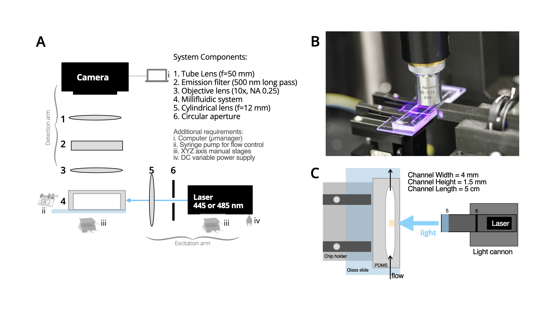
The excitation arm included a compact laser diode, an adjustable iris, and a cylindrical lens. To create the light sheet, similar to our prior efforts [8], we cut the central part of the elliptical beam of a laser diode (wavelength 445 nm or 488 nm) with the iris (Thor labs SM1D12), followed by a cylindrical lens (f=12 mm) that focused the light along one dimension to form a sheet of light at the observation plane. These components were housed inside a custom-designed 3D-printed enclosure. We used a cannon-like design (i.e., light-cannon) to bring the cylindrical lens close to the fluidic chip. The 3D-printed enclosure containing the optical components for excitation was then mounted on an XYZ micromanipulator. A light sheet of 7 µm thickness was created utilizing these components (see Supplemental for analysis). The excitation arm was mounted on an optical breadboard at a 90-degree angle relative to the detection arm. The detection arm included a custom-designed 3D-printed chip holder to bring the fluidic chip close to the light-cannon. Fluorescent signals were collected perpendicular to the excitation with an objective lens (10x, NA 0.25) followed by an emission filter (500 nm long pass) and a tube lens (f = 50 mm) for imaging with a CMOS camera (FLIR Blackfly S-U3-200S6C-C: 20 MP). SPIM-Flow’s system-level diagram is provided (Figure 1.A). SPIM-Flow was housed on a small optical breadboard (300 mm x 300 mm) and inside an opaque enclosure to shield the setup from external light. Micro-manager software was used to control the camera. Designs for the 3D parts will be available from a corresponding OSF page.
2.2 Millifluidic system
We used laser cutting to build the molds used in the study instead of conventional lithographic approaches. The size of Hydra (typically 0.5 -10 mm long) and our goal to investigate their unhindered hydrodynamics informed our choice. To this aim, channels were designed to be 4 mm wide and 5 cm long. We laser cut the design on acrylic sheets (1/16th inch, 1.5 mm thickness) via a desktop laser cutter. Plastic parts were permanently attached to a flat base (either a plastic part or a glass slide). The attachment prevented the unwanted PDMS accumulation under the mold during the silicone casting. After the mold fabrication, we used conventional protocols to cast silicone parts from the mold (1:10 linker to the base mass ratio followed by baking the molds on hotplates at 85°C until the PDMS were fully cured). Individual devices were cut from the mold using craft knives, and inlet/outlet holes were introduced using disposable (1 or 2 mm diameters) biopsy punches.
Next, each PDMS part was plasma bonded to a microscope glass slide via a Harrick Basic Plasma cleaner. We placed the edge of the PDMS part on the edge of the glass slide to minimize the distance between the channel and the light cannon (Figure 1.B and 1.C). Additionally, we took steps to remove any roughness from the outer edges of the devices to minimize potential imaging artifacts. Some roughness can be introduced to the outer edges of PDMS parts while cutting them via a craft knife. In a typical epifluorescent illumination, such roughness is of no concern,since the path of light is through the device top (typically PDMS) and glass bottom. In our system, however, the chamber is illuminated through a PDMS wall and viewed from the device top. We applied additional uncured PDMS to the device’s outer wall using a craft knife after plasma bonding and removed any roughness. We left the device (with the side with fresh PDMS facing up) at room temperature for three hours. This pause allowed the PDMS to spread evenly and partially cure. Then we backed the device at 95°C to ensure that the PDMS was entirely cured. During the early experiments, we also attached coverslips to the device roof. However, later experiments demonstrated that the coverslip did not significantly improve imaging.
2.3 Hydra culture
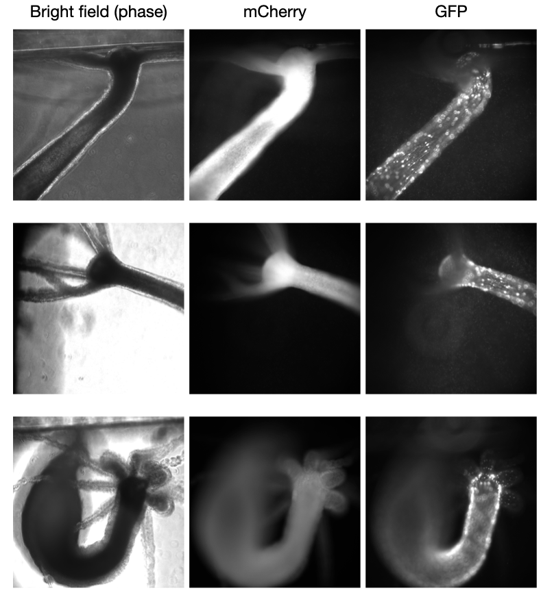
All experiments were carried out using the PT1 transgenic line of Hydra vulgaris [29]. PT1 contains two transgenes. One of the transgenes expresses green fluorescent protein under the control of the promoter from the gene encoding the Hym176B neuropeptide. The other transgene expresses the DsRed2 gene under the control of an actin gene promoter. This line expresses green fluorescent neurons and red fluorescent epithelial cells. PT1 was maintained in Hydra medium 4.0, which was prepared using house deionized water and contained 1 mM calcium chloride, 0.33 mM magnesium sulfate, 0.5 mM sodium bicarbonate, and 0.03 mM potassium chloride. The hydras were fed once a week with nauplii of Artemia franciscana from San Francisco Bay (Brine Shrimp Direct, Ogden, UT). Hydra cultures were kept in an incubator at 18°C on a 12 hour light/12 hour dark cycle.
3 Results
3.1 Loading the Hydra
We developed a simple protocol to minimize potential damage and increase the successful loading of animals. First, a device was partially (1/2) filled with media. We used a glass Pasteur pipette to retrieve an animal from a culture tube. Our biggest challenge was Hydra adhering to the glass transfer pipette since Hydra can rapidly adhere and remain firmly attached to surfaces. It is essential to transfer a Hydra quickly. Therefore, we released the animals directly inside a device inlet or on top of the inlet with excess media from the pipette to a device. Next, we used liquid withdrawal with a micropipette (1 mL) or a syringe to draw the animal with the additional liquid inside the chamber. We sought to position each Hydra toward the chamber’s center for ease of imaging. To enable flow visualization, we added red fluorescent beads (1 µm diameter, Fluoro-Max Polymer microspheres) to the medium. We note that beads are also visible in GFP emission wavelength.
After loading, the chamber was placed and secured on the 3D-printed chip holder. Hydra medium with beads was delivered to the chamber using an external syringe pump (KDS Legato, 210P series) via 3 or 10 mL syringes. Typical of all microfluidics experiments, care was taken to ensure there were no air bubbles upstream of the device. However, small bubbles were not a problem. For example, early in the studies, we accidentally introduced air bubbles into two chambers. In each case, the animal rapidly responded to the air bubble and the pressure by contracting/bending. Animals were able to survive these exposures. However, we chose to replace these animals with fresh ones.
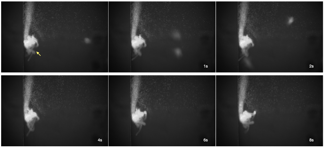
At the start of the study, we sought to observe feeding behavior of Hydra in the system to examine health. After loading a Hydra on a chip, multiple prey animals (Artemia franciscana) were added to the chamber. We first used the imaging without its light sheet capabilities to independently investigate the potential chamber effects. Hydra could successfully capture and eat multiple (2 to 3) Artemia inside the chamber. Next, we verified similar feeding behavior using the SPIM imaging capabilities across independent animals (Supplemental Figure 1, and Supplemental Video 1) - video and images are from two different animals. After feeding, Hydra ignored the remaining prey in the chamber. The feeding results (across N=4 different animals) suggested that the fluid chambers and the light sheet did not negatively impact the organisms’ health. Experiments presented in the study took 2-3 hours; however, we have confirmed that a Hydra can be kept alive on the same chip for multiple days.
3.2 Static conditions and baseline behaviors
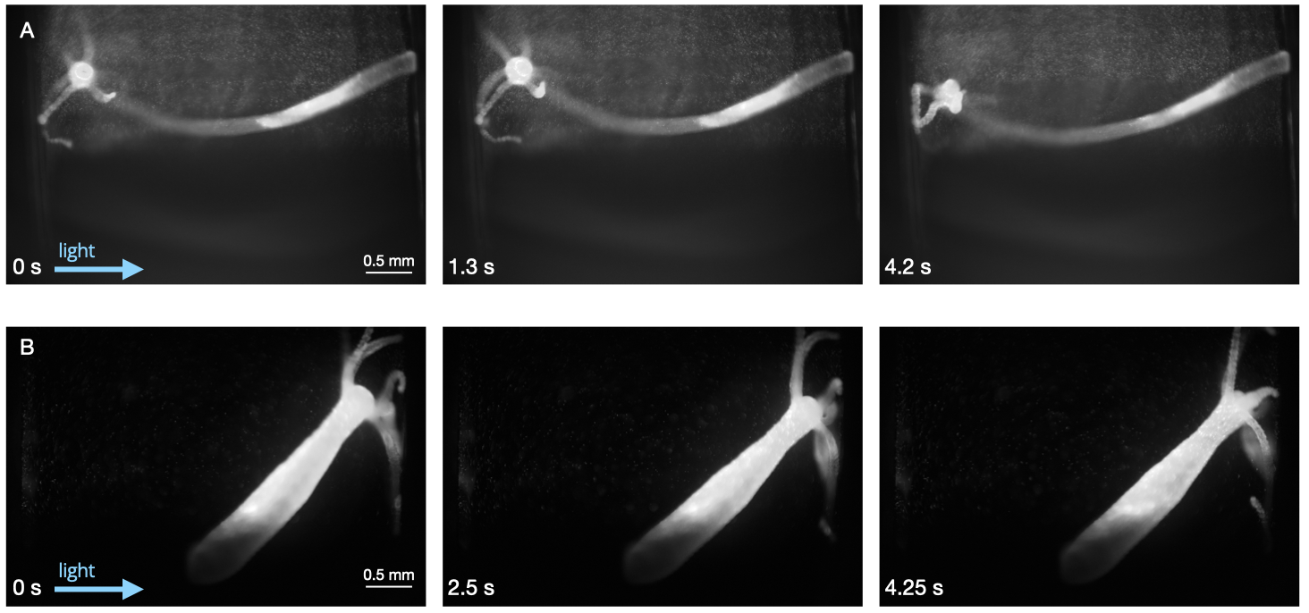
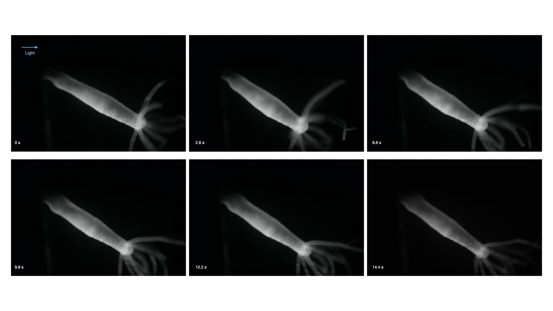
Next, we examined the animals’ health and sought to demonstrate the feasibility of observing various behaviors via SPIM-Flow without flow. Given the device’s dimension, the animals’ overall movements were unhindered. However, some animals could stretch across the entire 4 mm width of the chamber. Using the light sheet, we illuminated different regions of the freely moving animals. Hydra responds to light as a stimulus [30, 31, 32, 33].To minimize the role of light as a sudden stimulus, we started recording an experiment 5 minutes after the light introduction. A typical recording session would last between 2-3 hours. SPIM’s capability to limit the light exposure (only to the illuminated sheet) prevented photobleaching or phototoxicity. However, given the role of light as a stimulus, additional studies are required, and SPIM-Flow could provide a versatile tool for this aim. We observed that most animals typically adhered to the surfaces (PDMS wall or the glass slide) closer to the direction of excitation light (wavelength 485 nm or 455 nm). Without flow, we observed behaviors ranging from elongation, and tentacle movement/swaying. (Figures 2 and 3, Supplemental Figure 2, Supplemental Video 3). Most animals would remain in the chambers elongated or in a tentacle swaying state. Despite the chambers’ large size, we did not observe somersaulting behaviors. These observations further supported animals’ overall health and helped demonstrate that various repertoires of animal movements can be captured via SPIM-Flow.
3.3 Response to flow
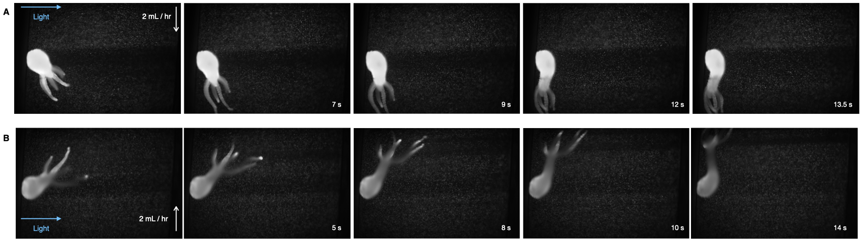
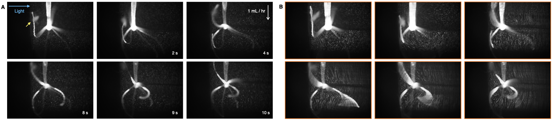
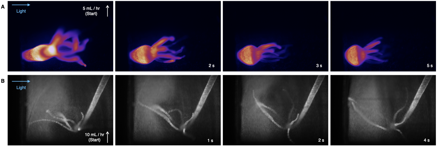
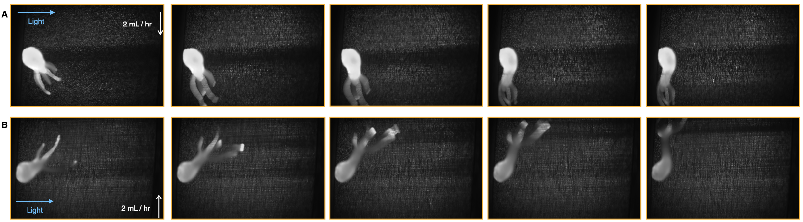
Next, we investigated Hydra response to flow. Hydra lives in bodies of freshwater that may experience local flow patterns due to environmental perturbations or currents in streams and rivers. Therefore, tools to investigate their hydrodynamic response to flow could provide valuable insights into the organisms’ biomechanical and hydrodynamic lifestyle. In their study, Badhiwala et al. [27] commented that animals typically bend in the flow direction. We sought to investigate this observation by utilizing our larger channels (1.5 mm in height, 4 mm wide). Similar to the Badhiwala et al. study, our results (across four independent animals) suggest that Hydra typically responds to flow by bending and swaying their tentacles in the flow direction. Reversing the direction of flow resulted in Hydra redirecting accordingly (Figure 4). For example, in results from Figure 4, the animal responded to the volumetric flow rate of 2 mL/hr. Larger animals could withstand flow rates as large as 50 mL/hr. Our results also suggest that an animal could immediately respond to the flow initiation via rapid contraction or tentacles swaying in the direction of the flow (Supplemental Figure 3). However, additional experiments are required to better understand the immediate response to flow initiation. Utilizing SPIM-Flow, we briefly explored a proof of concept experiment, to investigate Hydra’s response to pulsatile flow. Our preliminary results suggest that pulsatile flow resulted in an animal stretching towards the direction of the flow.
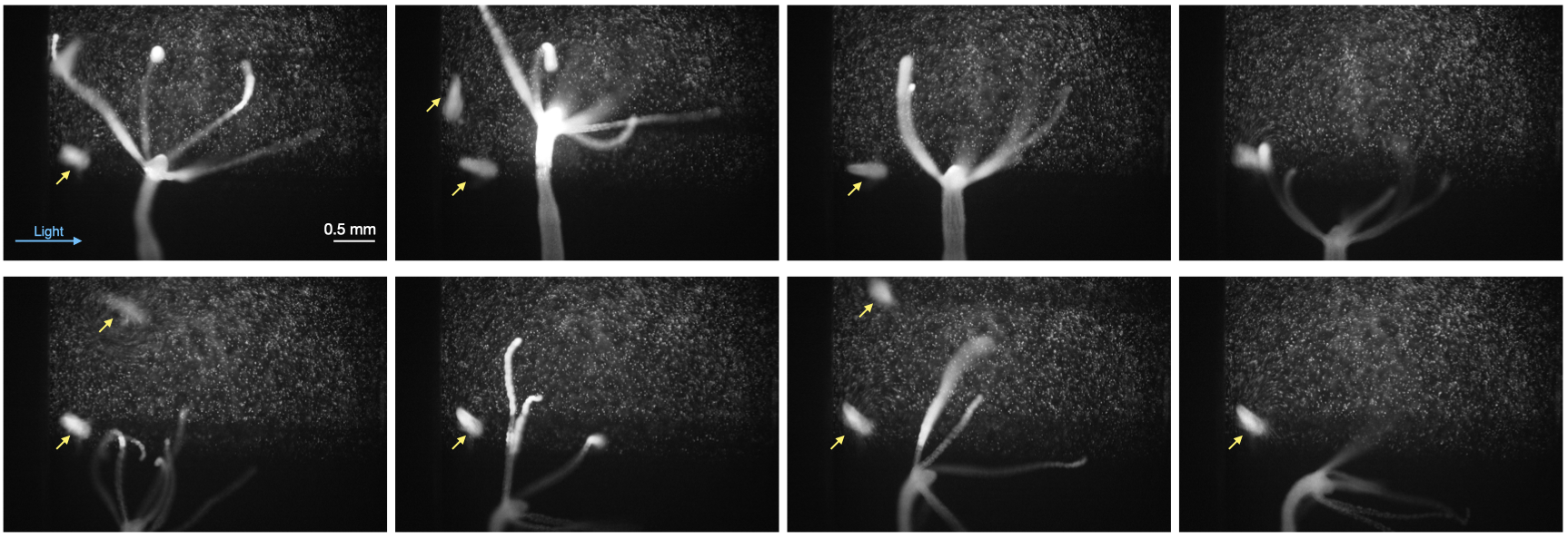
Finally, we used a previously described flow visualization algorithm, FlowTrace, to help better visualize the hydrodynamic patterns observed via SPIM-Flow. FlowTrace is a powerful yet simple algorithm that enables the extraction of flow features from video recordings [34, 35]. Unlike typical pipelines for particle image velocimetry, FlowTrace is not computationally taxing. FlowTrace has been used to extract pathlines to visualize the time-varying flow fields generated by starfish larvae and other organisms. We used FlowTrace analysis of the SPIM-Flow videos to visualize flow fields (e.g., vortex generated by the tentacle sways) and pathlines generated by and around Hydra. For this, we used the algorithm as a FIJI plugin [34]. Figure 5 demonstrates the original recording (A), and the corresponding FlowTrace visualizations (B). Pathline visualizations corresponding to Figure 4 and FlowTrace videos are provided as part of the supplemental material.
4 Discussion and Conclusion
Despite tremendous progress, access to SPIM imaging and interfacing the conventional systems with modern fluidics have remained challenging. We sought to address this gap by establishing SPIM-Flow. Our platform is an inexpensive light sheet microscope readily designed for integration with microfluidics. To validate the usefulness of SPIM-Flow, we used the platform to study the hydrodynamics of freely moving Hydra in static and dynamic conditions. SPIM-Flow could enable various applications; for example, we explored its use as an image-based flow cytometer. However, as demonstrated, it could serve as a powerful tool to visualize and manipulate whole organisms and their microenvironment. Light sheet imaging specifically enabled easy visualization of tracer beads to extract hydrodynamic features. As a comparison, we also imaged the animals inside the chambers via a standard fluorescent microscope (Nikon Eclipse Ti-E microscope 10X objective - Supplemental Figure 5). Simultaneously visualizing the beads and the animal (due to the high signal from the animal and the background volume) were difficult with a standard microscope. Indeed SPIM-Flow is not a replacement for a standard fluorescent microscope. Instead, it is a complementary and inexpensive tool which enables hydrodynamic studies of organisms, such as Hydra. Our initial experiments (feeding and observing various behaviors) supported that the platform did not impact animals’ health. Next, we used microfluidics to investigate Hydra’s response to continuous flow. Our results across multiple animals (N=4) suggest Hydra responds to flow by bending in the flow direction. Reversing the direction of flow led to animals’ reorientation. Utilizing the light sheet imaging, we visualized pathlines (via fluorescent beads) throughout the study. Moreover, using the FlowTrace algorithm, we produced visualizations highlighting the complex hydrodynamics of an animal movement.
Our study demonstrated the platform’s capability for studying freely-moving model organisms. In the future, we seek to explore the biomechanics of Hydra movement further (e.g., quantifying displacements generated by the animals or systematically exploring animals’ response to pulsatile flow). Microfluidics combined with conventional imaging has provided excellent platforms to study model systems such as zebrafish, Drosophila embryos, and C. elegans [36, 37]. Other emerging model systems could (and indeed have started to) benefit from need-driven systematic integration of micro and millifluidics and novel imaging techniques [38, 39, 40]. SPIM-Flow, as a simple and inexpensive platform, can contribute to these studies with a focus on hydrodynamics of small animals.
Acknowledgments
The authors express gratitude to Prof. Albert Siryaporn for his support.
This work was supported in part by CSULB startup funds and a CSUPRB research grant to SA.
Additionally, this work was supported by National Institutes of General Medical Sciences R21GM135493 to PNH.
LD is supported by an NSF GRFP (base number 2021317020).
Corresponding Authors Addresses
Siavash Ahrar (Ph.D.)
Mail: Department of Biomedical Engineering, CSU Long Beach,
1250 Bellflower Blvd.
VEC-404.A, Long Beach, CA 90840,
E-mail: siavash.ahrar@csulb.edu
Per Niklas Hedde (Ph.D.)
E-mail: phedde@uci.edu
Affiliations
-
•
PNH: Beckman Laser Institute and Medical Clinic, University of California Irvine, CA, US.
-
•
ELG: Department of Biomedical Engineering. CSU Long Beach, CA, US.
-
•
LD: Department of Molecular Biology and Biochemistry,University of California Irvine, CA, US.
-
•
RES: Department of Biological Chemistry, University of California Irvine, CA, US.
-
•
SA: Department of Biomedical Engineering. CSU Long Beach, CA, US.
Department of Physics and and Astronomy, University of California Irvine, CA, US.
List of Figures:
-
•
Figure 1: SPIM-Flow system diagram.
-
•
Figure 2: Baseline behaviors during static conditions as an indicator for overall health.
-
•
Figure 3: Baseline behaviors, bending and tentacle movement, during static conditions.
-
•
Figure 4: Response to flow.
-
•
Figure 5: Hydrodynamic visualization via FlowTrace.
List of Supplementary Figures:
-
•
Sup Figure 1: Hydra feeding behavior as an indicator for health.
-
•
Sup Figure 2: Hydra baseline behavior, elongation, during static conditions as an indicator for overall health.
-
•
Sup Figure 3: Hydra responses to the initiation of flow.
-
•
Sup Figure 4: FlowTrace visualizations - response to flow.
-
•
Sup Figure 5: Imaging Hydra via a standard fluorescent microscope.
List of Supplementary Videos:
-
•
Movie S1: Hydra feeding behavior
-
•
Movie S2: Hydra tentacle sway behavior
-
•
Movie S3: Hydra response to flow
-
•
Movie S4: FlowTrace - example 1 (vortex)
-
•
Movie S5: FlowTrace - response to flow example 2
References
- [1] Ernst HK Stelzer, Frederic Strobl, Bo-Jui Chang, Friedrich Preusser, Stephan Preibisch, Katie McDole, and Reto Fiolka. Light sheet fluorescence microscopy. Nature Reviews Methods Primers, 1(1):1–25, 2021.
- [2] Jan Huisken, Jim Swoger, Filippo Del Bene, Joachim Wittbrodt, and Ernst HK Stelzer. Optical sectioning deep inside live embryos by selective plane illumination microscopy. Science, 305(5686):1007–1009, 2004.
- [3] Omar E Olarte, Jordi Andilla, Emilio J Gualda, and Pablo Loza-Alvarez. Light-sheet microscopy: a tutorial. Advances in Optics and Photonics, 10(1):111–179, 2018.
- [4] Fabian F Voigt, Daniel Kirschenbaum, Evgenia Platonova, Robert AA Campbell, Rahel Kastli, Martina Schaettin, Ladan Egolf, Alexander Van der Bourg, Philipp Bethge, Karen Haenraets, et al. The mesospim initiative: open-source light-sheet microscopes for imaging cleared tissue. Nature methods, 16(11):1105–1108, 2019.
- [5] Emilio J Gualda, Hugo Pereira, Tiago Vale, Marta Falcão Estrada, Catarina Brito, and Nuno Moreno. Spim-fluid: open source light-sheet based platform for high-throughput imaging. Biomedical optics express, 6(11):4447–4456, 2015.
- [6] Bin Yang, Merlin Lange, Alfred Millett-Sikking, Xiang Zhao, Jordão Bragantini, Shruthi VijayKumar, Mason Kamb, Rafael Gómez-Sjöberg, Ahmet Can Solak, Wanpeng Wang, et al. Daxi—high-resolution, large imaging volume and multi-view single-objective light-sheet microscopy. Nature methods, 19(4):461–469, 2022.
- [7] Per Niklas Hedde. minispim—a miniaturized light-sheet microscope. ACS sensors, 6(7):2654–2663, 2021.
- [8] Per Niklas Hedde, Milka Stakic, and Enrico Gratton. Rapid measurement of molecular transport and interaction inside living cells using single plane illumination. Scientific reports, 4(1):1–6, 2014.
- [9] Etai Sapoznik, Bo-Jui Chang, Jaewon Huh, Robert J Ju, Evgenia V Azarova, Theresa Pohlkamp, Erik S Welf, David Broadbent, Alexandre F Carisey, Samantha J Stehbens, et al. A versatile oblique plane microscope for large-scale and high-resolution imaging of subcellular dynamics. Elife, 9:e57681, 2020.
- [10] William C Lemon, Stefan R Pulver, Burkhard Höckendorf, Katie McDole, Kristin Branson, Jeremy Freeman, and Philipp J Keller. Whole-central nervous system functional imaging in larval drosophila. Nature communications, 6(1):1–16, 2015.
- [11] Ludovico Silvestri, Alessandro Bria, Irene Costantini, Leonardo Sacconi, Hanchuan Peng, Giulio Iannello, and Francesco Saverio Pavone. Micron-scale resolution optical tomography of entire mouse brains with confocal light sheet microscopy. JoVE (Journal of Visualized Experiments), (80):e50696, 2013.
- [12] Daniel Von Wangenheim, Robert Hauschild, and Jiří Friml. Light sheet fluorescence microscopy of plant roots growing on the surface of a gel. JoVE (Journal of Visualized Experiments), (119):e55044, 2017.
- [13] Hiroki R Ueda, Hans-Ulrich Dodt, Pavel Osten, Michael N Economo, Jayaram Chandrashekar, and Philipp J Keller. Whole-brain profiling of cells and circuits in mammals by tissue clearing and light-sheet microscopy. Neuron, 106(3):369–387, 2020.
- [14] Pan Zhou, Haipeng He, Hanbin Ma, Shurong Wang, and Siyi Hu. A review of optical imaging technologies for microfluidics. Micromachines, 13(2):274, 2022.
- [15] Ignacio Albert-Smet, Asier Marcos-Vidal, Juan José Vaquero, Manuel Desco, Arrate Muñoz-Barrutia, and Jorge Ripoll. Applications of light-sheet microscopy in microdevices. Frontiers in neuroanatomy, 13:1, 2019.
- [16] Raju Regmi, Kavya Mohan, and Partha P Mondal. Light sheet based imaging flow cytometry on a microfluidic platform. Microscopy research and technique, 76(11):1101–1107, 2013.
- [17] Meiai Lin, Qiao Liu, Chao Liu, Xu Qiao, Changshun Shao, and Xuantao Su. Label-free light-sheet microfluidic cytometry for the automatic identification of senescent cells. Biomedical optics express, 9(4):1692–1703, 2018.
- [18] Yu-Jui Fan, Han-Yun Hsieh, Sheng-Fang Tsai, Cheng-Hsuan Wu, Chia-Ming Lee, Yen-Ting Liu, Chieh-Han Lu, Shu-Wei Chang, and Bi-Chang Chen. Microfluidic channel integrated with a lattice lightsheet microscopic system for continuous cell imaging. Lab on a Chip, 21(2):344–354, 2021.
- [19] Hao Jiang, Tingting Zhu, Hao Zhang, Jun Nie, Zeyi Guan, Chi-Ming Ho, Sheng Liu, and Peng Fei. Droplet-based light-sheet fluorescence microscopy for high-throughput sample preparation, 3-d imaging and quantitative analysis on a chip. Lab on a Chip, 17(13):2193–2197, 2017.
- [20] Gilles Vanwalleghem, Kevin Schuster, Michael A Taylor, Itia A Favre-Bulle, and Ethan K Scott. Brain-wide mapping of water flow perception in zebrafish. Journal of Neuroscience, 40(21):4130–4144, 2020.
- [21] Per Niklas Hedde, Leonel Malacrida, Siavash Ahrar, Albert Siryaporn, and Enrico Gratton. sidespim–selective plane illumination based on a conventional inverted microscope. Biomedical Optics Express, 8(9):3918–3937, 2017.
- [22] Matthias C Vogg, Brigitte Galliot, and Charisios D Tsiairis. Model systems for regeneration: Hydra. Development, 146(21):dev177212, 2019.
- [23] William M Kier. The diversity of hydrostatic skeletons. Journal of Experimental Biology, 215(8):1247–1257, 2012.
- [24] Shuting Han, Ekaterina Taralova, Christophe Dupre, and Rafael Yuste. Comprehensive machine learning analysis of hydra behavior reveals a stable basal behavioral repertoire. Elife, 7:e32605, 2018.
- [25] Jason A Carter, Callen Hyland, Robert E Steele, and Eva-Maria S Collins. Dynamics of mouth opening in hydra. Biophysical journal, 110(5):1191–1201, 2016.
- [26] Richard D Campbell. Structure of the mouth of hydra spp. a breach in the epithelium that disappears when it closes. Cell and tissue research, 249(1):189–197, 1987.
- [27] Krishna N Badhiwala, Daniel L Gonzales, Daniel G Vercosa, Benjamin W Avants, and Jacob T Robinson. Microfluidics for electrophysiology, imaging, and behavioral analysis of hydra. Lab on a Chip, 18(17):2523–2539, 2018.
- [28] Krishna N Badhiwala, Abby S Primack, Celina E Juliano, and Jacob T Robinson. Multiple neuronal networks coordinate hydra mechanosensory behavior. Elife, 10:e64108, 2021.
- [29] Robert E Steele, Megan D Updegrove, Sara A Kirolos, Lucas Mowery, Daniel E Martinez, and Peter J Bryant. Reproductive bet-hedging and existence in vernal pools as components of hydra life history. The Biological Bulletin, 237(2):111–118, 2019.
- [30] Abraham Trembley. Mémoires pour servir à l’histoire d’un genre de ploypes d’eau douce, à bras en forme de cornes, volume 1. Durand, 1744.
- [31] Edmund B Wilson. The heliotropism of hydra. The American Naturalist, 25(293):413–433, 1891.
- [32] Gottfried Haug. Die lichtreaktionen der hydren. Zeitschrift für vergleichende Physiologie, 19(2):246–303, 1933.
- [33] Robert H Singer, Norman B Rushforth, and Allison L Burnett. The photodynamic action of light on hydra. Journal of Experimental Zoology, 154(2):169–173, 1963.
- [34] William Gilpin, Vivek N Prakash, and Manu Prakash. Flowtrace: simple visualization of coherent structures in biological fluid flows. Journal of Experimental Biology, 220(19):3411–3418, 2017.
- [35] William Gilpin, Vivek N Prakash, and Manu Prakash. Dynamic vortex arrays created by starfish larvae. Physical Review Fluids, 2(9):090501, 2017.
- [36] Anna A Kim, Adam L Nekimken, Sylvia Fechner, Lucy E O’Brien, and Beth L Pruitt. Microfluidics for mechanobiology of model organisms. In Methods in cell biology, volume 146, pages 217–259. Elsevier, 2018.
- [37] Hyundoo Hwang and Hang Lu. Microfluidic tools for developmental studies of small model organisms–nematodes, fruit flies, and zebrafish. Biotechnology journal, 8(2):192–205, 2013.
- [38] Deepak Krishnamurthy, Hongquan Li, François Benoit du Rey, Pierre Cambournac, Adam G Larson, Ethan Li, and Manu Prakash. Scale-free vertical tracking microscopy. Nature Methods, 17(10):1040–1051, 2020.
- [39] Felix JH Hol, Louis Lambrechts, and Manu Prakash. Biteoscope, an open platform to study mosquito biting behavior. Elife, 9:e56829, 2020.
- [40] Shailabh Kumar, Felix JH Hol, Sujit Pujhari, Clayton Ellington, Haripriya Vaidehi Narayanan, Hongquan Li, Jason L Rasgon, and Manu Prakash. A microfluidic platform for highly parallel bite by bite profiling of mosquito-borne pathogen transmission. Nature communications, 12(1):1–12, 2021.
Supplemental section - analysis of light sheet properties
The Rayleigh criterion defines the optical resolution, r, as:
r = 0.61
where is the wavelength and NA the numerical aperture of the lens.
For small aperture lenses, the NA can be approximated by:
NA =
where D is the lens diameter, fthe lens focal length, and n the refractive index of the surrounding medium, which in our case was air (n = 1).
Combining the two equations yields:
r = 1.22
With an excitation wavelength of 488 nm, a beam diameter of 1 mm, and the focal length of the cylindrical lens of 12 mm, the light sheet minimum thickness was 7.1 µm.