Electron paramagnetic resonance of alkali metal atoms and dimers on ultrathin MgO
Abstract
Electron paramagnetic resonance (EPR) can provide unique insight into the chemical structure and magnetic properties of dopants in oxide and semiconducting materials that are of interest for applications in electronics, catalysis, and quantum sensing. Here, we demonstrate that EPR in combination with scanning tunneling microscopy (STM) allows for probing the bonding and charge state of alkali metal atoms on an ultrathin magnesium oxide layer on Ag substrate. We observe a magnetic moment of for Li2, LiNa, and Na2 dimers corresponding to spin radicals with a charge state of . Single alkali atoms have the same charge state and no magnetic moment. The ionization of the adsorbates is attributed to charge transfer through the oxide to the metal substrate. Our work highlights the potential of EPR-STM to provide insight into dopant atoms that are relevant for the control of electrical properties of surfaces and nanodevices.
keywords:
Scanning tunneling microscopy, electron spin resonance, alkali metal dopants, catalysisDepartment of Physics, Freie Universität Berlin, 14195 Berlin, Germany \altaffiliationDonostia International Physics Center (DIPC), Paseo Manuel de Lardizabal 4, 20018 San Sebastián, Spain
![[Uncaptioned image]](/html/2205.05998/assets/Figs/TOCGraphic.png)
1 Main text
Recent studies have shown that electron paramagnetic resonance (EPR) spectroscopy can be combined with scanning tunneling microscopy (STM) to achieve single spin sensitivity and sub-nanometer spatial resolution 1, 2, 3, 4, 5, 6. This unique combination of techniques enables the investigation of the low-energy magnetic excitations of individual atoms on surfaces by overcoming the thermal resolution limit of tunneling spectroscopy. EPR-STM has been used to gain insight into the interaction of the nuclear and electronic magnetic moments of single atoms 7, 8, the dipolar 2, 9 and exchange coupling between atoms and molecules 10, 11, 12, and the coherent spin dynamics of atoms and molecules on a surface 13, 14, 5, 15. These recent achievements highlight the potential of EPR-STM for the investigation of quantum phenomena on the atomic scale. However, the applicability of this technique to a broad range of systems remains an open question. Apart from early attempts to perform EPR spectroscopy on defect centers in Si 16, 17, which proved hard to reproduce, all of the EPR-STM studies mentioned above were performed on transition-metal atoms, i.e., Fe, Ti, and Cu1, 2, 10, 11, 7, 13, 8, 14, 3, 4, 6, 5, 12, 9 or an Fe ion in the iron phthalocyanine molecule15, 12 all adsorbed on the MgO surface. The application of this technique to other elemental systems and to a broader range of problems beyond single-atom magnetism remains to be demonstrated.
In this work, we show that EPR-STM can be applied to study the electronic configuration of alkali electron donor complexes on an oxide surface. Functionalized oxide surfaces are of particular importance for heterogeneous catalysis 18. One way of functionalization is to bring metal atoms to the surface of the oxides, which provide local binding sites and change in electron density and thus alter the chemical reactivity 19, 20. A widely used system for fundamental studies in heterogeneous catalysis is MgO(100) with a small amount of metal atoms or clusters added to the bulk or surface 21, 22. The doping of MgO with alkali metals transforms the bare MgO substrate into a catalyst suitable for industry-relevant reactions, e.g., oxidative coupling of methane to produce higher order hydrocarbons 23, 24, 25. In such systems, the chemical transformation is thought to occur through the formation of electron-rich oxygen sites in the vicinity of the alkali atoms 24. However, the electronic state and chemical bonds of the dopant atoms, which are crucial for the catalytic activity, can only be determined by means of density functional theory (DFT) 20. Here we use EPR-STM to investigate the charge state and binding configuration of individual Li and Na atoms as well as their dimers (Li2, Na2, and LiNa) deposited on ultrathin MgO(001) layers grown on a Ag substrate. Our measurements reveal a distinct EPR signal corresponding to a magnetic moment of µB for paramagnetic alkali metal dimers adsorbed near the top oxygen sites of MgO. In contrast, single alkali atoms adsorb on bridge O sites and show no magnetic moment. The absence of an EPR signal in the alkali atoms indicates a change of the orbital occupancy relative to the gas phase, where the valence -shell is singly occupied. This change in occupancy shows that the atoms and dimers have the same charge state and that both transfer exactly one electron to the Ag substrate. Our findings are supported by DFT calculations, which identify a single unpaired electron in the bonding molecular orbital as the origin of the magnetic moment of the alkali metal dimers.
The EPR-STM experiments were performed in ultra-high vacuum with an STM operating at 4.5 K. In the STM, we applied a bias voltage to the sample and detected the tunneling current by a transimpedance amplifier connected to the tip. The STM was equipped with a broadband (1-40 GHz) antenna that transmitted the signal with frequency from a microwave generator to the tunnel junction 4 to resonantly excite the probed magnetic moment in the external magnetic field applied perpendicular to the sample surface [see Figure 1 (a)]. Two monolayers of MgO were grown by depositing Mg in an oxygen atmosphere ( mbar) on a clean Ag(001) surface heated to 690 K. After the growth of MgO, the sample was inserted into the STM and cooled to 4.5 K, where Li, Na and Fe atoms were evaporated in sub-monolayer amounts. To achieve EPR sensitivity, we used spin-polarized tips prepared by transferring individual Fe atoms from MgO to the tip apex and applying an external magnetic field to polarize the magnetic cluster on the tip apex 1, 3.
The alkali metals adsorb mostly as single atoms on MgO [see Figure 1 (b)]. Individual atoms are imaged as round protrusions with an apparent height of 97(6) pm and 197(10) pm for Li and Na, respectively. Less than 1 % of the alkali atoms are found as dimers after deposition. The alkali metal dimers studied in this work were prepared by lateral manipulation of individual atoms with the STM tip stabilized at mV and nA. Both the as-grown and the assembled dimers can be disassembled into individual atoms using the same settings. All dimers appear as nearly round protrusions with an apparent height dependent on their composition, i.e., Li2 260(6) pm, LiNa 341(7) pm, and Na2 390(9) pm at mV, pA. Those height variations enable reliable identification of each dimer.
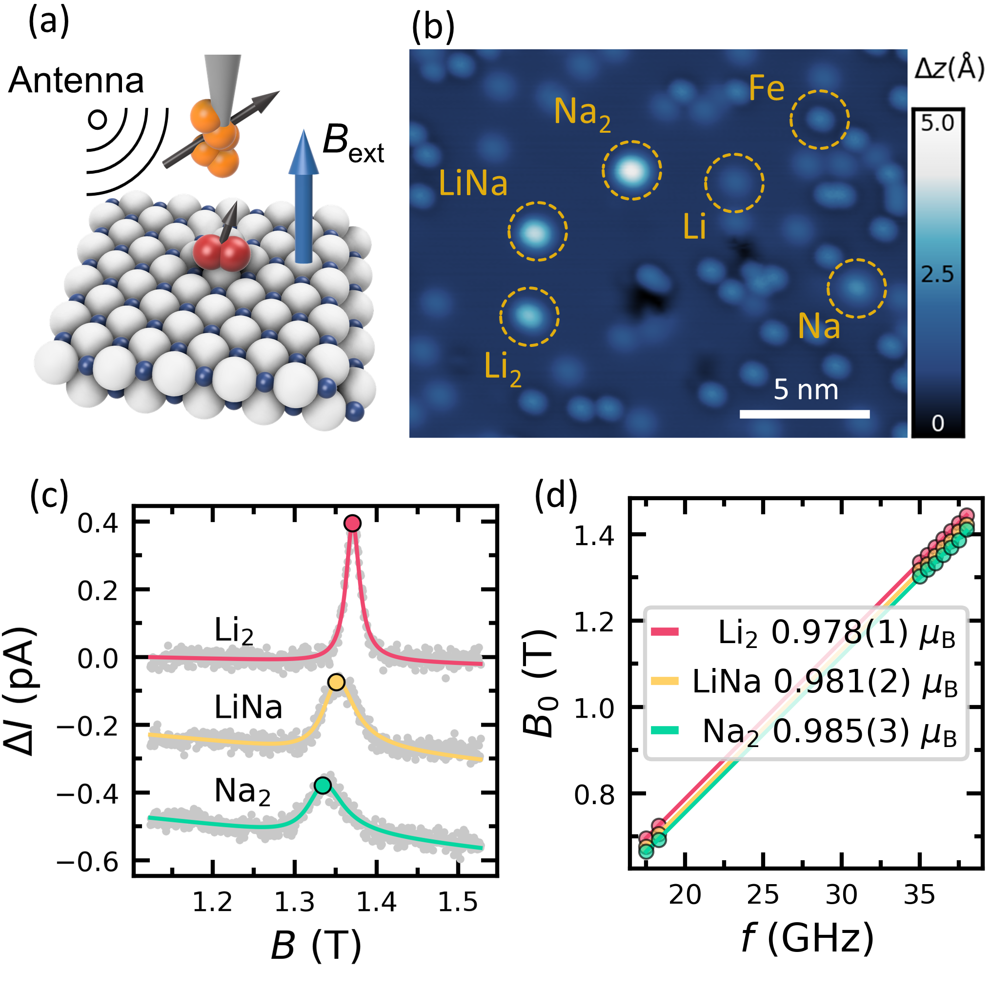
To gain insight into the charge state and associated magnetic moment of the alkali adsorbates, we use EPR-STM in magnetic field sweep mode. To this end, we place a magnetic tip above a species of interest, apply a bias voltage oscillating at gigahertz frequency with peak amplitude , and measure the time-averaged change in the tunneling current as a function of an external magnetic field 4. Typical EPR spectra are shown in Figure 1(c) for three different alkali metal dimers Li2, LiNa and Na2. EPR spectra recorded on individual alkali atoms (not shown) are featureless, suggesting the absence of a magnetic moment in the probed range from to . The observed resonances of alkali dimers are fit by a Fano lineshape to extract the external resonant magnetic field . Figure 1(d) presents the dependence of on , which allows us to extract the magnetic moment of the three alkali metal dimers. All configurations show a magnetic moment very close to 1 µB compatible with a spin , i.e., the presence of one unpaired electron in the outer electronic shell with no orbital magnetic moment. We probe the magnetic moment only in the out-of-plane direction, however, we do not expect the strong anisotropy of magnetic moment due to the low symmetry of molecular orbital and weak spin-orbit interaction in alkali metals. The absence of an EPR signal for the single atom species suggests that the alkali atoms are stable on the MgO/Ag substrate in the ionized state with no electrons in the valence shell.
The width of the EPR signals varies significantly between the different dimers. For the resonances presented in 1(c) the widths are 20.2(4) mT, 47(2) mT, and 51(2) mT for Li2, LiNa, and Na2, respectively. Upon closer inspection of the spectral fits, we identify a minor but systematic deviation of the data from the Fano lineshape commonly used to fit EPR-STM data. The relative deviation of the fits is presented in26. All alkali metals have a non-zero nuclear magnetic moment for the most abundant 7Li (92.4%) and 23Na (100%) species 27, leading to a splitting of the EPR line of alkali atoms in gas phase into four lines with similar intensity. The separation of the lines is given by the coupling strength between the nuclear and electronic magnetic moment which is stronger for Na atoms (885 MHz, 32 mT21) compared to Li (401 MHz, 14 mT 28) in the gas phase. Those coupling strengths are reduced upon adsorption on the MgO substrate by roughly 50% 20. Furthermore, we probe dimers instead of single atoms, which further decreases the coupling strength between the electronic and nuclear spins due to a reduced electron density of the bonding orbital at the position of the atomic nuclei 29. Also, the interaction of the electron spin with two identical nuclei in the homodimers causes a splitting of the EPR line into seven equidistant lines with relative intensities following the ratio 1:2:3:4:3:2:1. In our setup the minimum line width that can be resolved is 2 mT and we do not observe individual hyperfine-split resonant lines for the alkali dimers. However, the ratio of the atomic coupling strengths scales roughly as the observed line widths of the dimers. For the individual EPR transitions, we can only estimate the widths to be between one and tens of mT.
To further support this estimate, we have calculated the values of the isotropic hyperfine coupling constants following the method described in ref.30 to be MHz (4.2 mT) and MHz (10.4 mT) for the alkali atoms in the homodimers on MgO using DFT. Using the calculated coupling strengths and assuming seven equally spaced resonant lines, we estimate the total splitting of the EPR resonance line to be mT in the case of Li and mT for Na. These numbers are in good agreement with the observed linewidths, taking into account the relative intensities of the EPR lines in homodimers.
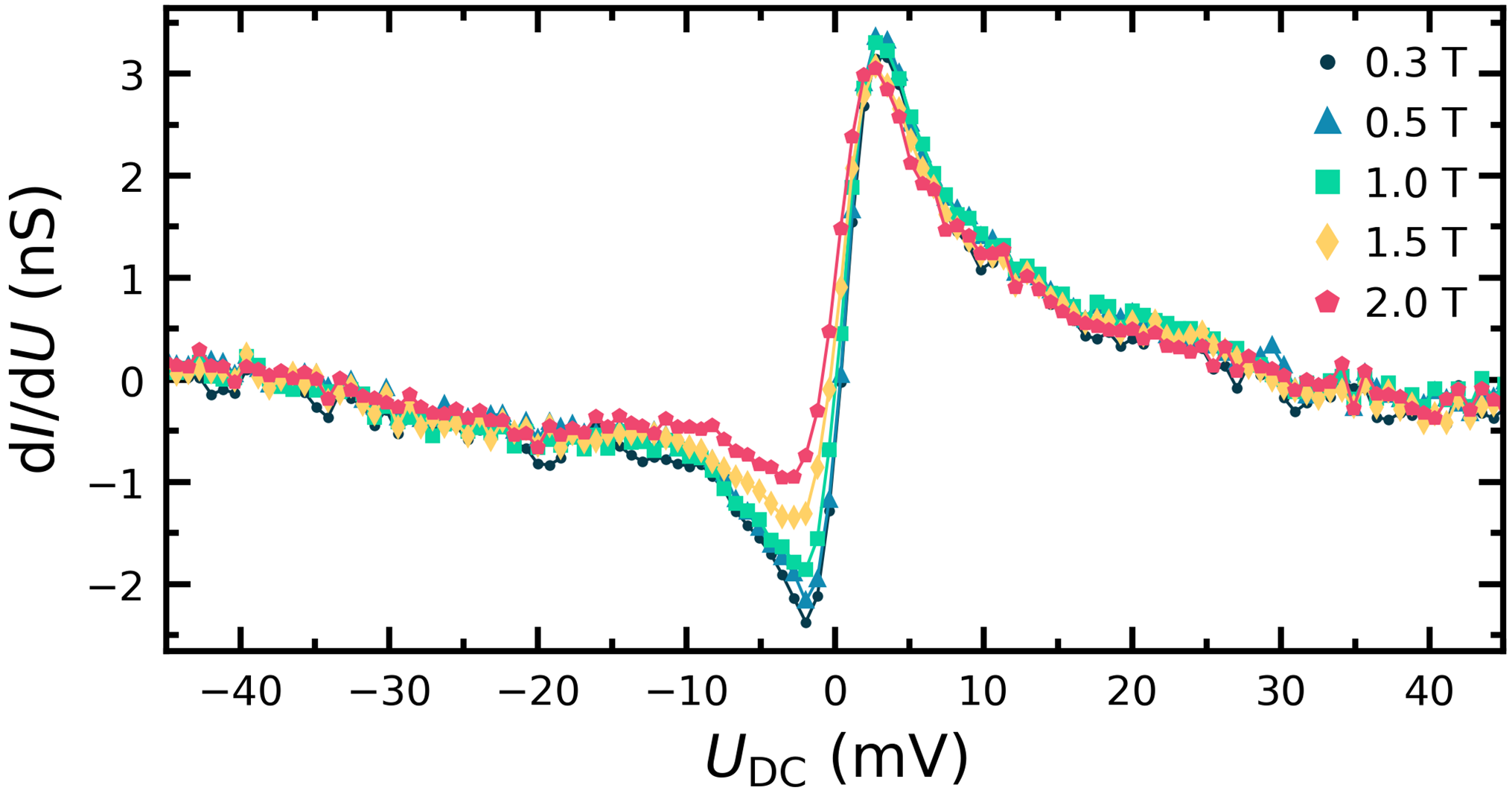
The presence of a magnetic moment in the alkali metal dimers is confirmed by measurements of the differential conductance of Li2 as a function of external magnetic field, which also allows getting insight into the spin relaxation time through a quantitative analysis using a rate equation model presented in 26. The spectra presented in Figure 2 show two noticeable features: a prominent negative-positive peak around zero bias and a magnetic field dependence of the peak at negative bias. We attribute the zero-bias structure to the spin-flip excitations of the magnetic moment of the dimer caused by the spin-polarized tunneling electrons, in analogy with previous measurements of transition-metal atoms on insulating substrates 31, 32, 10. The spin-polarization of the tip causes different excitation probabilities depending on the tunneling current direction, that leads to different conductance step heights for negative and positive bias 33, 34. The change in the polarisation of the tip with the applied magnetic field induces the change in the intensity of the dip at the negative bias in the data presented in 2. The dip and peak shape at negative and positive bias, respectively, are due to additional saturation effects of the spin-flip excitations that are observed in systems where the spin relaxation time is comparable to the average time between tunneling events 35, 34. Based on the quantitative analysis of the evolution of the conductance spectra presented in 26, we estimate the energy relaxation time ns for the experimental parameters used to perform the EPR-STM measurements presented in Figure 1(c).
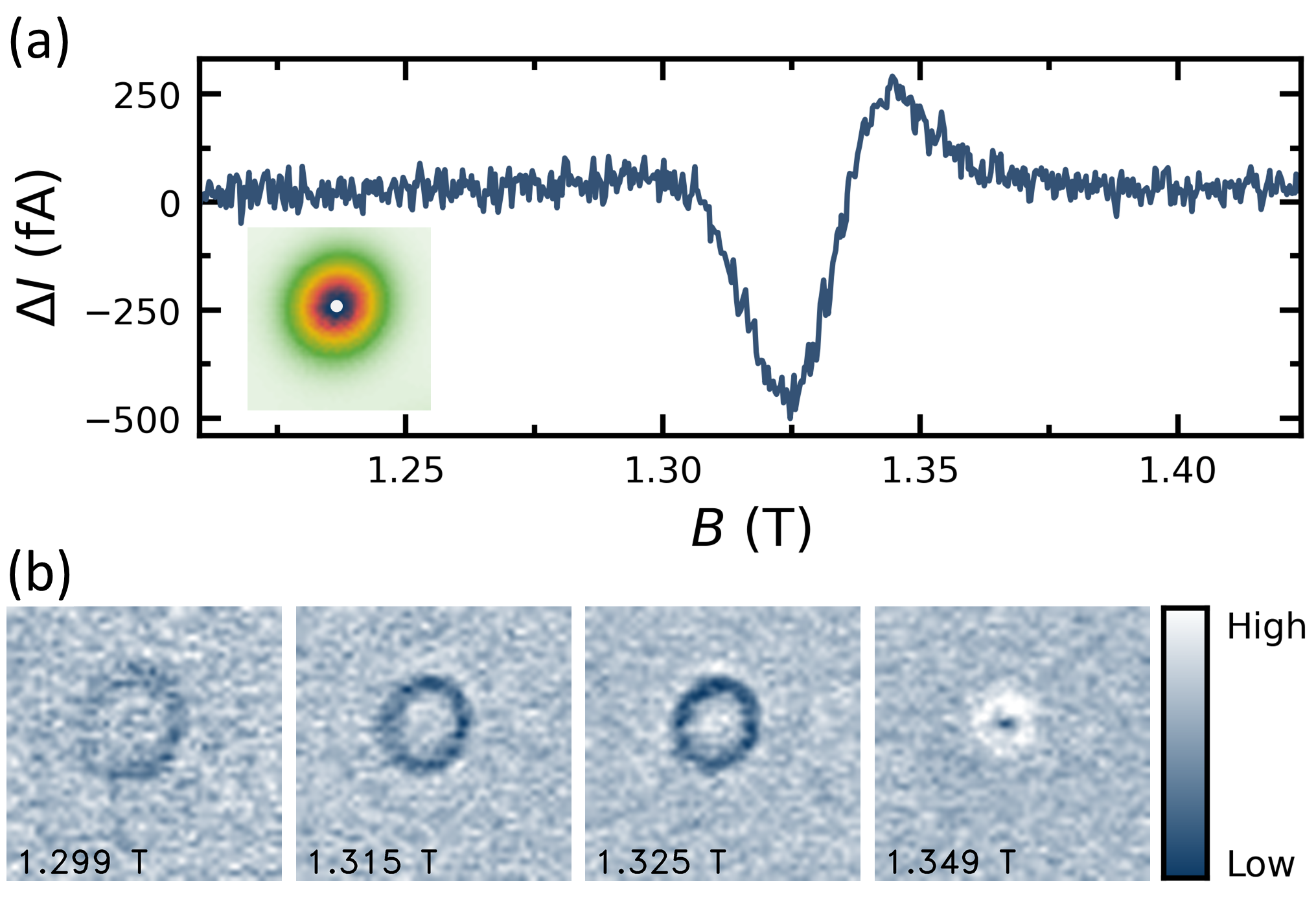
Besides probing unpaired electrons, EPR-STM also provides information on the spatial distribution of the spin density around the adsorbates. Figure 3 shows a spectrum and the spatial distribution of the EPR signal of Li2 measured using frequency modulation of the oscillating voltage. In contrast to the amplitude modulation used in Figure 1, frequency modulation allows us to obtain a spatial map of the EPR signal without the need to subtract a reference image to suppress the signal caused by the rectification of the excitation voltage 4. Moreover, the bipolar shape of the EPR signal recorded with the frequency modulation allows us to distinguish tip positions in which the probed species senses a magnetic field that is higher or lower than the resonant field. Since the local magnetic field at the position of the probed species is given by the external magnetic field and by interaction with the magnetic tip, the variation of the tip position causes detuning from the resonance 36. This is shown in the spatial maps of the EPR signal in Figure 3(b) obtained at several magnetic fields close to the resonant field. The EPR signal is distributed in rings with high values on the outer edge and low values toward the center of the ring. This behavior is due to a decreased energy splitting of the spin states as the tip approaches the center of the dimer, i.e. a shift of the EPR spectrum to higher external magnetic field values. Thus, the magnetic tip couples antiferromagnetically to the surface spin. From the almost circular shape of the signal, we conclude that the interaction of the tip with the dimer is dominated by the isotropic exchange interaction between the tip states and a nearly spherical orbital. This observation is consistent with the unpaired electron of Li2 residing in a bonding orbital.
To gain further insight into the binding and electronic configurations of the alkali adsorbates, we performed DFT calculations of Li and Na atoms and dimers on MgO/Ag(001). We find that the most stable binding site of Li and Na monomers is the bridge site of the oxygen sub-lattice. In contrast, the alkali atoms in the dimers bind near the oxygen top sites, with the axis of the dimer oriented diagonally across the unit-cell of the oxygen sub-lattice. The calculated binding configurations agree with STM measurements 26. The calculated bond length of Li2/MgO is stretched by 3% compared with the ionic molecules in the gas phase 37. In contrast, the bond lengths of LiNa and Na2 are compressed by 11% and 6%, respectively. The adsorption energies of the dimers are: 2.24 eV, 1.68 eV, and 1.08 eV for Li2, LiNa, and Na2, respectively.
The DFT calculations of individual Li and Na atoms identify a charge of around and no magnetic moment. The same charge state has been predicted earlier for K atoms adsorbed on 2 ML of MgO/Ag(001) 38. On ultrathin MgO the alkali atoms are positively charged due to the transfer of one electron to the nearby metal substrate via tunneling through the oxide 39, 22. This behavior is remarkably different from that of alkali atoms on the surface of bulk MgO, which maintain a neutral charge state and adsorb directly above oxygen 28.
In general, adsorbates on an oxide surface have a modified electronic structure that is affected by hybridization with the orbitals of the substrate, charge transfer, electrical polarization effects including dispersive forces as well as lattice deformations. For alkali metals adsorbed on bulk MgO, polarization effects dominate. Therefore, the alkali atoms remain electronically neutral but the electrical polarization modifies the orbital structure of the atom. The original spherically symmetric orbital becomes polarized by mixing with orbitals 19, 21. Our EPR maps and DFT calculations show that this does not happen for the ionized alkali dimers on ultrathin MgO, indicating that both the charge state and the binding site of the alkali species depend on the thickness of the oxide layer.
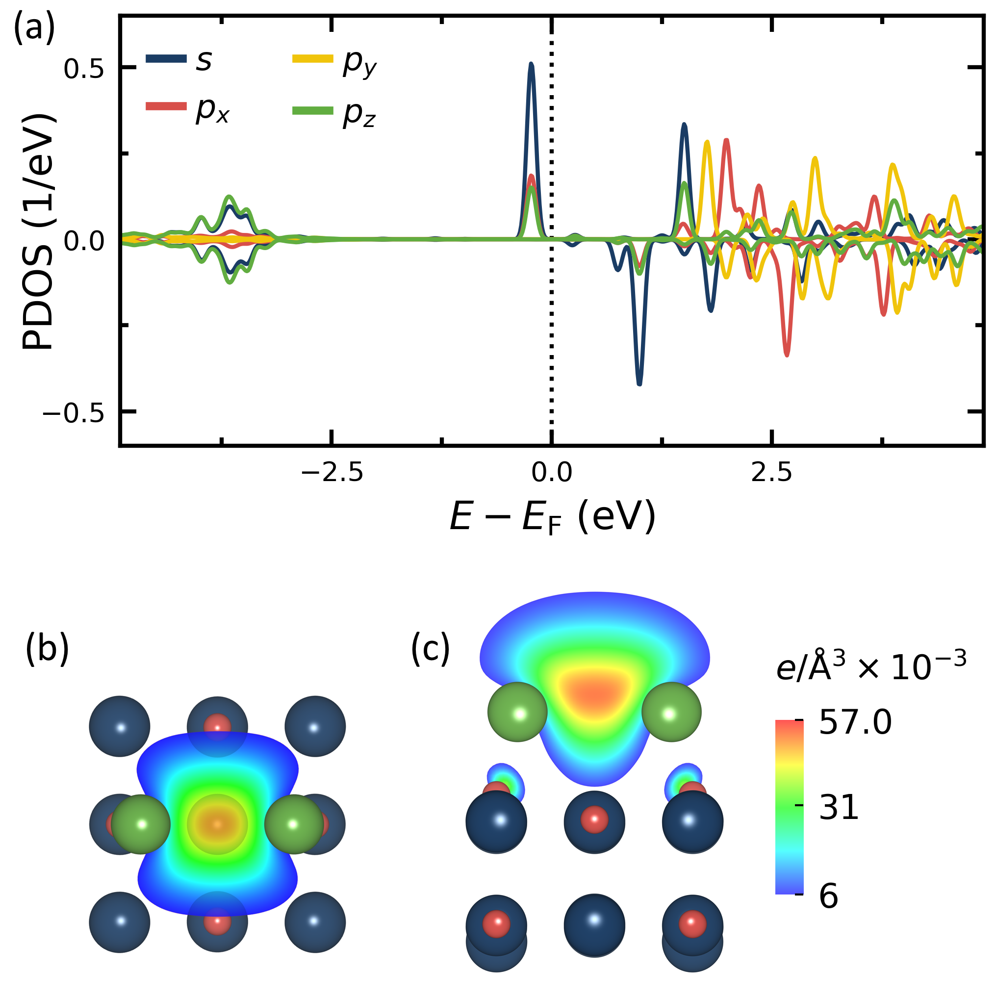
Unlike individual atoms, the calculated spin-dependent density of states (DOS) of the alkali metal dimers reveal a significant spin polarization of the electronic states close to the Fermi level. Figure 4(a) shows the calculated DOS of Li in Li2. The peak in the DOS just below the Fermi level corresponds to the bonding molecular orbital that arises from the hybridization of the and orbitals of the two Li atoms. This orbital is occupied by one electron, which gives rise to a finite spin-density [see Figure 4(b-c)]. In the gas phase, the Li2 molecule is electrically neutral and has no magnetic moment. The DFT results thus agree with the experimental observation of a magnetic moment of 1 µB and are consistent with the electropositive character of the alkali metals, which leads to a charge transfer from Li2 to the substrate resulting in the charged state .
Both homodimers Li2 and Na2 have the same magnetic moment as the heterodimer LiNa, indicating the same charge state. In contrast, the spin distribution of the heterodimer is qualitatively different compared to the homodimers, since it lacks mirror symmetry and has a maximum spin density closer to the Li atom 26. This asymmetry has not been resolved in the experiment, as we do not observe any prominent anisotropic feature of the electron density around the dimers in the STM images. This is attributed to the small spatial extension and almost round profile of the bonding orbital above the dimer [see Figure 4(c)].
Finally, we note that the synthesis of alkali trimers and more complex arrangements could not be achieved, probably due to a low dissociation energy of larger clusters. Preparing larger assemblies causes the immediate splitting of trimers into a dimer and a single atom, which further underscores the higher stability of the singly-charged dimers.
Our work sheds light on the bonding and charge state of alkali metal adsorbates on ultrathin MgO/Ag(001) and opens a new range of systems for EPR-STM investigations beyond transition metals 1, 2, 10, 11, 7, 13, 8, 14, 3, 4, 6, 5, 12, 9, 40. The ability to probe electron donor species using EPR-STM may be useful to perform atomic-scale characterization of silicon-based spin qubits 41, 42 and deterministic manipulation of dopants in semiconductors 43, 44, 41 and molecular adsorbates 45. Other systems of interests are alkali metal doped graphene 46 and organic semiconductors 47, 48, where charge transfer from the different alkali metal species induces a superconducting phase transition.
We acknowledge funding from the Swiss National Science Foundation, Project No. 200021_163225. RR and NL thank Grant RTI2018-097895-B-C44 funded by MCIN/AEI/10.13039/501100011033 and by ”ERDF A way of making Europe”.
Additional EPR-STM data, measurement of EPR-STM on Li2 at different experimental parameters, analysis of differential conductance spectra of Li2, identification of the binding configurations of alkali metals, additional details of DFT calculations.
References
- Baumann et al. 2015 Baumann, S.; Paul, W.; Choi, T.; Lutz, C. P.; Ardavan, A.; Heinrich, A. J. Electron paramagnetic resonance of individual atoms on a surface. Science 2015, 350, 417–420
- Choi et al. 2017 Choi, T.; Paul, W.; Rolf-Pissarczyk, S.; Macdonald, A. J.; Natterer, F. D.; Yang, K.; Willke, P.; Lutz, C. P.; Heinrich, A. J. Atomic-scale sensing of the magnetic dipolar field from single atoms. Nature Nanotechnology 2017, 12, 420–424
- Seifert et al. 2020 Seifert, T. S.; Kovarik, S.; Juraschek, D. M.; Spaldin, N. A.; Gambardella, P.; Stepanow, S. Longitudinal and transverse electron paramagnetic resonance in a scanning tunneling microscope. Science Advances 2020, 6, eabc5511
- Seifert et al. 2020 Seifert, T. S.; Kovarik, S.; Nistor, C.; Persichetti, L.; Stepanow, S.; Gambardella, P. Single-atom electron paramagnetic resonance in a scanning tunneling microscope driven by a radio-frequency antenna at 4 K. Physical Review Research 2020, 2, 013032
- Veldman et al. 2021 Veldman, L. M.; Farinacci, L.; Rejali, R.; Broekhoven, R.; Gobeil, J.; Coffey, D.; Ternes, M.; Otte, A. F. Free coherent evolution of a coupled atomic spin system initialized by electron scattering. Science 2021, 372, 964–968
- Steinbrecher et al. 2021 Steinbrecher, M.; van Weerdenburg, W. M. J.; Walraven, E. F.; van Mullekom, N. P. E.; Gerritsen, J. W.; Natterer, F. D.; Badrtdinov, D. I.; Rudenko, A. N.; Mazurenko, V. V.; Katsnelson, M. I.; van der Avoird, A.; Groenenboom, G. C.; Khajetoorians, A. A. Quantifying the interplay between fine structure and geometry of an individual molecule on a surface. Physical Review B 2021, 103, 155405
- Willke et al. 2018 Willke, P.; Bae, Y.; Yang, K.; Lado, J. L.; Ferrón, A.; Choi, T.; Ardavan, A.; Fernández-Rossier, J.; Heinrich, A. J.; Lutz, C. P. Hyperfine interaction of individual atoms on a surface. Science 2018, 362, 336–339
- Yang et al. 2018 Yang, K.; Willke, P.; Bae, Y.; Ferrón, A.; Lado, J. L.; Ardavan, A.; Fernández-Rossier, J.; Heinrich, A. J.; Lutz, C. P. Electrically controlled nuclear polarization of individual atoms. Nature Nanotechnology 2018, 13, 1120–1125
- Singha et al. 2021 Singha, A.; Willke, P.; Bilgeri, T.; Zhang, X.; Brune, H.; Donati, F.; Heinrich, A. J.; Choi, T. Engineering atomic-scale magnetic fields by dysprosium single atom magnets. Nature Communications 2021, 12, 4179
- Yang et al. 2017 Yang, K.; Bae, Y.; Paul, W.; Natterer, F. D.; Willke, P.; Lado, J. L.; Ferrón, A.; Choi, T.; Fernández-Rossier, J.; Heinrich, A. J.; Lutz, C. P. Engineering the Eigenstates of Coupled Spin- 1/2 Atoms on a Surface. Physical Review Letters 2017, 119, 1–5
- Bae et al. 2018 Bae, Y.; Yang, K.; Willke, P.; Choi, T.; Heinrich, A. J.; Lutz, C. P. Enhanced quantum coherence in exchange coupled spins via singlet-triplet transitions. Science Advances 2018, 4, eaau4159
- Zhang et al. 2022 Zhang, X.; Wolf, C.; Wang, Y.; Aubin, H.; Bilgeri, T.; Willke, P.; Heinrich, A. J.; Choi, T. Electron spin resonance of single iron phthalocyanine molecules and role of their non-localized spins in magnetic interactions. Nature Chemistry 2022, 14, 59–65
- Willke et al. 2018 Willke, P.; Paul, W.; Natterer, F. D.; Yang, K.; Bae, Y.; Choi, T.; Rossier, J. F.; Heinrich, A. J.; Lutz, C. P. Probing quantum coherence in single-atom electron spin resonance. Science Advances 2018, 4, eaaq1543
- Yang et al. 2019 Yang, K.; Paul, W.; Phark, S.-H. H.; Willke, P.; Bae, Y.; Choi, T.; Esat, T.; Ardavan, A.; Heinrich, A. J.; Lutz, C. P. Coherent spin manipulation of individual atoms on a surface. Science 2019, 366, 509–512
- Willke et al. 2021 Willke, P.; Bilgeri, T.; Zhang, X.; Wang, Y.; Wolf, C.; Aubin, H.; Heinrich, A.; Choi, T. Coherent Spin Control of Single Molecules on a Surface. ACS Nano 2021, 15, 17959–17965
- Manassen et al. 1989 Manassen, Y.; Hamers, R. J.; Demuth, J. E.; Castellano, A. J. Direct observation of the precession of individual paramagnetic spins on oxidized silicon surfaces. Physical Review Letters 1989, 62, 2531–2534
- Balatsky et al. 2012 Balatsky, A. V.; Nishijima, M.; Manassen, Y. Electron spin resonance-scanning tunneling microscopy. Advances in Physics 2012, 61, 117–152
- Freund and Pacchioni 2008 Freund, H. J.; Pacchioni, G. Oxide ultra-thin films on metals: New materials for the design of supported metal catalysts. Chemical Society Reviews 2008, 37, 2224–2242
- Pacchioni 2013 Pacchioni, G. Electronic interactions and charge transfers of metal atoms and clusters on oxide surfaces. Physical Chemistry Chemical Physics 2013, 15, 1737–1757
- Finazzi et al. 2008 Finazzi, E.; Di Valentin, C.; Pacchioni, G.; Chiesa, M.; Giamello, E.; Gao, H.; Lian, J.; Risse, T.; Freund, H.-J. J. Properties of Alkali Metal Atoms Deposited on a MgO Surface: A Systematic Experimental and Theoretical Study. Chemistry - A European Journal 2008, 14, 4404–4414
- Chiesa et al. 2007 Chiesa, M.; Napoli, F.; Giamello, E. The interaction of Na atoms with the surface of alkaline-earth oxides. Possible implications for a "magnetic basicity" scale. Journal of Physical Chemistry C 2007, 111, 5481–5485
- Giordano and Pacchioni 2011 Giordano, L.; Pacchioni, G. Oxide films at the nanoscale: New structures, new functions, and new materials. Accounts of Chemical Research 2011, 44, 1244–1252
- Ito and Lunsford 1985 Ito, T.; Lunsford, J. H. Synthesis of ethylene and ethane by partial oxidation of methane over lithium-doped magnesium oxide. Nature 1985, 314, 721–722
- Driscoll et al. 1985 Driscoll, D. J.; Martir, W.; Wang, J. X.; Lunsford, J. H. Formation of Gas-Phase Methyl Radicals Over MgO. Journal of the American Chemical Society 1985, 107, 58–63
- Qian et al. 2020 Qian, K.; You, R.; Guan, Y.; Wen, W.; Tian, Y.; Pan, Y.; Huang, W. Single-site catalysis of Li-MgO catalysts for oxidative coupling of methane reaction. ACS Catalysis 2020, 10, 15142–15148
- 26 See supplemental material for additional information
- Meija et al. 2016 Meija, J.; Coplen, T. B.; Berglund, M.; Brand, W. A.; Bièvre, P. D.; Gröning, M.; Holden, N. E.; Irrgeher, J.; Loss, R. D.; Walczyk, T.; Prohaska, T. Isotopic compositions of the elements 2013 (IUPAC Technical Report). Pure and Applied Chemistry 2016, 88, 293–306
- Lian et al. 2008 Lian, J. C.; Finazzi, E.; Di Valentin, C.; Risse, T.; Gao, H. J.; Pacchioni, G.; Freund, H. J. Li atoms deposited on single crystalline MgO(0 0 1) surface. A combined experimental and theoretical study. Chemical Physics Letters 2008, 450, 308–311
- Bruna and Grein 2002 Bruna, P. J.; Grein, F. Trends in hyperfine coupling constants and electron-spin g factors for X2(g,u)+ diatomics with 1,3, and 5 valence electrons. International Journal of Quantum Chemistry 2002, 90, 472–481
- Szász et al. 2013 Szász, K.; Hornos, T.; Marsman, M.; Gali, A. Hyperfine coupling of point defects in semiconductors by hybrid density functional calculations: The role of core spin polarization. Physical Review B 2013, 88, 075202
- Heinrich et al. 2004 Heinrich, A. J.; Gupta, J. A.; Lutz, C. P.; Eigler, D. M. Single-Atom Spin-Flip Spectroscopy. Science 2004, 306, 466–469
- Gauyacq et al. 2012 Gauyacq, J. P.; Lorente, N.; Novaes, F. D. Excitation of local magnetic moments by tunneling electrons. Progress in Surface Science 2012, 87, 63–107
- Loth et al. 2010 Loth, S.; Lutz, C. P.; Heinrich, A. J. Spin-polarized spin excitation spectroscopy. New Journal of Physics 2010, 12, 125021
- Loth et al. 2010 Loth, S.; von Bergmann, K.; Ternes, M.; Otte, A. F.; Lutz, C. P.; Heinrich, A. J. Controlling the state of quantum spins with electric currents. Nature Physics 2010, 6, 340–344
- Ternes 2015 Ternes, M. Spin excitations and correlations in scanning tunneling spectroscopy. New Journal of Physics 2015, 17, 63016
- Willke et al. 2019 Willke, P.; Yang, K.; Bae, Y.; Heinrich, A. J.; Lutz, C. P. Magnetic resonance imaging of single atoms on a surface. Nature Physics 2019, 15, 1005–1010
- De Sousa and Nascimento 2019 De Sousa, D. W. O.; Nascimento, M. A. C. One-electron bonds are not "half-bonds". Physical Chemistry Chemical Physics 2019, 21, 13340–13357
- Giordano and Pacchioni 2006 Giordano, L.; Pacchioni, G. Charge transfers at metal/oxide interfaces: a DFT study of formation of K\textdelta+ and Au\textdelta- species on MgO/Ag(100) ultra-thin films from deposition of neutral atoms. Physical Chemistry Chemical Physics 2006, 8, 3335–3341
- Freund 2007 Freund, H. J. Metal-supported ultrathin oxide film systems as designable catalysts and catalyst supports. Surface Science 2007, 601, 1438–1442
- Seifert et al. 2021 Seifert, T. S.; Kovarik, S.; Gambardella, P.; Stepanow, S. Accurate measurement of atomic magnetic moments by minimizing the tip magnetic field in STM-based electron paramagnetic resonance. Physical Review Research 2021, 3, 043185
- Mueller et al. 2021 Mueller, S. M.; Kim, D.; McMillan, S. R.; Tjung, S. J.; Repicky, J. J.; Gant, S.; Lang, E.; Bergmann, F.; Werner, K.; Chowdhury, E.; Asthagiri, A.; Flatté, M. E.; Gupta, J. A. Tunable tunnel barriers in a semiconductor via ionization of individual atoms. Journal of Physics Condensed Matter 2021, 33, 275002
- Morello et al. 2020 Morello, A.; Pla, J. J.; Bertet, P.; Jamieson, D. N. Donor Spins in Silicon for Quantum Technologies. Advanced Quantum Technologies 2020, 3, 2000005
- Koenraad and Flatté 2011 Koenraad, P. M.; Flatté, M. E. Single dopants in semiconductors. Nature Materials 2011, 10, 91–100
- Fuechsle et al. 2012 Fuechsle, M.; Miwa, J. A.; Mahapatra, S.; Ryu, H.; Lee, S.; Warschkow, O.; Hollenberg, L. C. L.; Klimeck, G.; Simmons, M. Y. A single-atom transistor. Nature Nanotechnology 2012, 7, 242–246
- Krull et al. 2013 Krull, C.; Robles, R.; Mugarza, A.; Gambardella, P. Site- and orbital-dependent charge donation and spin manipulation in electron-doped metal phthalocyanines. Nature Materials 2013, 12, 337–343
- Ludbrook et al. 2015 Ludbrook, B. M. et al. Evidence for superconductivity in Li-decorated monolayer graphene. Proceedings of the National Academy of Sciences of the United States of America 2015, 112, 11795–11799
- Tanigaki et al. 1991 Tanigaki, K.; Ebbesen, T. W.; Saito, S.; Mizuki, J.; Tsai, J. S.; Kubo, Y.; Kuroshima, S. Superconductivity at 33 K in CsxRbyC60. Nature 1991, 352, 222–223
- Mitsuhashi et al. 2010 Mitsuhashi, R.; Suzuki, Y.; Yamanari, Y.; Mitamura, H.; Kambe, T.; Ikeda, N.; Okamoto, H.; Fujiwara, A.; Yamaji, M.; Kawasaki, N.; Maniwa, Y.; Kubozono, Y. Superconductivity in alkali-metal-doped picene. Nature 2010, 464, 76–79