Computational Vibrational Spectroscopy
Abstract
Vibrational spectroscopy is a powerful technique to characterize the near-equilibrium dynamics of molecules in the gas- and the condensed-phase. This contribution summarizes efforts from computer-based methods to gain insight into the relationship between structure and spectroscopic response. Methods for this purpose include physics-based empirical energy functions, machine-learned force fields, and methods that separate sampling conformational space and determining the data for spectral analysis such as map-based approaches.
1 Introduction
Optical spectroscopy, and in particular infrared and Raman
spectroscopy, are versatile tools to determine the chemical
composition and structure of molecules. When extended into the
time-domain, techniques such as multidimensional infrared spectroscopy
also provide a structure-sensitive instrument to characterize the
environmental dynamics, couplings and energy transfer in solution. For
the interpretation of such experiments, simulations play an important
role. In the following, different approaches are summarized which aim
at providing a molecularly refined picture of the structural dynamics
underlying the experimentally observed spectroscopic features.
The present contribution revolves around dynamics-based approaches
which explicitly account for the structural dynamics in
solution. Methods based on electronic structure calculations can
typically only be applied to individual conformations in the gas
phase. This has been done, for example, for small peptides for which
experimentally measured conformer-specific spectra are available. The
underlying structures were assigned by comparing normal modes
determined from density functional theory (DFT) calculations for
optimized structures sampled from either finite-temperature MD or
Monte Carlo simulations.1, 2
The central quantity for dynamics-based approaches to vibrational spectroscopy is the potential energy surface (PES) which describes how the total energy of a system changes with geometry. Computing and representing a full-dimensional PES suitable for vibrational spectroscopy is a formidable task in itself. Several methods available for this are briefly mentioned and typical examples are highlighted. The approaches range from augmented empirical energy functions with physics-based input such a multipolar electrostatics3 to machine-learned energy functions.4
2 Molecular Dynamics Simulations with Physics-Based Energy Functions
Empirical energy functions - which are also called “force fields” -
have been extensively used to characterize the structure and dynamics
of macromolecules, including peptides and
proteins.5, 6, 7, 8, 9, 10, 11
The extensive parametrization of any “general purpose force field”
includes fitting to experimental structural and spectroscopic data for
equilibrium geometries and force constants, experimental results on
hydration free energies, heats of formation and other thermodynamic
properties for van der Waals parameters, and to electronic structure
data for atomic charges. As such, these energy functions are a useful
zeroth order model for a wide variety of problems in structural
biology and chemistry. However, for individual systems and specific
observables more refined parametrizations are required and possible.
One particularly informative observable is the infrared spectrum of a
solvated molecule. The solvent-induced red or blue shifts of the
spectral lines provide a quantitative measure of the solvent-solute
interaction. If time-resolved methods such as 2-dimensional IR
spectroscopy are used, the frequency-fluctuation correlation function
is an observable and reflects the characteristic time-scale(s) of the
solvent fluctuations to which the solute degrees of freedom are
coupled.12 These time scales are in the
picosecond range and at the core of the NCCR MUST. As such, they are
ideally suited for rigorous sampling by MD simulations and with
sufficiently long integration time the necessary averaging over
conformational substates can be achieved for direct comparison with
experiments. Typical time scales of such simulations are in the
nanosecond range which are sufficient for systems such as ions in
solution or ligands bound to proteins but not necessarily for ionic
liquids or deep eutectic solvent with considerably increased
viscosity.
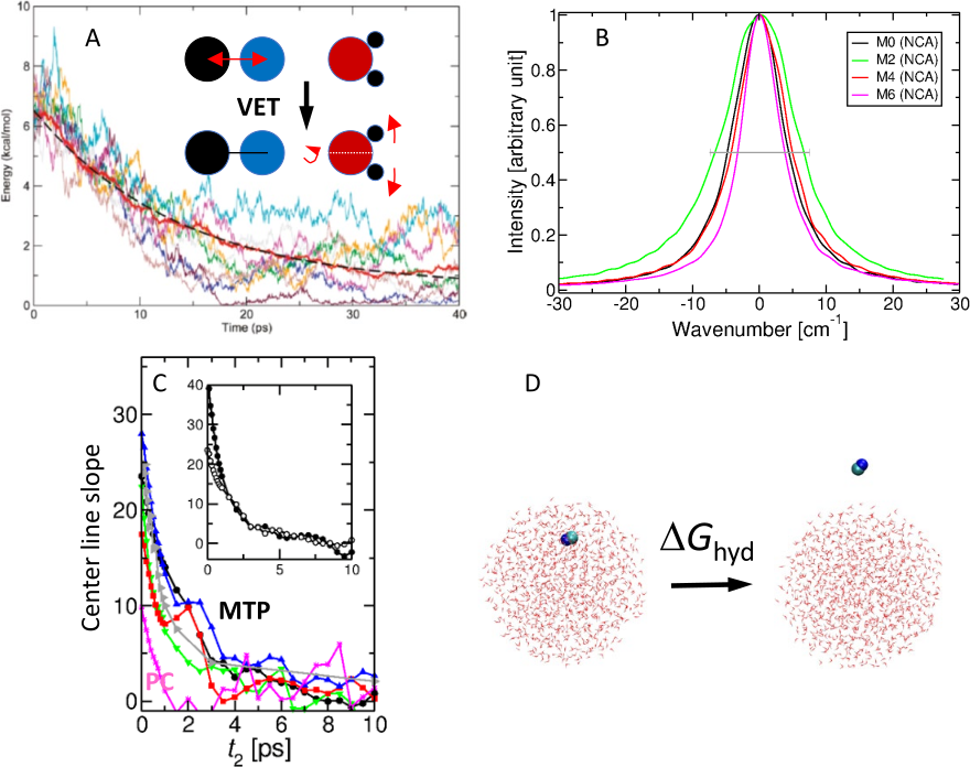
Atomistic simulations with multipolar force fields have demonstrated
that it is possible to realistically describe the 1d- and
2d-spectroscopy of small molecules in electrostatically demanding
environments such as in proteins or in
water.19, 20, 15 It was also shown
for cyanide (CN-) in water that the same energy function for the
solute is capable of correctly describing a range of condensed-phase
properties including the solvent-induced shift, the decay time of the
FFCF, the hydration free energy, and the vibrational energy relaxation
rate in water.15, 13, 17 This indicates
that physics-based refinements of such generic energy functions
provide a meaningful extension for molecularly resolved investigations
of complex systems.
The spectroscopy of photodissociated CO in myoglobin (Mb) was also
investigated by using a fluctuating multipolar representation for the
electrostatics.19, 20 This allowed to assign
for the first time the experimentally
observed21, 22 split infrared spectrum and to
identify the two peaks with two distinct conformational substates: one
in which the oxygen end of CO pointed towards the heme-iron atom and a
second state for which the carbon was closer to the Fe
atom.19, 20, 23 This model
correctly captures the red shift of the two bands relative to gas
phase CO, the splitting of the two peaks and their relative intensity.
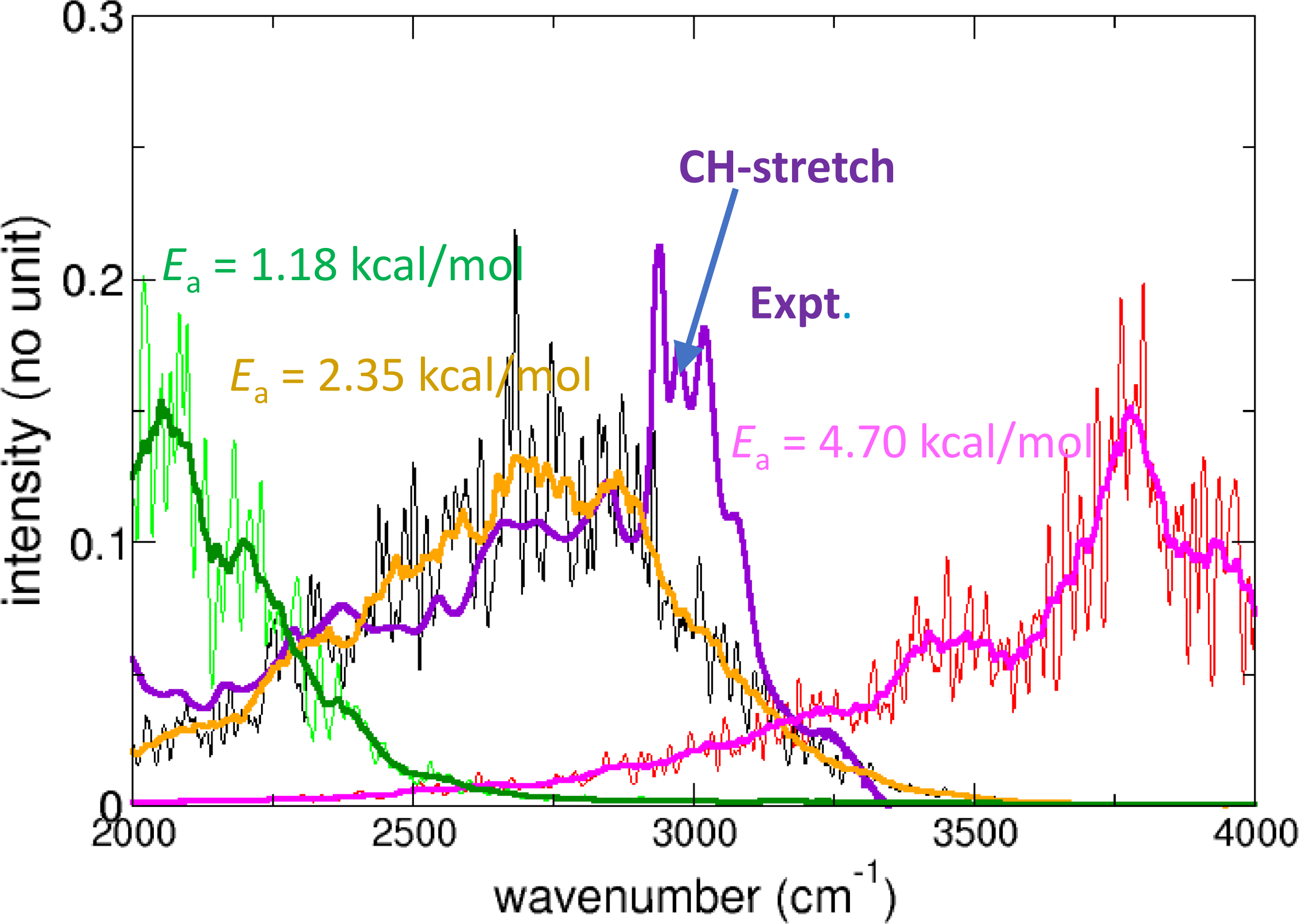
Finally, it is also possible to refine energy functions by comparing
experimentally determined IR spectra with those from computations. The
infrared spectrum of acetylacetone (doubly methylated malonaldehyde)
features a prominent band between 2000 and 3000 cm-1 which is due
to proton transfer across a low barrier.25
Morphing24, 26 a parametrized PES suitable
for following proton transfer and comparing the resulting infrared
spectrum with that from experiments yields an estimated barrier for
proton transfer of 2.35 kcal/mol; see Figure
2. Subsequent machine learning found a barrier of 3.25
kcal/mol from transfer learning to the PNO-LCCSDT(T)-F12 level of
theory.27
3 Molecular Dynamics Simulations with Machine-Learned Potentials
One of the major challenges in empirical force field development is to
find a suitable parametrized form of the energy function depending on
internal coordinates for a molecule. Various extensions to the generic
harmonic oscillator models for bonds and angles have been
considered. But all of them incur a considerably larger number of free
parameters to be determined.28, 29
In recent years an alternative approach has matured which is based on
using statistical models30 to represent precomputed
energies and forces from electronic structure calculations. Such
machine learning (ML) techniques do not necessarily require a
parametrized form to be used but rather represent data given a set of
kernel functions (kernel ridge regression) or by minimizing a loss
function of a neural network
(NN).31, 4, 32 For kernel-based methods
the long range physical shape of the PES can be encoded in the kernel
which guarantees correct extrapolation to large
separations.33, 34 No such procedure is known for
short range interactions.35 For NN-learned energy
functions extrapolation to geometries outside the training set needs
to be carefully assessed.4
One of the advantages of ML-based energy functions is that they
contain all couplings between the degrees of freedom. This is very
challenging for empirical energy functions. For instance, the CO bonds
in protonated oxalate change between single- and double-bond character
depending on where the proton is located.36 Although
such effects can be “encoded” in an empirical force field, capturing
such effects from a globally valid, machine-learned energy function is
more readily possible as has recently been done for formic acid
dimer.37, 38
As an example for the performance of state-of-the art ML-based methods
for vibrational spectroscopy, formic acid monomer and dimer (FAM and
FAD) in the gas phase is considered.39 Using
PhysNet40 a reference machine-learned PES was
determined at the MP2/aug-cc-pVTZ level of theory for FAM and FAD. The
mean averaged error between reference calculations and the statistical
model is 0.01 kcal/mol. Transfer learning the MP2-based PES to the
CCSD(T)/aug-cc-pVTZ level of theory yields normal mode frequencies
within 25 cm-1 on average compared with experiment for modes
below 2000 cm-1. Including anharmonic corrections within second
order vibrational perturbation theory
(VPT2)41 reduces this to 17 cm-1. For
the OH-stretch mode the VPT2 calculations yield 3011 cm-1,
compared with an experimentally reported, cm-1 broad
absorption band with center frequency at
cm-1. Finally, using diffusion Monte Carlo
(DMC)42 calculations for the full-dimensional
ground state potential energy and including corrections due to basis
set superposition and basis set completeness errors yield a
dissociation energy of kcal/mol compared with
an experimentally determined value of kcal/mol.43
It is of interest to note that experiment-guided refinement of an
advanced force field based on molecular mechanics with proton transfer
(MMPT)44 the barrier height for double proton
transfer in FAD could be inferred. For this, the height of the double
well potential was adjusted to match the experimentally observed broad
band associated with DPT in FAD. The resulting45
barrier height was 7.2 kcal/mol which compares with an experimentally
determined value from microwave spectroscopy of 7.3
kcal/mol.46 In this fashion, information from
vibrational spectroscopy can also be used to adapt (“morph”)
PESs.24
Finally, machine-learned energy functions can also be used in a mixed
quantum mechanics/molecular mechanics fashion to accurately describe
the bonded energetics for spectroscopic probes used in protein
2-dimensional IR spectroscopy.47 This was
successfully done using reproducing kernel-based representations for
the amide-I mode in insulin and trialanine or for azide attached to
all alanine residues in
Lysozyme.48, 49, 50 This provides
a positionally sensitive probe of the protein dynamics to follow
protein assembly or protein-ligand interactions.
4 Molecular Dynamics Simulations with Spectroscopic Maps
Determining the frequency trajectories required for
computing the FFCF and 2d-IR response can be computationally
prohibitive. One way to circumvent the expensive instantaneous normal
mode or reduced-dimensionality quantum bound state calculations is to
use spectroscopic
maps.51, 52, 53, 54
Such maps can be parametrized from electronic structure calculations
using model systems and exploit the fact that the frequency shift of
an oscillator in the field of surrounding point charges can be
approximately described by the Stark effect. Maps have been generated
for a range of spectroscopic probes, including the amide-I
stretch,52, the nitrile stretch, the azido
stretch, and others.54 Recently, machine learning has
been applied to refine the amide-I map.55
With such spectroscopic maps it is then quite straightforward to
determine the frequency trajectory for a particular oscillator from
conventional MD simulations. For every snapshot to be analyzed the
electric field at the position of the oscillator of interest is
determined and related to the frequency shift by evaluating the
spectroscopic map. This provides the information required to generate
the FFCF from which important information about the structural
dynamics around the spectroscopic reporter can be obtained.
One of the conceptual disadvantages of spectroscopic maps is the fact
that the energy function used to run the MD simulations typically
differs from the energy function used to evaluate the spectroscopic
response. This can be done from physics-based force
fields. Furthermore, some maps have been generated for rigid labels as
was the case for the amide-I maps. Hence, the MD simulations need to
be run with constrained CO distances for the maps to be valid. A
direct comparison between map-based analyses and results from
instantaneous normal modes and solutions of the nuclear Schrödinger
equation has been recently been given for insulin monomer and
dimer. For this system it was found that the maps perform inferior
compared with the other two approaches.48
5 Outlook
Molecular dynamics simulations with advanced energy functions provide
important information about the structural dynamics of molecules in
solution. Extensively sampling the conformational degrees of freedom
is essential and only possible with efficient implementations of the
energy functions. As with the spectroscopic probes to characterize
local protein dynamics simulations based on quantitatively accurate
energy functions provide information where to insert such probes in
order to be most sensitive to external perturbations such as binding
of a ligand. For such and other applications the combination of
experiment and simulation is indispensable and promises the necessary
molecular-level information to control and design chemical systems
with desired properties.
For the nuclear dynamics classical MD simulations have proven adequate for the purposes outlined in the present contribution. It is of interest to note that already 40 years ago it was pointed out for CO in the gas phase and in argon classical MD simulations are even capable of capturing R- and P-branches similar to what quantum mechanical treatments provide.56 More recently, this has also been found for liquid water.55 Hence, it is anticipated that such MD simulations with improved energy functions can contribute even more to understanding of the vibrational spectroscopy of solvated species than was previously anticiptated. Nevertheless, approximate quantum treatments will be of interest in order to delineate the range of applicability of classical mechanics for vibrational spectroscopy. Also, quantum effects including zero-point energy and tunneling are outside the scope of any classical mechanics-based method. For this reason, developing quantum methods applicable to the dynamics in solution remain an important quest in this field.57
Acknowledgments
The author acknowledges financial support from the Swiss National Science Foundation (NCCR-MUST and Grant No. 200021-188724), the AFOSR, and the University of Basel.
References
- Zwier 2006 Zwier, T. S. Laser probes of conformational isomerization in flexible molecules and complexes. J. Phys. Chem. A 2006, 110, 4133–4150
- Rizzo et al. 2009 Rizzo, T. R.; Stearns, J. A.; Boyarkin, O. V. Spectroscopic studies of cold, gas-phase biomolecular ions. Int. Rev. Phys. Chem. 2009, 28, 481–515
- Koner et al. 2020 Koner, D.; Salehi, S. M.; Mondal, P.; Meuwly, M. Non-conventional force fields for applications in spectroscopy and chemical reaction dynamics. J. Chem. Phys. 2020, 153, 010901
- Unke et al. 2021 Unke, O. T.; Chmiela, S.; Sauceda, H. E.; Gastegger, M.; Poltavsky, I.; Schütt, K. T.; Tkatchenko, A.; Müller, K.-R. Machine learning force fields. Chem. Rev. 2021, 121, 10142–10186
- Lifson and Warshel 1968 Lifson, S.; Warshel, A. Consistent force field fpr calculations of conformations vibrational spectra and enthalpies of cycloalkane and n-alkane molecules. J. Chem. Phys. 1968, 49, 5116–5129
- Levitt and Lifson 1969 Levitt, M.; Lifson, S. Refinement of protein conformations using a macromolecular energy minimization procedure. J. Mol. Biol. 1969, 46, 269–279
- Hwang et al. 1994 Hwang, M. J.; Stockfisch, T. P.; Hagler, A. T. Derivation of Class II Force Fields: 2. Derivation and Characterization of a Class II Force Fi eld, CFF93, for the Alkyl Functional Group and Alkane Molecules. J. Am. Chem. Soc. 1994, 116, 2515–2525
- Maple et al. 1994 Maple, J. R.; Hwang, M. J.; Stockfisch, T. P.; Dinur, U.; Waldman, M.; a nd A. T. Hagler, C. S. E. Derivation of class-II force-fields .1. Methodology and quantum force-field for the alkyl tunctional group and alkane molecules. J. Comp. Chem. 1994, 15, 162–182
- Brooks et al. 1983 Brooks, B.; Bruccoleri, R.; Olafson, B.; States, D.; Swaminathan, S.; Karplus, M. CHARMM: A program for macromolecular energy, minimization, and dynamics calculations. J. Comput. Chem. 1983, 4, 18–217
- Weiner et al. 1984 Weiner, S. J.; Kollman, P. A.; Case, D. A.; Singh, U.; Ghio, C.; Alagona, G.; Profeta Jr, S.; Weiner, P. A new force-field for molecular mechanical simulation of nucleic-acids and proteins. J. Am. Chem. Soc. 1984, 106, 765–784
- Jorgensen and Tirado-Rives 1988 Jorgensen, W. L.; Tirado-Rives, J. The OPLS potential functions for proteins - energy minimizations for crystals of cyclic-peptides and crambin. J. Am. Chem. Soc. 1988, 110, 1657–1666
- Hamm and Zanni 2011 Hamm, P.; Zanni, M. Concepts and methods of 2D infrared spectroscopy; Cambridge University Press, 2011
- Lee and Meuwly 2011 Lee, M. W.; Meuwly, M. On the role of nonbonded interactions in vibrational energy relaxation of cyanide in water. J. Phys. Chem. A 2011, 115, 5053–5061
- Hamm et al. 1997 Hamm, P.; Lim, M.; Hochstrasser, R. M. Vibrational energy relaxation of the cyanide ion in water. J. Chem. Phys. 1997, 107, 10523–10531
- Lee et al. 2013 Lee, M. W.; Carr, J. K.; Goellner, M.; Hamm, P.; Meuwly, M. 2D IR spectra of cyanide in water investigated by molecular dynamics simulations. J. Chem. Phys. 2013, 139, 054506
- Koziński et al. 2007 Koziński, M.; Garrett-Roe, S.; Hamm, P. Vibrational spectral diffusion of CN- in water. Chem. Phys. 2007, 341, 5–10
- Lee and Meuwly 2013 Lee, M. W.; Meuwly, M. Hydration free energies of cyanide and hydroxide ions from molecular dynamics simulations with accurate force fields. Phys. Chem. Chem. Phys. 2013, 15, 20303–20312
- Pearson 1986 Pearson, R. G. Ionization potentials and electron affinities in aqueous solution. J. Am. Chem. Soc. 1986, 108, 6109–6114
- Nutt and Meuwly 2003 Nutt, D. R.; Meuwly, M. Theoretical investigation of infrared spectra and pocket dynamics of photodissociated carbonmonoxy myoglobin. Biophys. J. 2003, 85, 3612–3623
- Plattner and Meuwly 2008 Plattner, N.; Meuwly, M. The role of higher CO-multipole moments in understanding the dynamics of photodissociated carbonmonoxide in myoglobin. Biophys. J. 2008, 94, 2505–2515
- Alben et al. 1982 Alben, J.; Beece, D.; Bowne, S.; Doster, W.; Eisenstein, L.; Frauenfelder, H.; Good, D.; McDonald, J.; Marden, M.; Moh, P.; Reinisch, L.; Reynolds, A.; Shyamsunder, E. Infrared spectroscopy of photodissociated carboxymyoglobin at low temperatures. Proc. Natl. Acad. Sci. 1982, 79, 3744–3748
- Lim et al. 1995 Lim, M.; Jackson, T. A.; Anfinrud, P. A. Mid-infrared vibrational spectrum of CO after photodissociation from heme: Evidence for a ligand docking site in the heme pocket of hemoglobin and myoglobin. J. Chem. Phys. 1995, 102, 4355–4366
- Meuwly 2006 Meuwly, M. On the Influence of the Local Environment on the CO Stretching Frequencies in Native Myoglobin: Assignment of the B-States in MbCO. Chem. Phys. Chem. 2006, 7, 2061–2063
- Meuwly and Hutson 1999 Meuwly, M.; Hutson, J. M. Morphing ab initio potentials: A systematic study of Ne–HF. J. Chem. Phys. 1999, 110, 8338–8347
- Howard et al. 2015 Howard, D. L.; Kjaergaard, H. G.; Huang, J.; Meuwly, M. Infrared and near-infrared spectroscopy of acetylacetone and hexafluoroacetylacetone. J. Phys. Chem. A 2015, 119, 7980–7990
- Gazdy and Bowman 1991 Gazdy, B.; Bowman, J. An adjusted potential surface for HCN based on rigorous vibrational calculations. J. Chem. Phys. 1991, 95, 6309–6316
- Käser et al. 2020 Käser, S.; Unke, O. T.; Meuwly, M. Reactive Dynamics and Spectroscopy of Hydrogen Transfer from Neural Network-based Reactive Potential Energy Surfaces. New J. Phys. 2020, 22, 055002
- Halgren 1996 Halgren, T. A. Merck molecular force field. I. Basis, form, scope, parameterization, and performance of MMFF94. J. Comp. Chem. 1996, 17, 490–519
- Hagler 2015 Hagler, A. Quantum derivative fitting and biomolecular force fields: functional form, coupling terms, charge flux, nonbond anharmonicity, and individual dihedral potentials. J. Chem. Theor. Comp. 2015, 11, 5555–5572
- Vapnik 1998 Vapnik, V. N. Statistical Learning Theory; Wiley-Interscience, 1998
- Manzhos and Carrington Jr 2021 Manzhos, S.; Carrington Jr, T. Neural network potential energy surfaces for small molecules and reactions. Chem. Rev. 2021, 121, 10187–10217
- Meuwly 2021 Meuwly, M. Machine Learning for Chemical Reactions. Chem. Rev. 2021, 121, 10218–10239
- Ho and Rabitz 1996 Ho, T.-S.; Rabitz, H. A general method for constructing multidimensional molecular potential energy surfaces from ab initio calculations. J. Chem. Phys. 1996, 104, 2584
- Unke and Meuwly 2017 Unke, O. T.; Meuwly, M. Toolkit for the Construction of Reproducing Kernel-Based Representations of Data: Application to Multidimensional Potential Energy Surfaces. J. Chem. Inf. Model 2017, 57, 1923–1931
- Soldán and Hutson 2000 Soldán, P.; Hutson, J. M. On the long-range and short-range behavior of potentials from reproducing kernel Hilbert space interpolation. J. Chem. Phys. 2000, 112, 4415–4416
- Xu and Meuwly 2017 Xu, Z.-H.; Meuwly, M. Vibrational spectroscopy and proton transfer dynamics in protonated oxalate. J. Phys. Chem. A 2017, 121, 5389–5398
- Käser and Meuwly 2022 Käser, S.; Meuwly, M. Transfer learned potential energy surfaces: accurate anharmonic vibrational dynamics and dissociation energies for the formic acid monomer and dimer. Phys. Chem. Chem. Phys. 2022, doi.org/10.1039/D1CP04393E
- Töpfer et al. 2022 Töpfer, K.; Käser, S.; Meuwly, M. Double Proton Transfer in Hydrated Formic Acid Dimer: Interplay of Spatial Symmetry and Solvent-Generated Force on Reactivity. arXiv preprint arXiv:2110.11785 2022,
- Käser and Meuwly 2022 Käser, S.; Meuwly, M. Transfer learned potential energy surfaces: accurate anharmonic vibrational dynamics and dissociation energies for the formic acid monomer and dimer. Phys. Chem. Chem. Phys. 2022,
- Unke and Meuwly 2019 Unke, O. T.; Meuwly, M. PhysNet: A Neural Network for Predicting Energies, Forces, Dipole Moments, and Partial Charges. J. Chem. Theor. Comp. 2019, 15, 3678–3693
- Barone 2005 Barone, V. Anharmonic vibrational properties by a fully automated second-order perturbative approach. J. Chem. Phys. 2005, 122, 014108
- Anderson 1975 Anderson, J. B. A random-walk simulation of the Schrödinger equation: H. J. Chem. Phys. 1975, 63, 1499–1503
- Kollipost et al. 2012 Kollipost, F.; Larsen, R. W.; Domanskaya, A. V.; Nörenberg, M.; Suhm, M. A. Communication: The highest frequency hydrogen bond vibration and an experimental value for the dissociation energy of formic acid dimer. J. Chem. Phys. 2012, 136, 151101
- Lammers et al. 2008 Lammers, S.; Lutz, S.; Meuwly, M. Reactive force fields for proton transfer dynamics. J. Comp. Chem. 2008, 29, 1048–1063
- Mackeprang et al. 2016 Mackeprang, K.; Xu, Z.-H.; Maroun, Z.; Meuwly, M.; Kjaergaard, H. G. Spectroscopy and dynamics of double proton transfer in formic acid dimer. Phys. Chem. Chem. Phys. 2016, 18, 24654–24662
- Li et al. 2019 Li, W.; Evangelisti, L.; Gou, Q.; Caminati, W.; Meyer, R. The barrier to proton transfer in the dimer of formic acid: a pure rotational study. Angew. Chem. Int. Ed. Engl. 2019, 58, 859–865
- Johnson et al. 2017 Johnson, P. J.; Koziol, K. L.; Hamm, P. Quantifying biomolecular recognition with site-specific 2D infrared probes. The Journal of Physical Chemistry Letters 2017, 8, 2280–2284
- Salehi et al. 2020 Salehi, S. M.; Koner, D.; Meuwly, M. Dynamics and infrared spectrocopy of monomeric and dimeric wild type and mutant insulin. J. Phys. Chem. B 2020, 124, 11882–11894
- Salehi and Meuwly 2021 Salehi, S. M.; Meuwly, M. Site-selective dynamics of azidolysozyme. J. Chem. Phys. 2021, 154, 165101
- Mondal et al. 2021 Mondal, P.; Cazade, P.-A.; Das, A. K.; Bereau, T.; Meuwly, M. Multipolar Force Fields for Amide-I Spectroscopy from Conformational Dynamics of the Alanine Trimer. J. Phys. Chem. B 2021, 125, 10928–10938
- Jansen et al. 2006 Jansen, T. l. C.; Dijkstra, A. G.; Watson, T. M.; Hirst, J. D.; Knoester, J. Modeling the amide I bands of small peptides. J. Chem. Phys. 2006, 125, 044312
- Reppert and Tokmakoff 2013 Reppert, M.; Tokmakoff, A. Electrostatic frequency shifts in amide I vibrational spectra: Direct parameterization against experiment. J. Chem. Phys. 2013, 138, 134116
- Wang et al. 2011 Wang, L.; Middleton, C. T.; Zanni, M. T.; Skinner, J. L. Development and Validation of Transferable Amide I Vibrational Frequency Maps for Peptides. J. Phys. Chem. B 2011, 115, 3713–3724
- Baiz et al. 2020 Baiz, C. R. et al. Vibrational spectroscopic map, vibrational spectroscopy, and intermolecular interaction. Chem. Rev. 2020, 120, 7152–7218
- Kananenka et al. 2019 Kananenka, A. A.; Yao, K.; Corcelli, S. A.; Skinner, J. Machine learning for vibrational spectroscopic maps. J. Chem. Theor. Comp. 2019, 15, 6850–6858
- Berens and Wilson 1981 Berens, P. H.; Wilson, K. R. Molecular dynamics and spectra. I. Diatomic rotation and vibration. J. Chem. Phys. 1981, 74, 4872–4882
- Rossi 2021 Rossi, M. Progress and challenges in ab initio simulations of quantum nuclei in weakly bonded systems. J. Chem. Phys. 2021, 154, 170902