Learning Incrementally to Segment Multiple Organs in a CT Image
Abstract
There exists a large number of datasets for organ segmentation, which are partially annotated and sequentially constructed. A typical dataset is constructed at a certain time by curating medical images and annotating the organs of interest. In other words, new datasets with annotations of new organ categories are built over time. To unleash the potential behind these partially labeled, sequentially-constructed datasets, we propose to incrementally learn a multi-organ segmentation model. In each incremental learning (IL) stage, we lose the access to previous data and annotations, whose knowledge is assumingly captured by the current model, and gain the access to a new dataset with annotations of new organ categories, from which we learn to update the organ segmentation model to include the new organs. While IL is notorious for its ‘catastrophic forgetting’ weakness in the context of natural image analysis, we experimentally discover that such a weakness mostly disappears for CT multi-organ segmentation. To further stabilize the model performance across the IL stages, we introduce a light memory module and some loss functions to restrain the representation of different categories in feature space, aggregating feature representation of the same class and separating feature representation of different classes. Extensive experiments on five open-sourced datasets are conducted to illustrate the effectiveness of our method.
Keywords:
Incremental learning Partially labeled datasets Multi-organ segmentation.1 Introduction
While most natural image datasets [imagenet, coco] are completely labeled for common categories, fully annotated medical image datasets are scarce, especially for a multi-organ segmentation (MOS) task [zhou2019handbook] that requires pixel-wise annotations, as constructing such a dataset requires professional knowledge of different anatomical structures [zhou2019handbook, zhou2021review]. Fortunately, there exist many partially labeled datasets [MSD, matlas, kits19_url3] for organ segmentation. Another dimension associated with these datasets is that they are constructed sequentially at different sites. Our goal is to train a single multi-organ segmentation model from partially labelled, sequentially constructed datasets.
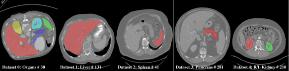
To achieve such a goal, we have to address two issues. (i) The first issue arising from partial labeling is knowledge conflict, that is, labels in different datasets have conflicts, e.g., the liver is marked as foreground in Dataset 1 but as background in Datasets 2-4, as shown in Fig. 1. (ii) The second issue arising from sequential construction is data availability, that is, the datasets are not simultaneously available for learning. What could be even worse is that, due to security concern, these datasets are not allowed to be transferred across the border of the curating institutes; only the model parameters are sharable.
There has been some emerging research [cvpr2021dodnet, PIPO, PaNN, ShiMargExc] that successfully handles knowledge conflict and trains a single model from pooled datasets for improved performance in multi-organ segmentation, proving that the unlabeled data in partially labeled datasets is also helpful for learning. However, these approaches conduct model learning in a batch model based and hence unable to be applied to deal with sequential construction. To deal with both issues, we hereby propose a novel multi-organ segmentation approach based on the principle of incremental learning (IL), which is a staged learning method that has an access to the data available at current learning stage, while losing the access to the data available in previous stages.
Our main contributions are summarized as below:
-
•
We make the first attempt in the literature to merge partially labeled datasets in medical image scenario using IL method, addressing the issues of knowledge conflict and data availability, and possibly security concern.
-
•
To combat the ‘catastrophic forgetting’ problem that commonly plagues IL, we introduce a light memory module to store the prototypical representation of different organ categories and corresponding loss functions to make different organs more distinguishable in feature space.
-
•
Our extensive experiments on five open-source organ datasets achieve comparable performance to state-of-the-art (SOTA) batch methods which can access all datasets in training phase, unleashing the great potential of IL in multiple organ segmentation.
2 Related work
MOS with Partially Labelled Datasets. Zhou et al. [PaNN] learn a segmentation model in the case of partial labeling by adding a prior-aware loss in the learning objective to match the distribution between the unlabeled and labeled datasets. In [PIPO], first multi-scale features at various depths are hierarchically incorporated for image segmentation and then a unified segmentation strategy is developed to train three separate datasets together, and finally multi-organ segmentation is achieved by learning from the union of partially labeled and fully labeled datasets. Zhang et al. [cvpr2021dodnet] propose a dynamic on-demand network (DoDNet) that learns to segment multiple organs and tumors on partially labeled datasets, which embedded dynamically generated filter by a task encoding module into an encoder-decoder architecture. Shi et al. [ShiMargExc] encode knowledge from different organs into a single multi-class segmentation model by introducing two simple but effective loss functions, Marginal loss and Exclusion loss.
Incremental Learning. IL has been studied for object recognition [lwf, kirkpatrick2017overcoming, icarl2017, LUCIR2019, meta2020] and detection [9035099, 8288446, shmelkov2017incremental], also segmentation [sensing2019, ILT, MiB, ozdemir2019extending]. The main challenge in IL is the so-called ‘catastrophic forgetting’ [mccloskey1989catastrophic]: how to keep the performance on old classes while learning new ones? Methods based on parameter isolation [rusu2016progressive, xu2018reinforced] and data replay [icarl2017, lopez2017gradient] are all with limited scalability or privacy issues. Regularization based method is the most ideal direction in IL community. In natural image segmentation, Cermelli et al. [MiB] solved knowledge conflicts existing in other IL methods [lwf, ILT] by remodeling old and new categories into background in loss functions, achieving a performance improvement. In 2D medical image segmentation, Ozdemir and Goksel [ozdemir2019extending] made some attempts using the IL methods used in natural images directly, with only two categories, and it mainly focuses on verifying the possibility of transferring the knowledge learned in the first category with more images to a second category with less images. In this paper, we apply IL to multiple organ segmentation for the first time.
3 Method
3.1 IL for MOS
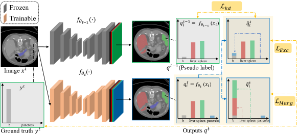
Framework of IL. The overview of the stage of IL in our method is shown in Fig. 2. Given a pair of 3D input image and ground truth, , we firstly process by the model in current stage, with trainable parameters , getting the output . And we assume that each image is composed by a set of voxels with constant cardinality . The whole label space cross all stages is expanded from with new classes added in current stage (), . Note that the annotations of the old categories will be inaccessible in the new stage under ideal IL settings. For preserving the knowledge of old categories in regularization based method, we process by the saved old model with frozen parameters and get as the pseudo label. Knowledge distillation loss, , is introduced in IL setting to keep old knowledge learned from previous stages. Trainable in the stage is expanded from with to segment new categories, .
Avoiding Knowledge Conflict in IL. The structures of old classes in , are marked as background in . And the new structures also do not exist in , that is new structures are marked as background in pseudo label. If we directly use to compute segmentation loss for new classes, and knowledge distillation loss for old classes, these conflicts between prediction and ground truth break the whole training process. So referring to marginal loss in MargExc [ShiMargExc], we modify the prediction to and , as shown in Fig. 2 and Eqs. (3) and (7).
| (3) | ||||
| (7) |
Then the probability of classes not marked in ground truth or pseudo label will not be broken during training.
3.2 Memory Module
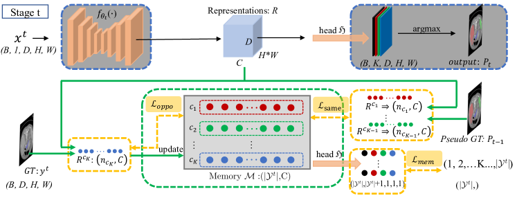
As shown in Fig. 3, representation is feature maps out of decoder, with shape of CDHW, where C means the number of channels in . To further mitigate ‘knowledge forgetting’ in IL setting, we introduce a light memory module of size C in feature space between decoder and segmentation head, , to remember the representation of each class. The size of is updated by more C on C after the stage. Then based on we can add some constraints in feature space to improve the IL learning progress.
During training of each stage, with the position supplied by ground truth, we can acquire the voxel representation of corresponding new organs in feature map . Then new class in can be updated via moving average after each iteration:
| (8) |
where is the momentum, denotes the current number of iterations, and is the total number of iterations of training. and are set as 0.9 empirically. After each stage of training ends, the mean representation of new organ of category in that stage is saved into the memory as .
When we have to save the mean representation of each class, we can introduce more regularization to constrain the learning of feature space. In this paper, we introduce , and :
| (9) | ||||
| (10) | ||||
| (11) |
In Eq. (9), is used to change to the size of , which can be regarded as voxels belong to classes. can be seen as corresponding ground truth. Through the shared segmentation head , features of classes in current stage are going to center around the mean representation in . Through , we constrain the learned feature of different classes in different stages more stable. The mean representation of old classes are treated as a kind of replay without privacy concerns. In Eqs. (10) and (11), and refer to old and new classes, respectively, and means background. Using Cosine Embedding Loss, , we can explicitly restrain the feature of old class close to , and the feature of new class away from all .
4 Experiments
4.1 Setup
Datasets and preprocessing. To compare with our base method, MargExc [ShiMargExc], we choose the same five organs and datasets in our experiments, including liver, spleen, pancreas, right kidney and left kidney. In addition, we find three more independent datasets for testing to give a comprehensive evaluation. The details of these datasets are shown in Table 4.1.
We preprocess all datasets to a unified spacing (2.41, 1.63, 1.63) and normalize them with mean and std of 90.9 and 65.5 respectively. We respectively split five training datasets into 5 folds and randomly select one fold as validation set. For our main IL setting, five organs are learned in four stages: liver (F+) spleen (F+) pancreas (F+) R/L kidney (F+). The annotations of different organs in dataset F are used separately in our IL setting.
Implementation details. We implement our experiments based on 3D lowres version of nnU-Net111github.com/mic-dkfz/nnunet [FabianNNUnet_nm] and also refer to MONAI222https://monai.io/ during our algorithm development. The patch-size and batch-size are set as (80, 160, 128) and 2, respectively, in our experiments. We train the network with the same optimizer and learning rate policy as nnU-Net for 350 epochs. The initial learning rate of the first stage and followed stages are set to 3e-4 and 15e-5.