– Under Review
\jmlryear2022 \jmlrworkshopFull Paper – MIDL 2022
\midlauthor\NameRuining Deng\nametag1 \Emailr.deng@vanderbilt.edu
\NameQuan Liu\nametag1 \Emailquan.liu@vanderbilt.edu
\NameCan Cui\nametag1 \Emailcan.cui.1@vanderbilt.edu
\NameZuhayr Asad\nametag1 \Emailzuhayr.asad@vanderbilt.edu
\NameHaichun Yang\nametag2 \Emailhaichun.yang@vumc.org
\NameYuankai Huo\nametag1 \EmailYuankai.huo@vanderbilt.edu
\addr1 Vanderbilt University, Department of Computer Science, Nashville, TN, USA 37215
\addr2 Vanderbilt University Medical Center, Department of Pathology, Nashville, TN, USA 37232
Omni-Seg: A Single Dynamic Network for Multi-label Renal Pathology Image Segmentation using Partially Labeled Data
Abstract
Computer-assisted quantitative analysis on Giga-pixel pathology images has provided a new avenue in histology examination. The innovations have been largely focused on cancer pathology (i.e., tumor segmentation and characterization). In non-cancer pathology, the learning algorithms can be asked to examine more comprehensive tissue types simultaneously, as a multi-label setting. The prior arts typically needed to train multiple segmentation networks in order to match the domain-specific knowledge for heterogeneous tissue types (e.g., glomerular tuft, glomerular unit, proximal tubular, distal tubular, peritubular capillaries, and arteries). In this paper, we propose a dynamic single segmentation network (Omni-Seg) that learns to segment multiple tissue types using partially labeled images (i.e., only one tissue type is labeled for each training image) for renal pathology. By learning from 150,000 patch-wise pathological images from six tissue types, the proposed Omni-Seg network achieved superior segmentation accuracy and less resource consumption when compared to the previous the multiple-network and multi-head design. In the testing stage, the proposed method obtains “completely labeled” tissue segmentation results using only “partially labeled” training images. The source code is available at \urlhttps://github.com/ddrrnn123/Omni-Seg.
keywords:
Renal pathology, Image segmentation, Multi-label, Self-supervised Learning1 Introduction
The recent advances in digital pathology, and particularly the approach of whole slide imaging (WSI), have led to a paradigm shift in pathology [Bandari et al.(2016)Bandari, Fuller, Turner, and D’Agostino]. Computer-assisted tumor segmentation has been broadly used in cancer pathology [Nguyen et al.(2021)Nguyen, Asad, and Huo]. However, there are increasing demands in analyzing more comprehensive tissue types beyond the tumor, including investigating tumor micro-environments and non-cancer pathology [Rangan and Tesch(2007)] rather than cancer pathology. Briefly, the learning algorithms can be asked to examine more comprehensive tissue types simultaneously, as a multi-label setting. Moreover, the recent advances in data sharing are providing increasingly large amounts of publicly available data, which requires a more robust computer-assisted analytics tool for large-scale multi-center and multi-stain WSIs [Ginley et al.(2019)Ginley, Lutnick, Jen, Fogo, Jain, Rosenberg, Walavalkar, Wilding, Tomaszewski, Yacoub, et al.]. Ideally, the computer-assisted segmentation method should be able to segment all types of tissue on large-scale pathological images in order to alleviate the labor-intensive, time-consuming manual annotation [Barisoni et al.(2017)Barisoni, Gimpel, Kain, Laurinavicius, Bueno, Zeng, Liu, Schaefer, Kretzler, Holzman, et al., Wernick et al.(1993)Wernick, Smith, Houghton, Phillips, Booth, Runckel, Johnson, Brown, and Gaboury].
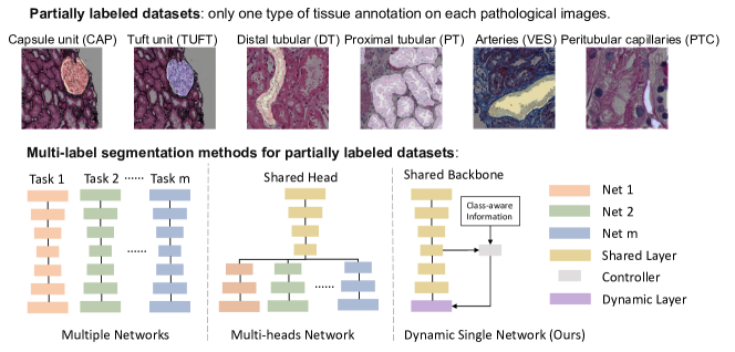
One major hurdle in achieving automated multi-label pathological image segmentation is the so-called partially labeling issue. This issue is caused by intensive labor and resource cost in pixel-level manual annotations on Giga-pixel pathological images, so that many datasets contain annotations of only one type of tissue. Mainstream approaches tackle the partial label issue by splitting the partially labeled cohorts into several fully labeled subsets and training “multiple networks” for different tasks [Yu et al.(2019)Yu, Shi, Sun, Gao, Zhu, and Dai, Isensee et al.(2019)Isensee, Jäger, Kohl, Petersen, and Maier-Hein, Zhang et al.()Zhang, Xie, Zhang, Chen, Xia, and Shen, Myronenko and Hatamizadeh(2019), Zhu et al.(2019)Zhu, Xia, Xie, Fishman, and Yuille]. This intuitive strategy, however, increases the computational complexity dramatically. Another more recent family of solutions is to employ multi-head networks. A multi-head network typically consists of a shared encoder, and several task-specific decoders (heads) [Chen et al.(2019)Chen, Ma, and Zheng, Fang and Yan(2020), Shi et al.(2021)Shi, Xiao, Chen, and Zhou]. Moreover, the multi-class segmentation method with a single network [González et al.(2018)González, Washko, and San José Estépar, Cerrolaza et al.(2019)Cerrolaza, Picazo, Humbert, Sato, Rueckert, Ballester, and Linguraru] shows the promising performance for partially labeled datasets. However, the redundant implementation of heads is not only wasteful but also inflexible, especially when it is extended to a new task. To mitigate the redundancy, Zhang et al. [Zhang et al.(2021)Zhang, Xie, Xia, and Shen] proposed a dynamic on-demand network (DoDNet) for radiological image analysis, which is an encoder-decoder network with a dynamic head that segments multiple organs and tumors as previously done by multiple networks or heads. However, these proposed methods are designed for 3D radiological data with multiple pre-trained models available. Unfortunately, it is still an unsolved problem on how to design a single multi-label segmentation model to obtain completely annotated pathological images only using partially annotated images.
In this paper, we propose a single segmentation network (Omni-Seg) which is optimal for pathology that learns to segment multiple tissue types at their optimal pyramid scales using partially labeled images (i.e., only one tissue type is labeled for each training image) in order to provide an efficient multi-label segmentation approach for pathology quantification. Briefly, our proposed method innovative contribution is three-fold: (1) To our knowledge, Omni-Seg is the first “single” dense object segmentation network for renal pathology using only a partially labeled dataset with large variations in magnification; (2) The proposed dynamic segmentation network achieves superior segmentation performances with fewer parameters compared with current multi-network approaches; (3) In the testing stage, the proposed method obtains “completely labeled” tissue segmentation results using only “partially labeled” training images. We benchmarked the existing methods on the latest publicly available tissue segmentation dataset in renal pathology to enable reproducible research, quick adaptations for other groups, and a new state-of-the-art performance as a reference for future studies. The source code has been made publicly available at \urlhttps://github.com/ddrrnn123/Omni-Seg.
2 METHOD
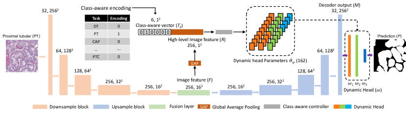
The overall framework of the proposed Omni-Seg method is presented in Fig.2.
2.1 Dynamic multi-label modeling
In partially labeled datasets [Jayapandian et al.(2021)Jayapandian, Chen, Janowczyk, Palmer, Cassol, Sekulic, Hodgin, Zee, Hewitt, O’Toole, et al.], only one tissue type might be provided by each training image, which cannot be directly used for training a canonical multi-label segmentation network. In the proposed method, we assign the class-aware knowledge with different tissue types to a -dimensional one-hot vector for class-aware encoding [Chen et al.(2017b)Chen, Xu, and Koltun]. The is the number of tissue types, The encoding calculation is presented in the following equation:
| (1) |
where is a class-aware vector of th tissue.
To encode domain-specific information to the embedded features, we merge the class-aware vector into the low dimensional feature embedding at the bottom of the residual U-Net architecture. The image feature is summarized by a global average pooling (GAP) via the feature vector , where is batch-size. The class-aware vector is reformed to be the same shape as the image features for the following fusion step. The high-level image features and the class-aware vector are concatenated. Then, a single 2D convolutional layer called the class-aware controller is employed as a feature-based fusion block to refine the fusion features as the final controller for the dynamic head mapping; this follows in the next equation:
| (2) |
The input is the combination of and with the fusion process , while the is the output channel of the class-aware controller, which also defines the parameter size for the dynamic head. Therefore, both the computational and spatial complexity of our task encoding strategy is lower than that seen in the multi-network strategy.
2.2 Dynamic head mapping
Inspired by DoDNet [Zhang et al.(2021)Zhang, Xie, Xia, and Shen], a binary segmentation network is employed to achieve multi-label segmentation via a dynamic filter. From the above multi-label modeling, we achieve joint low-dimensional image feature vectors and class-aware vectors at optimal segmentation magnification. Then, such information is mapped to control a light-weight dynamic head in order to specify the targeting tissue type from the inputs.
The dynamic head concludes with three layers. The first two have eight channels, while the last layer has two channels. The amount of the parameters for the dynamic head is 162. We directly map the parameters from the fusion-based feature controller to the kernels in the dynamic head to achieve precise segmentation by multi-modality features. As such, the filtering process can be defined in Eq.3
| (3) |
where is convolution, is the output from the decoder, is the final prediction, while , , and are the batch-size, width, and height of the dataset.
2.3 Residual U-Net backbone
For the implementation of the segmentation backbone, we utilize the residual U-Net as the segmentation unit according to DoDNet[Zhang et al.(2021)Zhang, Xie, Xia, and Shen], as shown in Fig.2. Different from the 3D network design in DoDNet, we develop the 2D convolutional blocks with kernel size 33 to be compatible with 2D pathological images. All the convolutional blocks are followed with ReLU activation after a group normalization [Wu and He(2018)]. A convolutional fusion layer with kernel size 33 is employed to learn high-level image features. Each downsampling block is implemented with a stride of 2 to halve the scale of input feature maps. Therefore, the feature maps are adjusted by multiple encoder-decoder blocks in pyramid scales. Symmetrically, the decoder upsamples the feature maps with an upsample factor of 2 and halves the channel number. In each upsample block, a low-level feature map (as a skip connection from the corresponding encoder layer) is summed with an upsampled feature map and then refined by a residual block. Following this feature-based learning, the output concludes high-level features for segmentation.
2.4 Testing stage aggregation with complete labels
In the testing stage, large-scale pathological images in 40 magnification are captured and split into 256 256 patches at the optimal scales [Jayapandian et al.(2021)Jayapandian, Chen, Janowczyk, Palmer, Cassol, Sekulic, Hodgin, Zee, Hewitt, O’Toole, et al.] with the downsampling process for different tissues. The same scales and methods (downsample and randomly crop) as those in [Jayapandian et al.(2021)Jayapandian, Chen, Janowczyk, Palmer, Cassol, Sekulic, Hodgin, Zee, Hewitt, O’Toole, et al.] were kept to ensure a consistent comparison. The class-aware controller is switched to receive six partially labeled segmentation for different tissue structures on patch-wise images. All of the segmentation is aggregated and mapping on the 40 pathological images as a completely labeled tissue segmentation for six tissue types.
3 Data and Experiments
3.1 Data
1751 regions of interest (ROIs) images were captured from 459 WSIs, obtained from 125 patients with minimal change diseases. The images were manually segmented for six structurally normal pathological primitives [Jayapandian et al.(2021)Jayapandian, Chen, Janowczyk, Palmer, Cassol, Sekulic, Hodgin, Zee, Hewitt, O’Toole, et al.], using the digital renal biopsies from a multi-center Nephrotic Syndrome Study Network (NEPTUNE) [Barisoni et al.(2013)Barisoni, Nast, Jennette, Hodgin, Herzenberg, Lemley, Conway, Kopp, Kretzler, Lienczewski, et al.]. All of the images were 30003000 pixels in 40 magnification, including glomerular tuft (TUFT); glomerular unit (CAP); proximal tubular (PT); distal tubular (DT); peritubular capillaries (PTC); and arteries (VES) in Hematoxylin and Eosin (H&E), Periodic-acid-Schiff (PAS), Silver(SIL), and Trichrome (TRI) stain. Four stain methods were regarded as color augmentations in each type of tissue. We followed [Jayapandian et al.(2021)Jayapandian, Chen, Janowczyk, Palmer, Cassol, Sekulic, Hodgin, Zee, Hewitt, O’Toole, et al.] to randomly crop all the images and downsample them into 256256 patches for the optimal magnifications. To resolve the unbalanced issue, we calculated the amount of each type of tissue and normalized the cropped patch numbers from the original images. Using the publicly available dataset, we kept the same splits as its original release in [Jayapandian et al.(2021)Jayapandian, Chen, Janowczyk, Palmer, Cassol, Sekulic, Hodgin, Zee, Hewitt, O’Toole, et al.] that randomly split the entire dataset into training, validation, and testing sets, following the ratio of 6:1:3. The splits were performed at the patient level to avoid data contamination. One sample randomly selected in-house samples from human nephrectomy tissues with 3 m thickness sections cut and stained with Periodic acid–Schiff (PAS) were used for completely labeled segmentation aggregation in the testing stage.
3.2 Experimental details
Six image pools were designed for different tissues to organize training batches with the same tissue type patches by following the image pool from Cycle-GAN [Zhu et al.(2017)Zhu, Park, Isola, and Efros]. The batch size was four, while the image pool size was eight. Once one pool stacked the number of images to more than the batch size, the images in the pool were queried out from the pool and fed into the network. Binary Dice loss and cross-entropy loss were combined during each backpropagation for all the tasks as the loss function. The boundaries of ground truth were assigned a higher weight with 1.2 when calculating the loss to emphasize the boundary prediction accuracy, which is widely implemented in segmentation [Ronneberger et al.(2015)Ronneberger, Fischer, and Brox]. Stochastic Gradient Descent (SGD) was used for weight update with a learning rate of 0.001 and 0.99 decay. The general data augmentation including Affine, Flip, Contrast, Brightness adjustment, Coarse Dropout, Gaussian Blur, and Gaussian Noise in the imagaug package [Jung et al.(2020)Jung, Wada, Crall, Tanaka, Graving, Reinders, Yadav, Banerjee, Vecsei, Kraft, Rui, Borovec, Vallentin, Zhydenko, Pfeiffer, Cook, Fernández, De Rainville, Weng, Ayala-Acevedo, Meudec, Laporte, et al.] was implemented for the whole training dataset in all of the methods, with a probability of 0.5.
The Dice similarity coefficient (Dice), Hausdorff distance (HD), and Mean Surface Distance (MSD) were used as performance metrics for this study. The distance metrics are in the Micron unit. For 40 images, each pixel is equal to 0.25 Micron. The mean of the Dice similarity coefficient from six tasks was used to select the optimal model in the validation dataset, and the performances were evaluated in the testing dataset. The performances selected the best models in testing within 100 epochs. All the experiments were completed on the same workstation, with the NVIDIA Quadro P5000 GPU.
| Method | DT | PT | CAP | ||||||
|---|---|---|---|---|---|---|---|---|---|
| Dice | HD | MSD | Dice | HD | MSD | Dice | HD | MSD | |
| Multi U-Net [Jayapandian et al.(2021)Jayapandian, Chen, Janowczyk, Palmer, Cassol, Sekulic, Hodgin, Zee, Hewitt, O’Toole, et al.] | 78.51 | 107.63 | 36.05 | 88.25 | 63.84 | 8.53 | 95.42 | 54.38 | 9.34 |
| Multi DeepLabV3 [Chen et al.(2017a)Chen, Papandreou, Schroff, and Adam] | 77.92 | 107.61 | 35.45 | 88.49 | 60.24 | 9.05 | 95.78 | \textcolorred50.42 | 9.76 |
| TAL [Fang and Yan(2020)] | 47.76 | 280.44 | 198.07 | 48.49 | 179.41 | 84.91 | 51.82 | 402.76 | 272.76 |
| Med3D [Chen et al.(2019)Chen, Ma, and Zheng] | 47.73 | 194.55 | 110.09 | 35.80 | 217.41 | 109.51 | 89.49 | 89.76 | 19.92 |
| Multi-class [González et al.(2018)González, Washko, and San José Estépar] | 47.76 | 280.44 | 198.07 | 88.36 | 64.84 | 8.85 | 95.93 | 85.64 | 11.1 |
| Omni-Seg(Ours) | \textcolorred81.01 | \textcolorred97.27 | \textcolorred24.19 | \textcolorred89.80 | \textcolorred57.11 | \textcolorred6.86 | \textcolorred96.50 | 53.08 | \textcolorred7.30 |
| Method | TUFT | VES | PTC | Average | ||||||||
|---|---|---|---|---|---|---|---|---|---|---|---|---|
| Dice | HD | MSD | Dice | HD | MSD | Dice | HD | MSD | Dice | HD | MSD | |
| Multi U-Net [Jayapandian et al.(2021)Jayapandian, Chen, Janowczyk, Palmer, Cassol, Sekulic, Hodgin, Zee, Hewitt, O’Toole, et al.] | 96.05 | 63.16 | 9.72 | 77.66 | 101.59 | 54.30 | 72.73 | 31.80 | 13.53 | 84.77 | 70.4 | 21.91 |
| Multi DeepLabV3 [Chen et al.(2017a)Chen, Papandreou, Schroff, and Adam] | 96.45 | 51.34 | 7.16 | 81.08 | 84.31 | 44.69 | 72.69 | 30.75 | 14.27 | 85.40 | 64.11 | 20.06 |
| TAL [Fang and Yan(2020)] | 76.95 | 137.18 | 65.58 | 47.67 | 244.11 | 191.25 | 49.37 | 52.15 | 35.79 | 53.67 | 216.01 | 141.39 |
| Med3D [Chen et al.(2019)Chen, Ma, and Zheng] | 92.80 | 80.84 | 14.80 | 58.46 | 150.71 | 78.77 | 49.78 | 44.53 | 27.24 | 62.34 | 129.63 | 60.06 |
| Multi-class [González et al.(2018)González, Washko, and San José Estépar] | 46.63 | 486.30 | 359.04 | 47.67 | 244.12 | 191.20 | 49.28 | 64.38 | 49.41 | 62.69 | 204.28 | 136.27 |
| Omni-Seg(Ours) | \textcolorred96.59 | \textcolorred44.36 | \textcolorred6.20 | \textcolorred85.05 | \textcolorred71.13 | \textcolorred21.99 | \textcolorred77.23 | \textcolorred26.49 | \textcolorred8.15 | \textcolorred87.70 | \textcolorred58.24 | \textcolorred12.45 |
4 Results
We compared the proposed Omni-Seg with baseline models, including (1) multiple individual U-Net models (Multi U-Net) [Jayapandian et al.(2021)Jayapandian, Chen, Janowczyk, Palmer, Cassol, Sekulic, Hodgin, Zee, Hewitt, O’Toole, et al.], (2) multiple individual DeepLabv3 models (Multi DeepLabv3) [Chen et al.(2017a)Chen, Papandreou, Schroff, and Adam] trained on each partially labeled dataset to solve the multi-label issue independently, (3) One multi-head model with target adaptive loss (TAL) for multi-class segmentation [Fang and Yan(2020)], (4) one multi-head 3D model (Med3D) for multiple partially labeled datasets [Chen et al.(2019)Chen, Ma, and Zheng], one multi-class segmentation model for partially labeled datasets [González et al.(2018)González, Washko, and San José Estépar].
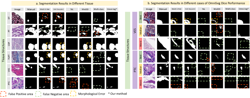
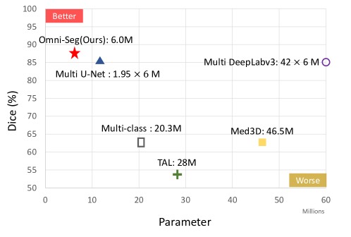
Table 1 shows the performance metrics for the segmentation of each tissue over the whole dataset. Figure 3.(a) shows the performance of different methods on the multi-label dataset. Figure 3.(b) demonstrates the cases of the best, the median, and the worst dice score in OmniSeg in two tissue types with consistent results from different methods. Figure 4 displays the relationship between parameter number and Dice score in each method.
As a result, training the whole dataset on a single multi-label network aggregated more domain-specific knowledge on pathological images, achieving better results than individual networks (i.e., Multi U-Net, Multi DeepLabv3). Due to missing the perspectives from other tissues, the results from individual networks had more false-positive predictions (the red bounding box) for the similar structures among different tissues and more false-negative predictions (the green box) from the incomplete dataset.
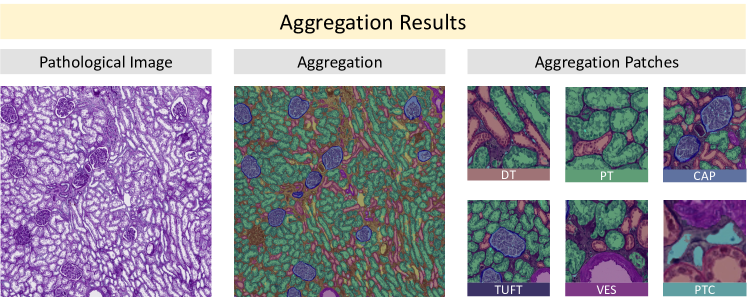
Our single dynamic network also led to “completely labeled” tissue segmentation results from only “partially labeled” training images on each histological image in Figure 5. The overlap areas of tufts and capsules will be displayed as tufts. For the remaining multiple foreground pixels, we labeled them as background. Only qualitative results are presented since the manual annotation is not available. The large-scale multi-label segmentation aggregation generates quantitative estimation for clinical examination.
5 Conclusion
In this paper, we propose a holistic segmentation network (Omni-Seg) that segments multiple tissue types using partially labeled images via a single dynamic head. The proposed method achieves superior segmentation performance with less computational resource consumption. In the testing stage, our single dynamic network achieved “completely labeled” tissue segmentation aggregation from only “partially labeled” training images.
References
- [Bandari et al.(2016)Bandari, Fuller, Turner, and D’Agostino] Jathin Bandari, Thomas W Fuller, Robert M Turner, and Louis A D’Agostino. Renal biopsy for medical renal disease: indications and contraindications. Can J Urol, 23(1):8121–6, 2016.
- [Barisoni et al.(2013)Barisoni, Nast, Jennette, Hodgin, Herzenberg, Lemley, Conway, Kopp, Kretzler, Lienczewski, et al.] Laura Barisoni, Cynthia C Nast, J Charles Jennette, Jeffrey B Hodgin, Andrew M Herzenberg, Kevin V Lemley, Catherine M Conway, Jeffrey B Kopp, Matthias Kretzler, Christa Lienczewski, et al. Digital pathology evaluation in the multicenter nephrotic syndrome study network (neptune). Clinical Journal of the American Society of Nephrology, 8(8):1449–1459, 2013.
- [Barisoni et al.(2017)Barisoni, Gimpel, Kain, Laurinavicius, Bueno, Zeng, Liu, Schaefer, Kretzler, Holzman, et al.] Laura Barisoni, Charlotte Gimpel, Renate Kain, Arvydas Laurinavicius, Gloria Bueno, Caihong Zeng, Zhihong Liu, Franz Schaefer, Matthias Kretzler, Lawrence B Holzman, et al. Digital pathology imaging as a novel platform for standardization and globalization of quantitative nephropathology. Clinical kidney journal, 10(2):176–187, 2017.
- [Cerrolaza et al.(2019)Cerrolaza, Picazo, Humbert, Sato, Rueckert, Ballester, and Linguraru] Juan J Cerrolaza, Mirella López Picazo, Ludovic Humbert, Yoshinobu Sato, Daniel Rueckert, Miguel Ángel González Ballester, and Marius George Linguraru. Computational anatomy for multi-organ analysis in medical imaging: A review. Medical Image Analysis, 56:44–67, 2019.
- [Chen et al.(2017a)Chen, Papandreou, Schroff, and Adam] Liang-Chieh Chen, George Papandreou, Florian Schroff, and Hartwig Adam. Rethinking atrous convolution for semantic image segmentation. arXiv preprint arXiv:1706.05587, 2017a.
- [Chen et al.(2017b)Chen, Xu, and Koltun] Qifeng Chen, Jia Xu, and Vladlen Koltun. Fast image processing with fully-convolutional networks. In Proceedings of the IEEE International Conference on Computer Vision, pages 2497–2506, 2017b.
- [Chen et al.(2019)Chen, Ma, and Zheng] Sihong Chen, Kai Ma, and Yefeng Zheng. Med3d: Transfer learning for 3d medical image analysis. arXiv preprint arXiv:1904.00625, 2019.
- [Fang and Yan(2020)] Xi Fang and Pingkun Yan. Multi-organ segmentation over partially labeled datasets with multi-scale feature abstraction. IEEE Transactions on Medical Imaging, 39(11):3619–3629, 2020.
- [Ginley et al.(2019)Ginley, Lutnick, Jen, Fogo, Jain, Rosenberg, Walavalkar, Wilding, Tomaszewski, Yacoub, et al.] Brandon Ginley, Brendon Lutnick, Kuang-Yu Jen, Agnes B Fogo, Sanjay Jain, Avi Rosenberg, Vighnesh Walavalkar, Gregory Wilding, John E Tomaszewski, Rabi Yacoub, et al. Computational segmentation and classification of diabetic glomerulosclerosis. Journal of the American Society of Nephrology, 30(10):1953–1967, 2019.
- [González et al.(2018)González, Washko, and San José Estépar] Germán González, George R Washko, and Raúl San José Estépar. Multi-structure segmentation from partially labeled datasets. application to body composition measurements on ct scans. In Image Analysis for Moving Organ, Breast, and Thoracic Images, pages 215–224. Springer, 2018.
- [Isensee et al.(2019)Isensee, Jäger, Kohl, Petersen, and Maier-Hein] Fabian Isensee, Paul F Jäger, Simon AA Kohl, Jens Petersen, and Klaus H Maier-Hein. Automated design of deep learning methods for biomedical image segmentation. arXiv preprint arXiv:1904.08128, 2019.
- [Jayapandian et al.(2021)Jayapandian, Chen, Janowczyk, Palmer, Cassol, Sekulic, Hodgin, Zee, Hewitt, O’Toole, et al.] Catherine P Jayapandian, Yijiang Chen, Andrew R Janowczyk, Matthew B Palmer, Clarissa A Cassol, Miroslav Sekulic, Jeffrey B Hodgin, Jarcy Zee, Stephen M Hewitt, John O’Toole, et al. Development and evaluation of deep learning–based segmentation of histologic structures in the kidney cortex with multiple histologic stains. Kidney international, 99(1):86–101, 2021.
- [Jung et al.(2020)Jung, Wada, Crall, Tanaka, Graving, Reinders, Yadav, Banerjee, Vecsei, Kraft, Rui, Borovec, Vallentin, Zhydenko, Pfeiffer, Cook, Fernández, De Rainville, Weng, Ayala-Acevedo, Meudec, Laporte, et al.] Alexander B. Jung, Kentaro Wada, Jon Crall, Satoshi Tanaka, Jake Graving, Christoph Reinders, Sarthak Yadav, Joy Banerjee, Gábor Vecsei, Adam Kraft, Zheng Rui, Jirka Borovec, Christian Vallentin, Semen Zhydenko, Kilian Pfeiffer, Ben Cook, Ismael Fernández, François-Michel De Rainville, Chi-Hung Weng, Abner Ayala-Acevedo, Raphael Meudec, Matias Laporte, et al. imgaug. \urlhttps://github.com/aleju/imgaug, 2020. Online; accessed 01-Feb-2020.
- [Myronenko and Hatamizadeh(2019)] Andriy Myronenko and Ali Hatamizadeh. 3d kidneys and kidney tumor semantic segmentation using boundary-aware networks. arXiv preprint arXiv:1909.06684, 2019.
- [Nguyen et al.(2021)Nguyen, Asad, and Huo] Cam Nguyen, Zuhayr Asad, and Yuankai Huo. Evaluating transformer-based semantic segmentation networks for pathological image segmentation. arXiv preprint arXiv:2108.11993, 2021.
- [Rangan and Tesch(2007)] Gopala K Rangan and Greg H Tesch. Quantification of renal pathology by image analysis (methods in renal research). Nephrology, 12(6):553–558, 2007.
- [Ronneberger et al.(2015)Ronneberger, Fischer, and Brox] Olaf Ronneberger, Philipp Fischer, and Thomas Brox. U-net: Convolutional networks for biomedical image segmentation. In International Conference on Medical image computing and computer-assisted intervention, pages 234–241. Springer, 2015.
- [Shi et al.(2021)Shi, Xiao, Chen, and Zhou] Gonglei Shi, Li Xiao, Yang Chen, and S Kevin Zhou. Marginal loss and exclusion loss for partially supervised multi-organ segmentation. Medical Image Analysis, 70:101979, 2021.
- [Wernick et al.(1993)Wernick, Smith, Houghton, Phillips, Booth, Runckel, Johnson, Brown, and Gaboury] Richard M Wernick, David L Smith, Donald C Houghton, David S Phillips, James L Booth, Douglas N Runckel, David S Johnson, Kevin K Brown, and Cynthia L Gaboury. Reliability of histologic scoring for lupus nephritis: a community-based evaluation. Annals of internal medicine, 119(8):805–811, 1993.
- [Wu and He(2018)] Yuxin Wu and Kaiming He. Group normalization. In Proceedings of the European conference on computer vision (ECCV), pages 3–19, 2018.
- [Yu et al.(2019)Yu, Shi, Sun, Gao, Zhu, and Dai] Qian Yu, Yinghuan Shi, Jinquan Sun, Yang Gao, Jianbing Zhu, and Yakang Dai. Crossbar-net: A novel convolutional neural network for kidney tumor segmentation in ct images. IEEE transactions on image processing, 28(8):4060–4074, 2019.
- [Zhang et al.()Zhang, Xie, Zhang, Chen, Xia, and Shen] Jianpeng Zhang, Yutong Xie, Pingping Zhang, Hao Chen, Yong Xia, and Chunhua Shen. Light-weight hybrid convolutional network for liver tumor segmentation.
- [Zhang et al.(2021)Zhang, Xie, Xia, and Shen] Jianpeng Zhang, Yutong Xie, Yong Xia, and Chunhua Shen. Dodnet: Learning to segment multi-organ and tumors from multiple partially labeled datasets. In Proceedings of the IEEE/CVF Conference on Computer Vision and Pattern Recognition, pages 1195–1204, 2021.
- [Zhu et al.(2017)Zhu, Park, Isola, and Efros] Jun-Yan Zhu, Taesung Park, Phillip Isola, and Alexei A Efros. Unpaired image-to-image translation using cycle-consistent adversarial networks. In Proceedings of the IEEE international conference on computer vision, pages 2223–2232, 2017.
- [Zhu et al.(2019)Zhu, Xia, Xie, Fishman, and Yuille] Zhuotun Zhu, Yingda Xia, Lingxi Xie, Elliot K Fishman, and Alan L Yuille. Multi-scale coarse-to-fine segmentation for screening pancreatic ductal adenocarcinoma. In International conference on medical image computing and computer-assisted intervention, pages 3–12. Springer, 2019.