Herding of proteins by the ends of shrinking polymers
Abstract
The control of biopolymer length is mediated by proteins that localize to polymer ends and regulate polymerization dynamics. Several mechanisms have been proposed to achieve end localization. Here, we propose a novel mechanism by which a protein that binds to a shrinking polymer and slows its shrinkage will be spontaneously enriched at the shrinking end through a “herding” effect. We formalize this process using both a lattice-gas model and a continuum description, and we present experimental evidence that the microtubule regulator spastin employs this mechanism. Our findings extend to more general problems involving diffusion within shrinking domains.
Regulating the length of biological polymers is essential for myriad cellular processes such as mitosis, muscle contraction and ciliary motility [1]. This regulation is often mediated by proteins that localize to polymer ends, where they promote or inhibit polymerization or depolymerization [2]. The mechanisms of protein end-localization on growing polymers such as microtubules and actin filaments have been well documented, both experimentally and theoretically. Some proteins bind directly to ends from solution [3, 4], some bind the polymer near the end and undergo one-dimensional diffusion to reach the end [5, 6, 7], and others are carried to ends by motor proteins [8, 9, 10]. Much less is known, however, about the localization of proteins to shrinking ends.
Microtubule severing proteins such as spastin and katanin cut microtubules along their lengths using the energy of ATP hydrolysis [11]. In addition to severing, both spastin and katanin possess an ATP-independent activity that promotes “rescue”, the conversion of shrinking microtubule ends to growing ones. This activity leads to the amplification of microtubule mass [12, 13], which is critical to spastin’s biological function in cells (reviewed in [14]). Furthermore, it has recently been reported that spastin is significantly enriched on shrinking microtubule ends, where it slows the shrinkage rate and facilitates the regrowth-promoting activity [15]. The mechanism of this enrichment is unknown.
End localization by spastin is not likely to involve direct binding or diffusion-and-capture: such mechanisms would require very high attachment rates and diffusion coefficients to target shrinking microtubule ends, which move 10-40-fold faster than growing ones [15]. A potential mechanism is that spastin uses multivalent interactions (avidity) with the microtubule to maintain contact with the depolymerizing end, as is thought to occur for end-tracking multiprotein complexes. For example, NDC80 forms a large sleeve that ensheathes shrinking microtubules in the mitotic spindle [16], the DASH/Dam1 complex forms a ring that encircles microtubules[17], and Kar9 with associated proteins remains on the ends of shrinking cytoplasmic microtubules [18]. Engineered [19] and synthetic oligomers [20] can also robustly track shrinking ends. While the avidity mechanism cannot be ruled out for spastin, whose hexameric form cuts microtubules, there is no evidence that spastin end-localization requires oligomerization. Indeed, spastin binds to and diffuses along microtubules as a monomer and localizes to their ends at low concentration and in the absence of ATP [15].
In this Letter, we present a novel theoretical mechanism of shrinking-end localization that requires neither multivalent interactions, diffusive capturing, nor directed motility. We show that a protein that binds to a shrinking polymer and slows its shrinkage will be spontaneously enriched at the shrinking end due to a purely kinetic “herding” effect. This behavior is counter-intuitive because one might expect that slowdown is driven by end enrichment, not the other way around. We experimentally demonstrate that spastin’s end enrichment is quantitatively explained by this herding theory. In addition to providing insight into spastin’s regulatory function, our results extend to a more general class of problems in which diffusive particles hinder the boundaries of a shrinking domain.
Mathematical Model.
Lattice-gas models have been successfully employed to explain the collective dynamics of polymer-associated proteins [21, 22], including depolymerizing kinesins [9] and end-binding microtubule polymerases [5]. In this vein, we describe the shrinking polymer as a semi-infinite one-dimensional lattice situated in a reservoir of the soluble protein (figure 1). We index the lattice sites where is the shrinking end. The lattice represents protein binding sites, which can bind only one protein at a time. Proteins from solution bind vacant lattice sites at a rate , where is the concentration of the protein in solution. Lattice-bound proteins can detach at a rate , or hop to adjacent vacant sites at a rate (in either direction). Shrinkage of the polymer occurs via the loss of lattice sites from one end at a rate or , depending on whether the site at the end is vacant or occupied, respectively. We are interested in the case , corresponding to a slowing of shrinkage by the protein.
The state of the system is described by the set of occupancy numbers where if the th site is occupied. Under these definitions, the time evolution of the mean occupancy at the end obeys
| (1) | |||||
Similarly, the mean occupancy of all other sites follows
| (2) | |||||
Equations 1 and 2 are derived in section I of the Supplementary Information (SI). Due to the moving frame, there is an apparent advection of particles towards the shrinking end at .
In the case (no slowdown by end-occupancy), equations 1 and 2 have the stationary solution
for all . This is the Langmuir isotherm, the stationary density that would be achieved if the polymer was in equilibrium with the protein bath and constant in length. For , direct solution of equations 1 and 2 is complicated by the presence of correlations between and the other sites, but we can invoke a mean field (MF) approximation . The continuum limit of the lattice model is then
| (3) | |||||
where we have defined the continuous spatial variable as well as the dimensionless time , which normalizes the natural polymer shrinkage rate to unity. The mean linear density of protein on the polymer is . We further define , the ratio of the hopping rate to the natural shrinkage rate, , the dimensionless detachment rate of the protein, and , which determines the position of Langmuir equilibrium . Lastly, we define a slowdown parameter controlling the extent to which the protein slows shrinkage. When , depolymerization is only possible when the terminal site is vacant, and when , the depolymerization rate is unaffected by end occupancy. From equation 3, we identify the (dimensionless) measured shrinkage velocity of the microtubule
Far from the shrinking end, the density of the protein should be the equilibrium Langmuir density . With this boundary condition, stationary solutions to equation 3 take the form of an exponential decay with the distance from the end,
| (4) |
where
To set the boundary condition at the end, we note that the microscopic model has no binding sites to the left of , and so the end must be reflecting. This gives
| (5) |
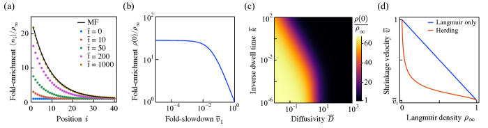
where we have imposed that the total flux (diffusive and advective) through the end must equal the net binding or detachment at the end. This condition can be used together with equation 4 to express as the root of a cubic polynomial (SI, section II). Notably, the solutions predict , implying that there is an enrichment of the protein at the shrinking filament end. Simulations of the microscopic model using the Gillespie algorithm predict that the mean lattice occupancies approach a steady state that is in good agreement with the stationary solution of the mean field model (figure 2a). This agreement is robust against changes in the parameters (figure S1).
The gradual accumulation at the end is due to a “herding” effect, where the shrinking end slows when it encounters a protein and then speeds up again when the protein hops away from the end along the lattice. This results in a build up of the protein near the reflecting end. This unexpected behavior, that slowdown causes end enrichment and not vice versa, depends sharply on the extent of slowdown: the enrichment is maximum when (figure 2b); that is, when a protein in the site completely pauses depolymerization. Since every lattice site is indistinguishable to the protein, the herding is a purely kinetic effect and is not due to a higher affinity to the polymer end. If or are made too large, the proteins achieve diffusive or Langmuir equilibrium between lattice depolymerization events, preventing end enrichment. That is, no build up at the end would be possible if the protein were to hop much faster than the end moves or if the lifetime of the protein on the polymer were small compared to the time between depolymerization events (figure 2c). This is in contrast to diffusion-and-capture mechanisms, in which a large diffusion coefficient increases the end localization rather than decreasing it [5], or to direct-binding mechanisms, in which fast lattice turnover promotes end enrichment.
Herding has two biological implications. First, far less protein is needed on the lattice in order to impart a certain level of shrinkage slowdown (figure 2d). This is due to a feedback effect where slowdown causes end enrichment and end enrichment in turn promotes slowdown. Second, a high end occupancy is achievable at low solution concentrations. This can facilitate the regulation of polymer ends through mechanisms additional to the slowdown itself, such as the promotion of rescue by spastin.
Application to spastin.
To validate this model, we investigated the behavior of spastin labeled with green fluorescent protein (GFP-spastin) on dynamic microtubules using the in vitro reconstitution assay described in [23]. Experimental details are provided in section IV of the SI. Briefly: We grew GMPCPP-stabilized microtubule “seeds”, which do not shrink, and immobilized them on a functionalized surface within a microfluidic flow chamber. We perfused a tubulin and GTP solution, which causes dynamic microtubule extensions to grow from the ends of the seeds. We then perfused a tubulin, GTP, and spastin solution, allowing spastin to decorate the dynamic microtubules and reach its equilibrium binding density. To prevent severing, no ATP was included in the solution. Finally, we perfused a solution containing spastin but no tubulin. The removal of tubulin induces “catastrophe” events, forcing the microtubule ends to begin shrinking. GFP-spastin was visualized using total-internal-reflection fluorescence (TIRF)-microscopy [24]. In all microtubules observed (), robust end enrichment was evident, as seen in the sample time series and intensity traces in figure 3a. The enriched region coincides with the position of the microtubule end, and occurs on both ends of a shrinking microtubule (figure S2). This experiment confirms that spastin concentrates on depolymerizing microtubule ends.
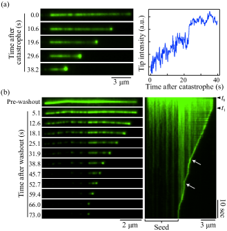
We then asked whether the end enrichment is due to spastin preferentially binding to the shrinking end from solution or if it is due to the sweeping of lattice-bound spastin. To this end, we employed a “wash-out” assay, where spastin was loaded onto microtubules in the presence of tubulin, and then both tubulin and spastin were washed out. This induces catastrophe within a spastin-free solution. We found that spastin was still enriched at shrinking ends, as seen in the representative time series in figure 3b (left), and in the corresponding kymograph (right). This result conclusively demonstrates that a direct-binding EB1-like mechanism [3] is not necessary for end enrichment. Indicated on the kymograph (white arrows) are points where the end slows down when encountering regions of high spastin density and then speeds up again when spastin detaches from the end. This indicates a correlation between end occupancy and shrinkage speed, supporting the herding effect. These results show that the enrichment of spastin on shrinking ends does not require spastin in solution and provide direct evidence that the shrinking end sweeps spastin with it as it moves.
To quantitatively validate our theory, we directly measured spastin’s attachment rate , detachment rate , and diffusion coefficient . The conversion between the macroscopic experimental parameters (, and ) and the model parameters ( and ) is given in Table S2 (SI). The attachment rate was estimated by counting the frequency of single-molecule landing events at GFP-spastin. This yielded the estimate (mean SEM; 153 events). To measure the diffusion coefficient and detachment rate, we performed a washout assay using a low initial spastin concentration to visualize single-molecule trajectories (figure S3). The spastin dwell time displayed the expected exponential distribution (figure S4a), with an estimated mean (158 events). Diffusion coefficients were estimated on a per-trajectory basis and fell into a broad range (figure S4b), with a median of (interquartile range: 0.008). These parameter values are similar to those measured for human spastin [25].
We next measured the microtubule shrinkage velocity as a function of the concentration of spastin in solution (figure 4, circles). The curve allows an estimation of the slowdown parameter as the ratio , where is the saturating shrinkage velocity at high concentrations. Direct measurement of is complicated by spastin’s tendency to aggregate at high concentrations. However, using the measurement at 1000 nM, we can place an upper bound at . Curve fitting suggests that the true value is even closer to zero. Thus, we were able to measure the key parameters in the model.
To make model predictions, we calculated the dimensionless parameters: , , and . This places spastin in a region where herding is expected to be significant (figure 2b and c). At 50 nM, which is close to the physiological concentration [26, 27], the enrichment at the end is at least 21-fold and likely closer to 200-fold. The width of the enriched region is on the order , which is below the diffraction limit and in agreement with experiments.
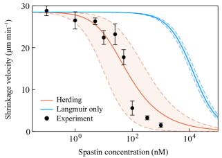
To compare the predictions directly to the data, figure 4 shows the measured values along with theoretical curves from our herding theory and for the case (Langmuir kinetics without end enrichment). The shaded regions represent 95% confidence intervals for the parameter estimates. It is evident that Langmuir kinetics alone are insufficient to explain the measured slowdown in the shrinkage velocity, even for . This suggests that spastin’s end-enrichment is likely to be biologically significant, in that it allows slowdown at much lower cellular concentrations. According to experiments, a 50% slowdown in the shrinkage velocity is achieved at around 55nM, which is close to the physiological concentration [26, 27]. Without end-enrichment, this level of slowdown would be achievable only at micromolar concentrations (figure 4, blue curve). The experimental data are all within the confidence bounds of the herding theory (orange region), and it is clear that the theory is a good quantitative predictor of the measured shrinkage velocity.
Other applications.
Although we have framed the herding theory in the context of regulatory proteins on shrinking polymers, the central principle is more general: particles that diffuse in a shrinking domain will enrich at the shrinking boundary if they hinder its shrinkage. The essence of this effect is contained within the following simplified version of the mean-field model,
| (6) |
where the Langmuir kinetics are removed and is set to zero. Equation 6 serves as a generic theory of herding phenomena, independently of the specific microscopic model from which it was derived. To illustrate this, and to motivate our terminology, we consider the following toy model of sheepherding (figure 5a): a one-dimensional domain is bounded on one end by a sheepdog, which moves with unit velocity in the positive direction when it is unimpeded. There are sheep in the domain, each behaving as an independent run-and-tumble particle with run velocity and tumble rate . The sheep are reflected by the sheepdog, and the sheepdog pauses when there is a sheep within one unit of length ahead of it.
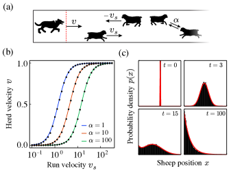
The problem of determining the resultant average velocity of the sheepdog (and the herd) appears complicated at first glance. However, numeric simulations suggest that the behavior is well predicted by equation 6 with , which is the known effective diffusion coefficient for a run-and-tumble particle. With a reflecting boundary at and the condition , the stationary solution to equation 6 predicts a steady-state herd velocity of
| (7) |
This result is in good agreement with simulations (figure 5b). The dynamics of this process are also well-described by equation 6: Although time-dependent analytic solution is difficult due to the nonlocal nonlinearity in the advection term, numeric integration is straightforward with a nonlocal finite-difference scheme (SI, section III). This reveals that equation 6 is a nearly exact Fokker-Planck equation for the sheep position when (figure 5c).
Discussion.
We have shown that a protein that slows a polymer’s shrinkage will enrich near the shrinking end, leading to a feedback between slowdown and end enrichment. In our model, the binding sites on the polymer are energetically equivalent, and the equilibrium density is uniform across the lattice. What drives the density distribution out of equilibrium is the polymer’s shrinkage (figure 1), which breaks detailed balance and is a dissipative process. For microtubules, the energy source is GTP hydrolysis, for actin filaments it is ATP hydrolysis [28] and for exonuclease-mediate DNA depolymerization it is nucleotide triphosphate hydrolysis. It is known that such processes can generate mechanical work [29, 30]; here, we show that they can also build protein density gradients with regulatory functions.
A central aspect of our model is that the build up near the end is due to a sweeping of lattice-bound protein. Although many MAPs diffuse on MTs and are reflected by MT ends, most fall off with depolymerizing subunits and are not swept by the end. Examples include tau [31, 32] and kinesins 8 and 13 [33]. Sweeping requires a mechanism that stops proteins at the end from falling off with the subunits that they occupy. One possibility is to processively track the end as a multivalent complex [34, 35, 18]. Processive tracking has been explored theoretically by Klein et al. in the context of depolymerizing kinesin motors [9]: using a formalism similar to ours, it was shown that accumulation at shrinking ends occurs if there is a sufficient probability that a protein at the end moves onto the next site when the terminal site dissociates. Our herding theory provides an alternative and distinct mechanism for sweeping – by slowing depolymerization when the end is occupied, proteins are not lost with depolymerizing subunits. This means that the protein can be swept without any processivity.
We applied our herding theory to spastin’s end enrichment and slowing of depolymerization. Our experimental results show that the shrinking microtubule end sweeps lattice-bound spastin, eliminating direct end-binding as an alternative mechanism (figure 3). We directly measured spastin’s diffusion coefficient, dwell time, and slowdown activity, and determined that they imply significant herding (figure 2b and c). The herding theory quantitatively predicts the microtubule shrinkage rate as a function of spastin concentration; it explains how significant slowdown can be achieved at concentrations of 10 nM despite the average lattice occupancy being very low (figure 4). Although it is difficult to rule out processive tracking by oligomers as an alternative mechanism, we note that processive motion by single puncta was never observed in our single-molecule measurements. We further note that oligomerization and herding are not mutually exclusive; in fact, if oligomers slow depolymerization, then this will lead to herding.
Our findings suggest that spastin’s slowdown activity is self-catalyzing because it concentrates the protein at the shrinking end. This effect is what allows regulation of the microtubule shrinkage velocity at spastin concentrations within the physiological range. Furthermore, herding has implications for spastin’s regulation of the rescue frequency: although the precise mechanism of spastin’s ATP-independent promotion of rescue is unclear, it is reasonable that the activity depends on an interaction with the end. For example, if spastin accelerates the binding of GTP-tubulin to the shrinking end, then its accumulation at the end can restore the GTP cap and thus the growing of that end.
Lastly, we showed how a simplified version of our mean field model, Equation 6, can describe more macroscopic herding phenomena. The dynamics described by equation 6 may arise in diverse contexts. An example from biology is the motion of molecular motors under the hindrance of diffusive roadblocks. This includes microtubule motors slowed by other MAPs [36], as well DNA replication forks and RNA-polymerases moving against DNA-binding proteins [37, 38]. Outside of biology, such dynamics could arise in transport processes where a density buildup at the boundary slows the flow in the bulk. For example, the flow of a particle suspension through a filter.
References
- Marshall [2015] W. F. Marshall, How cells measure length on subcellular scales, Trends in Cell Biology 25, 10.1016/j.tcb.2015.08.008 (2015).
- Howard and Hyman [2007] J. Howard and A. A. Hyman, Microtubule polymerases and depolymerases, Current Opinion in Cell Biology 19 (2007).
- Bieling et al. [2007] P. Bieling, L. Laan, H. Schek, E. L. Munteanu, L. Sandblad, M. Dogterom, D. Brunner, and T. Surrey, Reconstitution of a microtubule plus-end tracking system in vitro, Nature 450 (2007).
- Edwards et al. [2014] M. Edwards, A. Zwolak, D. A. Schafer, D. Sept, R. Dominguez, and J. A. Cooper, Capping protein regulators fine-tune actin assembly dynamics, Nature Reviews Molecular Cell Biology 15, 10.1038/nrm3869 (2014).
- Reithmann et al. [2016] E. Reithmann, L. Reese, and E. Frey, Nonequilibrium diffusion and capture mechanism ensures tip localization of regulating proteins on dynamic filaments, Physical Review Letters 117 (2016).
- Helenius et al. [2006] J. Helenius, G. Brouhard, Y. Kalaidzidis, S. Diez, and J. Howard, The depolymerizing kinesin MCAK uses lattice diffusion to rapidly target microtubule ends, Nature 441 (2006).
- Brouhard et al. [2008] G. J. Brouhard, J. H. Stear, T. L. Noetzel, J. Al-Bassam, K. Kinoshita, S. C. Harrison, J. Howard, and A. A. Hyman, XMAP215 is a processive microtubule polymerase, Cell 132 (2008).
- Varga et al. [2009] V. Varga, C. Leduc, V. Bormuth, S. Diez, and J. Howard, Kinesin-8 motors act cooperatively to mediate length-dependent microtubule depolymerization, Cell 138 (2009).
- Klein et al. [2005] G. A. Klein, K. Kruse, G. Cuniberti, and F. Jülicher, Filament depolymerization by motor molecules, Physical Review Letters 94 (2005).
- Chen et al. [2019] X. Chen, L. A. Widmer, M. M. Stangier, M. O. Steinmetz, J. Stelling, and Y. Barral, Remote control of microtubule plus-end dynamics and function from the minus-end, eLife 8, 10.7554/eLife.48627 (2019).
- McNally and Roll‑Mecak [2018] F. J. McNally and A. Roll‑Mecak, Microtubule-severing enzymes: From cellular functions to molecular mechanism, Journal of Cell Biology 217, 10.1083/jcb.201612104 (2018).
- Vemu et al. [2018] A. Vemu, E. Szczesna, E. A. Zehr, J. O. Spector, N. Grigorieff, A. M. Deaconescu, and A. Roll-Mecak, Severing enzymes amplify microtubule arrays through lattice GTP-tubulin incorporation, Science 361 (2018).
- Kuo et al. [2019a] Y. W. Kuo, O. Trottier, and J. Howard, Predicted effects of severing enzymes on the length distribution and total mass of microtubules, Biophysical Journal 117 (2019a).
- Kuo and Howard [2021] Y. W. Kuo and J. Howard, Cutting, amplifying, and aligning microtubules with severing enzymes, Trends in Cell Biology 31 (2021).
- Kuo et al. [2019b] Y. W. Kuo, O. Trottier, M. Mahamdeh, and J. Howard, Spastin is a dual-function enzyme that severs microtubules and promotes their regrowth to increase the number and mass of microtubules, Proceedings of the National Academy of Sciences of the United States of America 116 (2019b).
- Alushin et al. [2010] G. M. Alushin, V. H. Ramey, S. Pasqualato, D. A. Ball, N. Grigorieff, A. Musacchio, and E. Nogales, The ndc80 kinetochore complex forms oligomeric arrays along microtubules, Nature 467, 10.1038/nature09423 (2010).
- Westermann et al. [2006] S. Westermann, H. W. Wang, A. Avila-Sakar, D. G. Drubin, E. Nogales, and G. Barnes, The dam1 kinetochore ring complex moves processively on depolymerizing microtubule ends, Nature 440 (2006).
- Meier et al. [2021] S. M. Meier, A.-M. Farcas, A. Kumar, M. Ijavi, R. T. Bill, J. Stelling, E. Dufresne, M. O. Steinmetz, and Y. Barral, High interaction valency ensures cohesion and persistence of a microtubule +tip body at the plus-end of a single specialized microtubule in yeast, bioRxiv (2021).
- Lombillo et al. [1995] V. A. Lombillo, R. J. Stewart, and J. R. McIntosh, Minus-end-directed motion of kinesin–coated microspheres driven by microtubule depolymerization, Nature 373 (1995).
- Drechsler et al. [2019] H. Drechsler, Y. Xu, V. F. Geyer, Y. Zhang, and S. Diez, Multivalent electrostatic microtubule interactions of synthetic peptides are sufficient to mimic advanced map-like behavior, Molecular Biology of the Cell 30 (2019).
- Kolomeisky et al. [1998] A. B. Kolomeisky, G. M. Schütz, E. B. Kolomeisky, and J. P. Straley, Phase diagram of one-dimensional driven lattice gases with open boundaries, Journal of Physics A: Mathematical and General 31 (1998).
- Parmeggiani et al. [2003] A. Parmeggiani, T. Franosch, and E. Frey, Phase coexistence in driven one-dimensional transport, Physical Review Letters 90 (2003).
- Kuo and Howard [2022] Y.-W. Kuo and J. Howard, In vitro reconstitution of microtubule dynamics and severingsevering imaged by label-free interference-reflection microscopy, in Microtubules: Methods and Protocols, edited by H. Inaba (Springer US, New York, NY, 2022) pp. 73–91.
- Tuna et al. [2022] Y. Tuna, A. Al-Hiyasat, and J. Howard, Simultaneous interference reflection and total internal reflection fluorescence microscopy for imaging dynamic microtubules and associated proteins, Journal of Visualized Experiments , e63730 (2022).
- Eckert et al. [2012] T. Eckert, D. T. V. Le, S. Link, L. Friedmann, and G. Woehlke, Spastin’s microtubule-binding properties and comparison to katanin, PLoS ONE 7 (2012).
- Solowska et al. [2008] J. M. Solowska, G. Morfini, A. Falnikar, B. T. Himes, S. T. Brady, D. Huang, and P. W. Baas, Quantitative and functional analyses of spastin in the nervous system: Implications for hereditary spastic paraplegia, Journal of Neuroscience 28 (2008).
- Itzhak et al. [2016] D. N. Itzhak, S. Tyanova, J. Cox, and G. H. Borner, Global, quantitative and dynamic mapping of protein subcellular localization, eLife 5 (2016).
- Wang [1985] Y. L. Wang, Exchange of actin subunits at the leading edge of living fibroblasts: Possible role of treadmilling, Journal of Cell Biology 101 (1985).
- Koshland et al. [1988] D. E. Koshland, T. J. Mitchison, and M. W. Kirschner, Polewards chromosome movement driven by microtubule depolymerization in vitro, Nature 331 (1988).
- Coue et al. [1991] M. Coue, V. A. Lombillo, and J. R. McIntosh, Microtubule depolymerization promotes particle and chromosome movement in vitro, Journal of Cell Biology 112 (1991).
- Hinrichs et al. [2012] M. H. Hinrichs, A. Jalal, B. Brenner, E. Mandelkow, S. Kumar, and T. Scholz, Tau protein diffuses along the microtubule lattice, Journal of Biological Chemistry 287, 10.1074/jbc.M112.369785 (2012).
- Castle et al. [2020] B. T. Castle, K. M. McKibben, E. Rhoades, and D. J. Odde, Tau avoids the gtp cap at growing microtubule plus-ends, iScience 23, 10.1016/j.isci.2020.101782 (2020).
- Leong et al. [2020] S. Y. Leong, T. Edzuka, G. Goshima, and M. Yamada, Kinesin-13 and kinesin-8 function during cell growth and division in the moss physcomitrella patens[open], Plant Cell 32, 10.1105/tpc.19.00521 (2020).
- Volkov et al. [2018] V. A. Volkov, P. J. H. I. Veld, M. Dogterom, and A. Musacchio, Multivalency of ndc80 in the outer kinetochore is essential to track shortening microtubules and generate forces, eLife 7 (2018).
- Grishchuk [2017] E. L. Grishchuk, Biophysics of microtubule end coupling at the kinetochore, Progress in molecular and subcellular biology 56 (2017).
- Korten and Diez [2008] T. Korten and S. Diez, Setting up roadblocks for kinesin-1: Mechanism for the selective speed control of cargo carrying microtubules, Lab on a Chip 8 (2008).
- Epshtein et al. [2003] V. Epshtein, F. Toulmé, A. R. Rahmouni, S. Borukhov, and E. Nudler, Transcription through the roadblocks: The role of RNA polymerase cooperation, EMBO Journal 22 (2003).
- Weaver et al. [2019] G. M. Weaver, K. A. Mettrick, T. A. Corocher, A. Graham, and I. Grainge, Replication fork collapse at a protein-DNA roadblock leads to fork reversal, promoted by the RecQ helicase, Molecular Microbiology 111 (2019).