MIDLMedical Imaging with Deep Learning
\jmlrpages
\jmlryear2021
\jmlrworkshopShort Paper – MIDL 2021 submission
\jmlrvolume– Under Review
\midlauthor\NameHoang C. Nguyen\nametag1 \Email\textcolorbluecanhhoang30011999@gmail.com
\NameTung T. Le\nametag1 \Email\textcolorbluelethanhtung.tung06@gmail.com
\NameHieu H. Pham\nametag1,2 \Email\textcolorbluev.hieuph4@vinbigdata.org
\NameHa Q. Nguyen\nametag1,2 \Email\textcolorbluev.hanq3@vinbigdata.org
\addr1 Medical Imaging Department, Vingroup Big Data Institute, Hanoi, Vietnam
\addr2 College of Engineering and Computer Science, VinUniversity, Hanoi, Vietnam
VinDr-RibCXR: A Benchmark Dataset for Automatic Segmentation and Labeling of Individual Ribs on Chest X-rays
Abstract
We introduce a new benchmark dataset, namely VinDr-RibCXR, for automatic segmentation and labeling of individual ribs from chest X-ray (CXR) scans. The VinDr-RibCXR contains 245 CXRs with corresponding ground truth annotations provided by human experts. A set of state-of-the-art segmentation models are trained on 196 images from the VinDr-RibCXR to segment and label 20 individual ribs. Our best performing model obtains a Dice score of 0.834 (95% CI, 0.810–0.853) on an independent test set of 49 images. Our study, therefore, serves as a proof of concept and baseline performance for future research.
keywords:
Rib segmentation, CXR, Benchmark dataset, Deep learning.1 Introduction
A wide range of diagnostic tasks can benefit from an automatic system that is able to segment and label individual ribs on chest X-ray (CXR) images. To this end, traditional approaches [Candemir et al.(2016)Candemir, Jaeger, Antani, Bagci, Folio, Xu, and
Thoma] exploited hand-crafted features to identify the ribs, but failed with anterior ribs. Recently, deep learning (DL) has shown superior performance to other methods in the segmentation and labeling of individual ribs [Wessel et al.(2019)Wessel, Heinrich, von Berg, Franz, and
Saalbach]. However, developing DL algorithms for this task requires annotated images for each rib structure at pixel-level. To the best of our knowledge, there exists no such benchmark datasets and protocols. Hence, we present VinDr-RibCXR – a benchmark dataset for the automatic segmentation and labeling of individual ribs on CXRs. This work also reports performance of several state-of-the-art DL-based segmentation models on the VinDr-RibCXR dataset. The dataset111\urlhttps://vindr.ai/datasets/ribcxr and codes222\urlhttps://github.com/vinbigdata-medical/MIDL2021-VinDr-RibCXR are made publicly available to encourage new advances.
2 Material and Method
2.1 Dataset
We built a dataset, namely VinDr-RibCXR, of 245 AP/PA CXR images for segmentation and labeling of individual ribs. The raw images in DICOM format were sourced from VinDr-CXR dataset [Nguyen et al.(2020)Nguyen, Lam, Le, Pham, Tran, Nguyen, Le, Pham,
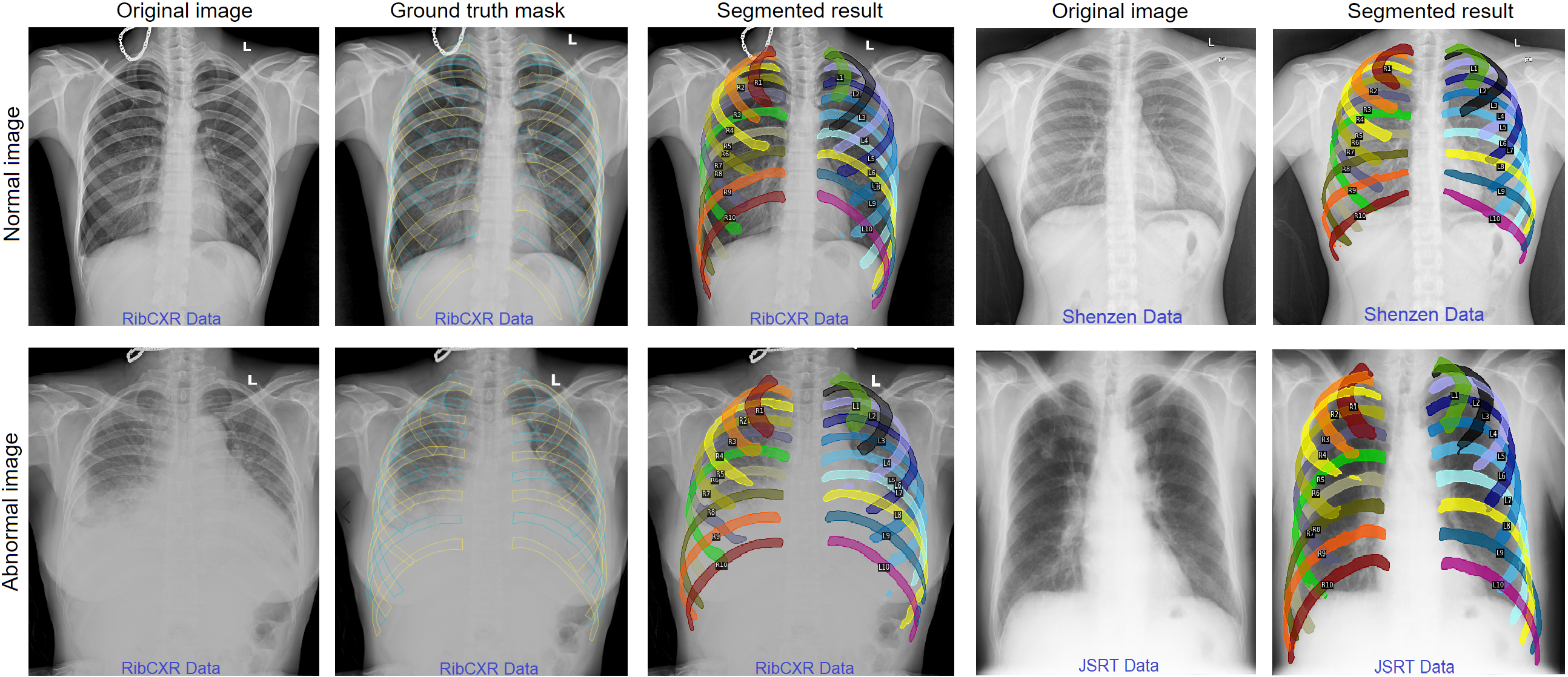
Tong, Dinh, et al.], for which all scans have been de-identified to protect patient privacy. We then designed an in-house web-based labeling tool called VinDr Lab (\urlhttps://vindr.ai/vindr-lab) that allows to segment individual ribs at pixel-level on DICOM scans. Each image was assigned to an expert, who manually segmented and annotated each of 20 ribs, denoted as L1L10 (left ribs) and R1R10 (right ribs). The masks of ribs (see Figure LABEL:fig:demo) were then stored in a JSON file that can later be used for training instance segmentation models. To the best of our knowledge, the VinDr-RibCXR is the first publicly released dataset that includes segmentation annotations of the individual ribs, and for both anterior and posterior ribs. To develop and evaluate segmentation algorithms, we divided the whole dataset into a training set of 196 images and a validation set of 49 images.
2.2 Model development
We deployed various state-of-the-art DL-based segmentation models for the task of individual rib segmentation and labeling, including U-Net [Ronneberger et al.(2015)Ronneberger, Fischer, and
Brox], U-Net with EfficientNet-B0 [Tan and Le(2019)], Feature Pyramid Network (FPN) [Lin et al.(2017)Lin, Dollár, Girshick, He, Hariharan, and
Belongie] with EfficientNet-B0, and U-Net++ with EfficientNet-B0. These models are well-known to be effective for many medical image segmentation tasks. To train the networks, we resized all training images to 512512 pixels. Dice loss and Adam optimizer were used to optimize the network’s parameters. All models were trained for 200 epochs with a learning rate of . Some augmentation techniques (i.e. horizontal flip, shift, scale, rotate, and random brightness contrast) were applied to avoid overfitting. The models were implemented using Pytorch (v1.7.1) and trained on a NVIDIA RTX 2080 Ti GPU.
3 Evaluation and Result
The effectiveness of the segmentation models was evaluated by Dice score, 95% Hausdorff distance (95% HD), sensitivity, and specificity. Experimental results reported by the proposed segmentation models are shown in Table 1. Our best performing model, which was Nested U-Net with EfficientNet-B0 encoder, achieved a Dice score of 0.834 (95% CI, 0.810–0.853), a 95% HD of 15.453 (95% CI, 13.340–17.450), a sensitivity of 0.841 (95% CI, 0.812–0.858), and a specificity 0.998 (95% CI, 0.997–0.998). Figure LABEL:fig:demo shows segmentation and labeling results of the Nested U-Net on some representative cases from the validation set of VinDr-RibCXR and external datasets including JSRT and Shenzen. We observe that the algorithm correctly segmented the individual ribs on both normal and abnormal CXR scans, for both the VinDr-RibCXR and two external datasets. Although the abnormal CXR contains ribs that are obscured by lung abnormalities, the proposed algorithm still produced a considerably accurate segmentation mask for all ribs.
| Model | Dice | 95% HD | Sensitivity | Specificity |
|---|---|---|---|---|
| U-Net | .765 (.737–.788) | 28.038 (22.449–34.604) | .773 (.738–.791) | .996 (.996–.997) |
| U-Net w. EfficientNet-B0 | .829 (.808–.847) | 16.807 (14.372–19.539) | .844 (.818–.858) | .998 (.997–.998) |
| FPN w. EfficientNet-B0 | .807 (.773–.824) | 15.049 (13.190–16.953) | .808 (.773–.828) | .997 (.997–.998) |
| U-Net++ w. EfficientNet-B0 | .834 (.810–.853) | 15.453 (13.340–17.450) | .841 (.812–.858) | .998 (.997–.998) |
4 Conclusion
In this work, we introduced a new benchmark dataset and baseline predictive models for automatically segmenting and labeling individual ribs from chest radiographs. Our experimental results have shown the effectiveness of the DL-based segmentation models for this task. This study serves as a proof of concept and baseline performance for future research. Future works include investigating more powerful DL models and extending the dataset to enhance the segmentation performance.
fig:demo
5 Acknowledgements
This research was supported by the Vingroup Big Data Institute. We are especially thankful to Tien D. Phan, Dung T. Le, and Chau T.B. Pham for their help during the data annotation process.
References
- [Candemir et al.(2016)Candemir, Jaeger, Antani, Bagci, Folio, Xu, and Thoma] Sema Candemir, Stefan Jaeger, Sameer Antani, Ulas Bagci, Les R Folio, Ziyue Xu, and George Thoma. Atlas-based rib-bone detection in chest X-rays. Computerized Medical Imaging and Graphics, 51:32–39, 2016.
- [Lin et al.(2017)Lin, Dollár, Girshick, He, Hariharan, and Belongie] Tsung-Yi Lin, Piotr Dollár, Ross Girshick, Kaiming He, Bharath Hariharan, and Serge Belongie. Feature pyramid networks for object detection. In IEEE Conference on Computer Vision and Pattern Recognition, pages 2117–2125, 2017.
- [Nguyen et al.(2020)Nguyen, Lam, Le, Pham, Tran, Nguyen, Le, Pham, Tong, Dinh, et al.] Ha Q Nguyen, Khanh Lam, Linh T Le, Hieu H Pham, Dat Q Tran, Dung B Nguyen, Dung D Le, Chi M Pham, Hang TT Tong, Diep H Dinh, et al. VinDr-CXR: An open dataset of chest X-rays with radiologist’s annotations. arXiv preprint arXiv:2012.15029, 2020.
- [Ronneberger et al.(2015)Ronneberger, Fischer, and Brox] Olaf Ronneberger, Philipp Fischer, and Thomas Brox. U-net: Convolutional networks for biomedical image segmentation. In International Conference on Medical Image Computing and Computer-assisted Intervention, pages 234–241. Springer, 2015.
- [Tan and Le(2019)] Mingxing Tan and Quoc Le. Efficientnet: Rethinking model scaling for convolutional neural networks. In International Conference on Machine Learning, pages 6105–6114, 2019.
- [Wessel et al.(2019)Wessel, Heinrich, von Berg, Franz, and Saalbach] Jöran Wessel, Mattias P Heinrich, Jens von Berg, Astrid Franz, and Axel Saalbach. Sequential rib labeling and segmentation in chest X-ray using Mask R-CNN. arXiv preprint arXiv:1908.08329, 2019.